V
*
بسم الله الرحمن الرحمن والصلاة والسلام على أشرف الخلق أجمعين
اللهم علمنا ما ينفعنا وانفعنا بما علمتنا وزدنا علماً
V
Surgical Management
>
Ethical considerations
>
Conflict of interest
*
COI exists when “professional judgement concerning a primary interest may be influenced by a secondary interest”
>
Types of COI
*
Financial: grants, personal fees, patents, employment…
>
Nonfinancial:
*
Intellectual: personal views; ideas; morals; strong opinions on theoretic approach
*
Professional: personal relationships; academic competition; peer recognition
*
A COI exists when an uninterested observer would interpret a situation as potentially sufficient to influence the judgement of the physician in question
*
The most important question is not whether a conflict of interest exists but whether it “presents an unacceptable risk of undue influence or bias”
>
Management strategies:
>
1. Disclosure
*
It cannot reduce or eliminate bias, but when complete and transparent, the scientific community has the opportunity to evaluate it
*
Disclosure of a large number of nonfinancial COIs may also distract from a more significant financial COI. For these reasons some authors have questioned the value of extensive disclosure of nonfinancial interests
>
2. Elimination
*
Unfortunately, nonfinancial interests cannot be divested from either the investigator or the research
*
Elimination is best performed a priori, preferably in the planning stage
>
3. Balance
*
It may be ideal for two groups with differing viewpoints to participate equally in trial design with a neutral party leading data collection and analysis
>
4. Exclusion
*
Individuals who might be influenced by a secondary interest should be excluded from evaluation and interpretation of results, although they may selectively participate in trial development
>
Surgical innovation
>
The Society of University Surgeons published a position statement on the responsible application of surgical innovation into clinical practice in 2008. This position statement provides guidance to the surgeon on what constitutes an innovation that should undergo a formal review.
>
If the innovation is planned, AND
*
the surgeon seeks to confirm a theory about the innovation, OR
*
the innovation differs significantly from currently accepted local practice, OR
*
the outcomes of the innovation have not been previously described, OR
*
the innovation entails potential risks for complication, OR
*
specific or additional patient consent appears appropriate, THEN
*
the innovation should be reviewed by a local surgical innovation committee (or IRB), submission to a national innovations registry should be done, and additional informed consent is required of the patient specific to the nature of the proposed innovation
>
End-of-Life & Surgical Care
V
Providing excellent & ethical end-of-life care requires:
>
Effective communication
*
As the most responsible practitioner, you should facilitate the meeting rather than run it
*
Prognostic information must be delivered clearly and compassionately with honesty avoiding false hope
*
Communication issues are magnified by cultural and language barriers.
*
While it may be convenient to use a family member, the translator may insert their own values and judgments or inaccurately describe a medical situation due to a lack of understanding or verbal facility
>
Prognostication
*
Use of a time-limited trial is appropriate and ethical when there is prognostic uncertainty or difficulty accepting outcomes. This may include a surgical procedure with time-limited postoperative ICU support, setting specific functional and physiologic parameters to be attained within a specified time frame
V
Sound decision-making
V
Principles of medical/surgical ethics
>
Respect for autonomy
*
The right of patients to determine their own goals of care and accept or decline treatments
>
Advanced directives enable a competent individual to determine and document their healthcare plan in advance of a future terminal illness or disability
*
Even when an advanced directive is completed, it may not provide guidance for a specific end-of-life situation such as when an acute potentially solvable problem is superimposed on a chronic terminal illness
*
When the care provider also functions as the surrogate decision-maker, it may be difficult to perform both roles due to exhaustion and burnout. A study of surrogate decision-makers in end-of-life care found that over 75% of them wished to eliminate or limit their role in medical decision-making
>
The concept of shared decision-making (SDM) ensures that the patient’s viewpoint is always considered
*
It is clear that SDM should not be limited to a discussion between a physician and patient/surrogate that is focused solely on choosing the best treatment option from a list of potential options
*
Another approach to SDM is the best case/worst case framework
>
Beneficience
*
Doing what is best for the patient includes actions that improve a situation and those that prevent or remove potential harms
>
What is typically recognized as routine and standard of care may not be appropriate or ethical at end of life
*
The surgeon must differentiate between ordinary “medicines, treatments and operations which offer a reasonable hope of benefit and which can be obtained and used without excessive expense, pain or other inconvenience” and extraordinary “that which cannot be obtained or used without excessive expense, pain or other inconvenience or which, if used, would not offer a reasonable hope of benefit”
*
Aggressive interventions to control symptoms may lead to an inadvertent decline in the patients’ overall well-being also called the double effect.
>
Nonmalificience
*
To do no harm has long been associated with therapeutic intervention and is at the core of surgical care: “… that it is only the second law of therapeutics to do good, its first law being this not to do harm” (Bartlett 1844)
V
Removal of life-sustaining interventions once they have been initiated or removal of care following a therapeutic trial is ethically justifiable if one of two conditions are met:
*
1. The patient/surrogate recognizes that the treatment is no longer consistent with previously identified goals and values.
*
2. The most responsible physician recognizes that the treatment is no longer medically indicated.
*
The term “no longer medically indicated” should be used for any treatment that will not or has ceased to accomplish the intended goal or deliver the benefit for which it was intended. The term “futility” has also been used in the past, but it has proven difficult to define as it is value based and requires contextual discussion
V
Justice
*
Justice is a form of fairness where each patient is entitled to the fair distribution of all available medical resources, based on equality and equity.
>
Equality means that everyone has equal access to all available treatments
*
Issues that factor into access:
*
Age
*
Social status
*
Ethnic background
*
Culture
*
Sexual preference
*
Disability
*
insurance coverage
V
Equity means that everyone gets the support they need when needed.
*
The goal is to ensure equal access to quality healthcare and good health, even if this requires giving some people more support and resources.
>
Informed consent
V
There are 7 elements in 3 categories for drafting an informed consent:
V
Threshold elements (preconditions to consent)
*
Competence (to understand and decide)
*
Voluntariness (in deciding) — and lack of coercion
V
Transformation elements
*
Disclosure (of material information) by the physician
*
Recommendation (of a plan) by the physician
*
Understanding by the patient (of the disclosure & plan)
V
Consent elements
*
Decision (in favour of a plan) by the patient
*
Authorization (of the chosen plan) — usually by a signing a document
>
Default surrogates in decision making (sequence):
*
1. Spouse
*
2. Adult child
*
3. Parent
*
4. Adult sibling
*
5. Nearest living relative
*
6. Close friend
>
Perioperative considerations & care
>
General Preoperative planning
*
Is the patient on Rx that need reversal?
*
Choice of incision or trocar placement
*
Is there a hernia, mesh or foreign body that require a change in the surgical approach?
>
Is there a need for:
*
Bowel preparation
*
Preoperative Abx
*
On-table endoscopy
*
Postoperative ICU monitoring
*
Has the anatomy been previously changed by surgery or disease?
*
Has the vasculature been previously compromised by surgery, radiation, or disease? What is the blood supply to the remaining bowel after transection of the desired part?
*
Is there any extra-organ tumor involvement?
>
Nutrition
>
2-Question validated nutrition screening tool
*
Assessment of phenotype: Have you lost weight in the past 6 months without trying to lose weight?
*
Assessment of etiology: Have you been eating less than usual for > 1 week?
*
Answering “Yes” to both questions indicate nutrition risk
>
Severe malnutrition is defined as
*
> 10% weight loss
*
Albumin < 35 g/L
*
Pre-albumin < 0.15 g/L
V
PeriOp Cardiac Assessment
*
Patients with less than 1% risk of perioperative death from cardiac disease do not require additional workup.
>
Emergency surgery?
>
>
Perioperative monitoring
>
Goldman’s criteria
>
Point system
*
S1+S2+S3 = 11
*
JVP = 11
*
Recent MI = 10
*
PAC or non-NSR = 7
*
>5 PVC = 7
*
Age > 70 = 5
*
Emergency surgery = 4
>
Cardiac complication rate
*
0-5 = 1%
*
6-12 = 7%
*
13-25 = 14%
*
≥26 = 78%
*
OR
*
Risk Management
>
V
>
Active cardiac condition?
Active Cardiac Conditions
• Acute Coronary Syndrome
• Significant arrhythmia
• 1. Heart block that is
• Mobitz
• 3rd Degree
• High-grade
• 2. Symptomatic ↓HR
• 3. SVT
• 4. Uncontrolled ventricular rate
• Decompensated CHF
• NYHA Class IV
• New CHF
• Worsening CHF
• Severe valvular disease
• Severe AS
• Symptomatic MS
>
>
Treat as per AHA & AAC
>
PCI
>
Stenting
>
Standard (recommended) dual antiplatelet therapy = 6-12 months
*
May be continued 6 more months if low risk for bleeding
>
Interrupting DAPT is associated with high risk of
*
Stent thrombosis
*
MI
*
Death
>
If surgery is needed in <12 months:
*
Try to continue DAPT perioperatively (depends on risk for bleeding)
*
At least, continue ASA perioperatively
*
For newer generation DES: ‘some experts will allow elective non-cardiac surgery as soon as 4-6 weeks after placement’
>
Minimum duration of uninterrupted therapy
*
1 Month for BMS
*
6 Months for DES
*
Interruption is associated with the highest risk of stent thrombosis
>
Balloon angioplasty
*
Continue DAPT for 14 days
>
MI
*
Re-infarction rate for non-cardiac surgery within 3 months = 5%
*
AHA: wait 4-6 weeks before elective non-cardiac surgery
>
B-blocker will
*
Adrenergic surge
*
Platelet activation thrombosis
>
Definitions
*
Acute MI = within 7 days
*
Recent MI = 7-10 days from evaluation
>
>
Low risk surgery?
>
*
OR
>
>
MET score?
*
≥4 + symptoms OR
>
Risk factor assessment
*
OR
>
1-2 RF
V
± B-blocker
*
Starting perioperatively without a strong indication increases the risk for stroke & death
>
Consider noninvasive testing
*
Exercise stress test
*
Echo
*
24h-Ambulatory monitoring
*
Cardiology consultation
V
≥3 RF
*
Consider vascular surgery
*
B-blocker
>
Duration of AntiPlatelet Therapy after PCI
V
*
Duration of APT after PCI for ACS
Screen Shot 2020-03-30 at 4.44.23 PM
V
*
Duration of APT after elective PCI
Screen Shot 2020-03-30 at 4.44.16 PM
>
Timing of surgery with Stents (CCS 2018)
*
Delay 14 days after balloon angioplasty (AHA/ACC 2014, no guidance in CCS 2018)
V
Nonemergency, non-urgent non-cardiac surgery:
*
Delay at least 6 month after BMS or DES
V
If semi-urgent (ex. malignancy, where feasible) that require cessation of one or both antiplatelet agents that cannot wait 6 months:
*
Delay at least 4-6w after DES or BMS
*
This is based in part on evidence suggesting that the increased risk of MI and cardiac death is highest within the first month after stent placement and no clear difference in risk between BMS and DES
*
Continue ASA wherever possible [CCS 2018 Antiplatelet Guideline]
V
Urgent/Emergent surgery
*
Do not delay surgery
*
Monitored bed, counsel patient, no neuraxial anesthesia on P2Y12 inhibitor
V
*
CCS 2016 medication management
Screen Shot 2020-03-30 at 3.58.05 PM
>
Automatic implantable cardioverter-defibrillators
*
Find out the make and model of the device, if possible
V
*
If not pacemaker dependent: magnet is used for surgery
Application of a magnet may be used to suspend anti-tachyarrhythmia therapy in an ICD or to produce asynchronous pacing in a PM. This alternative approach to reprogramming with a machine is recommended for selected patients by some agencies (eg, the Heart Rhythm Society [HRS] and Canadian Anesthesiologists' Society [CAS]/Canadian Cardiovascular Society [CCS]) in some settings
*
If pacemaker dependent: may need reprogramming for OR
*
Ensure external defibrillator and transcutaneous pacers are available
*
Utilize bipolar cautery device rather than monopolar, when available
>
Perioperative stroke after noncardiac, non carotid, non-neurologic surgery
V
Delay at least 6 months after ischemic stroke
*
67-fold increase in the risk of perioperative ischemic stroke in patients who had surgery within three months of an ischemic stroke, compared with patients without prior stroke
*
The incidence of perioperative stroke was 12 percent in patients who had surgery within three months of a stroke
*
Observational data suggest that the risk of recurrent stroke in the perioperative period continues to decline until nine months after an index ischemic stroke, at which point the risk remains elevated but stable at 2.5 to 3 times that of a patient without prior stroke
V
Patients at particular risk for perioperative stroke (below) warrant consideration for ongoing antiplatelet therapy perioperatively when the surgical procedure permits:
*
Recent ischemic stroke and/or transient ischemic attack
*
Existing intracranial stents
>
PeriOp PFT assessment
V
>
Increased risk is associated with:
Spirometry
*
FEV1 < 70% of predicted
*
FVC < 70% of predicted
*
FEV1/FVC < 65% — normally it’s 80%: 4L/5L
>
PeriOp anemia, bleeding the thrombosis
*
Pulmonary embolism, although not common, remains the most likely cause of potentially preventable death in surgical patients
>
Basics
>
Folate is needed for thymidine then DNA synthesis; B12 is needed to incorporate folate into RBCs
*
Folate absorption = proximal jejunum & ileum
*
B12 absorption = terminal ileum with the help of IF
*
MCV > 115 fL is seen exclusive with ↓B12 or ↓folate
*
Smokers & those exposed to CO have Hct higher than normal (false reading) → This may mask underlying anemia
>
For patients refusing blood transfusion or when there is risk of excessive bleeding, consider the following:
>
Prophylactic TXA prior to surgery & when bleeding occurs: 1g prior to procedure
*
Meta-analyses of RCTs indicate that TxA for prophylaxis of excessive bleeding is effective in reducing perioperative blood loss, the number of patients transfused, and the volume of blood products transfused
*
RCTs comparing TxA with placebo or no TxA controls report no differences for stroke, myocardial infarction, renal failure, reoperation for bleeding, or mortality
*
Consider TxA prophylaxis for certain orthopedic procedures (such as knee replacement surgery), in liver surgery and other clinical circumstances at high risk for excessive bleeding (Postpartum Hemorrhage etc)
*
Erythropoietin ± Fe at least 2-4w prior to surgery
>
If anemia is identified, treat as appropriate
V
*
In all patients, the cause of iron deficiency must be identified and addressed
An evaluation is not complete until a site of blood loss is found or until negative upper and lower endoscopy and capsule endoscopy results are found.
*
All patients with IDA and most with iron deficiency without anemia should be treated
*
The iron content of foods is unlikely to be sufficient to replete iron stores in an individual with iron deficiency
>
Lab findings
*
↓ Sr Fe — most of which is bound to the transport protein transferrin
*
↑ Sr transferrin (also reported as TIBC)
*
↓ Transferrin saturation (ratio of Sr. Fe to TIBC)
>
Older individuals
*
Have ↓ tolerance to PO Fe supplementation, especially with constipation
*
Have ↓ absorption of PO Fe, especially if taking antacids
*
PO Fe should be administered no more frequently than QD or QOD
>
IV Fe administration is advised for
*
Older individuals — The threshold for IV Fe administration should be low
*
IBD patients
*
Preoperatively (especially if estimated surgical blood loss is likely to be significant and that ↓Fe is demonstrated to be the cause of anemia)
V
Therapy
>
Oral replacement
>
It has been estimated that the maximum amount of elemental iron that can be absorbed with an oral iron preparation is 25 mg per day
*
FeSO4 contains ~20% elemental iron per mg of mineral salt (eg, each 325 mg tablet contains 65 mg elemental iron)
*
Advantages: generally effective, readily available, inexpensive, and safe
*
Disadvantage: ~ 70 percent report gastrointestinal side effects (especially with FeSO4)
>
PO route is preferred for:
*
Cost-effectiveness
*
Minimize invasiveness of therapy
*
Elimination of potential infusion reaction &/or anaphylaxis
*
Infants, children, adolescents
>
Considerations
*
Certain foods bind iron (phosphates, phytates, tannates, Ca-containing foods, and eggs)
V
Fe is best absorbed as ferrous (Fe++) salt in a mildly acidic medium (PPI may impair absorption)
*
Fe should be given 2h before or 4h after ingestion of antacids
>
Regimen
>
Oral iron should be administered no more frequently than once daily
*
There is not a reason to think higher doses improve absorption, and adverse effects are generally dose related
*
Excessive dosing is counterproductive, resulting in ↓absorption & ↑AE
*
Iron absorption is best when dosing was restricted to lower doses and less frequent administration (40 to 80 mg of iron no more than once a day)
V
*
Dosing once QOD is preferred as long as the schedule can be managed properly
Alternate-day dosing (taking the iron every other day rather than every day) appears to result in equivalent or better iron absorption than daily dosing, usually with fewer adverse effects
*
Most formulations are equally effective, as long as they are taken
*
Side effects are generally similar among different preparations
V
IV replacement
*
Allows near full-replacement doses in 1-2 infusions
>
Preferred for:
*
Patients unable to tolerate GI AE of PO Fe
*
Coexisting inflammatory state that interferes with Fe homeostasis
*
Older individuals
*
Patients with existing GI disorders which may be exacerbated by GI AE or PO Fe
*
Severe/ongoing blood loss (e.g telangiectasias, varices)
*
Bariatric surgery
*
Malabsorption (celiac disease, Whipple’s disease, bacterial overgrowth)
>
Pregnant women with:
*
GI symptoms
*
2nd trimester and Hgb < 10.5 g
*
3rd trimester (PO Fe unlikely to supply adequate iron to the developing fetus)
V
Regimen
*
All formulations are equally effective & safe
*
The dose is calculated based on body weight, current Hgb level, & amount of elemental Fe per mL of the iron product
*
There is no evidence that total doses > 1,000 mg of elemental Fe are clinically useful
>
UTD:
*
We often give a fixed dose of approximately 1000 mg, which is generally sufficient to treat anemia (typical RBC iron deficit between 500 and 1000 mg) and provide additional storage iron without causing iron overload
*
We do not give any premedications to patients without a history of asthma or more than one drug allergy
*
For patients with asthma or more than one drug allergy, who are at slightly increased risk of an allergic or infusion reaction, we routinely premedicate with 125 mg of methylprednisolone and an H2 blocker (eg, 10 mg of famotidine) given intravenously prior to administration of any IV iron product
*
We (and others) do not use H1 blockers (antihistamines) to prevent (or treat) infusion reactions
*
Erythropoietin ± Fe to reduce the need for allogeneic blood in selected populations (ESRD, anemia of chronic disease, refusal of transfusions) — requires a minimum of 2-4w to be effective
*
Administer Fe to patients with IDA if time permits — IV Fe requires a minimum of 3-4d for any effect
*
For in patients requiring iron sucrose (Venofer ®), the dose expressed is in mg of elemental iron and can be given as 300mg QD X 3d or 500mg QD X 2d
>
NOACs
>
Detecting NOAC effect
V
Apixaban (Eliquis ®), Rivaroxaban (Xeralto ®) and Endoxaban (Lixiana ®)
*
Calibrated anti-Xa > 30 ng/mL likely indicates significant anticoagulant effect, and vice versa
V
Dabigatran (Pradaxa ®)
*
Thrombin Time or Clotting Time can be used to indicated anticoagulant effect
V
*
Reversing NOAC effect may be done with PCC (Octaplex ®, Beriplex ®) or tranexamic acid (Cyclokapron ®)
Tranexamic acid: 1g IV bolus then 1g over 8h
Beriplex & Octaplex: 50 units/Kg, max 3000 units
*
Andexanet alfa - Recombinant factor Xa inhibitor antidote is approved by FDA for reversal of rivaroxaban & apixaban
*
Apixaban should be discontinued a minimum of 48 h prior to abdominal or anorectal surgery. There is a boxed warning regarding the use of neuraxial anesthesia and risk of spinal or epidural hematoma (which could result in temporary or permanent paralysis), as the optimal interval from drug discontinuation to intervention is not well-defined. Therefore we recommend not using this drug for perioperative anticoagulation if an epidural catheter or spinal anesthesia is planned
V
*
Considerations for bridging anticoagulation
DD845237-5377-432D-BBEE-D76C2278CDD8_1_105_c
>
Contraindications for ‘RBC salvage / Cell Saver’
*
Site-specific (e.g bone cement)
*
Sepsis
*
Malignancy
>
Modified Caprini risk assessment for VTE in general surgical patients
*
Screen Shot 2020-03-29 at 12.56.12 PM
*
For most patients in whom thromboprophylaxis is indicated and the risk of bleeding is low, experts agree that mechanical methods may commence just before surgery and that pharmacologic agents should ideally commence within 2 to 12 hours preoperatively
*
Very low thrombosis risk: Early ambulation
*
Low VTE risk: Mechanical methods
>
Moderate or high VTE risk
*
With low bleeding risk: Pharmacologic alone
*
With low bleeding risk: Combined prophylaxis — Combining pharmacologic and mechanical methods of prophylaxis are generally reserved for those considered to be at the highest risk of VTE
*
With high bleeding risk: Mechanical methods
*
A typical colorectal surgery patient will have at least 5-6 points (age + procedure + malignancy or IBD)
>
Fibrinolytics & thrombectomy
V
*
Contraindications to fibrinolytics
Screen Shot 2020-03-16 at 00.45.02
>
Embolectomy
>
Indications
*
Unstable PE patient in whom thrombolytic therapy is contraindicated
*
Failure of thrombolysis
>
Approach: depends upon available expertise, the presence or absence of a known diagnosis of PE, and the anticipated response to such therapies
*
Catheter-based
*
Surgical
*
Thrombolysis &/or catheter-based therapies may be considered on a case-by-case basis when the benefits are assessed by the clinician to outweigh the risk of hemorrhage. Examples: large clot burden, severe right ventricular enlargement/dysfunction, high oxygen requirement, &/or severe tachycardia
V
>
IR Embolization
Screen Shot 2019-01-28 at 22.39.12
V
*
Large vessel + temporary = gelfoam
Examples: vascular trauma
V
*
Small vessel + temporary = gelfoam powder, starch microsphere, Avitene ®
Example: tumor embolization
V
*
Large vessel + permanent = coils
Examples: traumatic pseudoaneurysm, aortic endoleaks, visceral aneurysms, GI bleeds
V
*
Small vessel + permanent + tissue death desired = absolute alcohol, small particles (< 300 um)
Examples: peripheral AVM, renal ablation, tumor embolization
V
*
Small vessel + permanent + tissue viability maintained = large particles (> 300 um)
Examples: GI bleed, uterine fibroids
>
Post-thrombotic syndrome may cause long-term disability
V
Etiology:
*
Impaired venous return
*
↓ calf muscle perfusion
*
Abnormal microvascular function
*
DVT can cause chronic venous hypertension by persistent venous obstruction and valvular reflux
V
Symptoms
*
Chronic pain limiting activity
*
Edema
*
Leg ulcers
>
PeriOp considerations in pregnancy
*
Document fetal HR preOp & postOp for all patients (fetal HR is detectable at GA 8w)
*
Continuous monitoring of fetal HR intraOp & in PACU is required for GA > 23-34w
*
Unfractionated heparin is safe in pregnancy
*
NSAIDs are not used in pregnancy because of the risk for premature closure of the ductus arteriosis
*
There is no role for prophylactic tocolytics
*
Elective surgical procedures are delayed until at least 6 weeks after delivery, when maternal physiology has returned to the nonpregnant state
>
PeriOp hypothalamic-pituitary-adrenal HPA axis assessment
*
ICUPocketGuide.pdf-page-53-of-63
>
Morning cortisol level
*
>10 mcg/dL = normal functioning HPA axis
*
5-10 mcg/dL = unclear adequacy of HPA axis
*
<5 mcg/dL = suppressed HPA axis
V
ACTH stimulation testing:
*
250mcg of ACTH is given
*
Serum cortisol is measured after 30 minutes
*
Cortisol > 180 mcg/dL = adequately functioning HPA axis
>
Frailty assessment
*
Frailty is the accumulation of deficits that result in an inability to tolerate stress = ↑ vulnerability
>
Phenotypic frailty (≥ 3 of the following traits):
*
Slow walking speed
*
Impaired grip strength
*
Self-report of declining activity levels
*
Unintended weight loss
*
Exhaustion
>
Frequently used indices
*
abbreviated Comprehensive Geriatric Assessment (aCGA)
*
Vulnerable Elders Survey-13 (VES-13)
*
Groningen Frailty Indicator (GFI)
*
Geriatric 8 (G8)
>
Accuracy of indices
*
Sensitivity: 67-87%
*
Specificity: 59-73%
*
G8 may be the most valuable for patients undergoing elective abdominal surgery
*
Carli et al JAMA Surgery 2020: In frail patients undergoing colorectal cancer resection (predominantly minimally invasive) within an enhanced recovery pathway, a multimodal prehabilitation program did not affect postoperative outcomes. Alternative strategies should be considered to optimize treatment of frail patients preoperatively.
>
Evidence driven controlling SSI
*
The development/application of care-bundles has been consistently shown to reduce the rate of SSI; the main issue remains in compliance to the bundle recommendations
V
What works
*
Supplemental oxygen: Level I: evidence that FiO2 80% vs 30% reduces SSI in CRS
>
Normothermia / Active rewarming: Level I: Active rewarming decreases SSI (but the ambient room temperature doesn’t affect the patient’s core temperature)
*
Warming should be started in the preOp holding bay; a duration of 30m is required
V
V
Triclosan coated sutures for fascia & subcuticular closure: Level IA
Untitled
*
Triclosan properties: It is an antiseptic, not antibiotic (less concern for bacterial resistance)
V
V
Recent studies show that this actually effective and may reduce the rate of SSI by 30%
Screen Shot 2022-05-15 at 17.51.41
*
2017 Meta-analysis including 21 RCT and 6,458 patients: SSIs were reduced significantly by the use of TCS vs a comparable non-coated suture (RR 0.72, 95%CI 0.60-0.86; p < 0.001) — moderate quality of evidence
*
The risk reduction is applicable to wound classifications I-III
*
Multiple guidelines now endorse triclosan coated sutures for fascia closure
V
Hibiclens shower (PreOp shower with 4% chlorhexidine): Level IA
*
When used, 4oz should be applied and left for 1m prior to washing it off. This is applied on the night prior to surgery and the day of surgery
*
4-week smoking cessation
*
Normoglycemia
*
Pfannenstiel has lower SSI than vertical midline
*
PreOp Abx: Level IA
*
PO Abx bowel preparation
*
Wound edge protector (effective for open surgery)
*
Purse-string stoma operature closure
*
Glove change prior to fascia / subcuticular closure
*
Separate wound closure set
>
What maybe doesn’t work so well
*
Masks: Surgeons wearing OR masks (Supported by Cochrane review)
*
Scrubs: No convincing evidence that wearing hospital scrubs (and having them laundered at the hospital) affects SSI
*
Hand-washing: no difference between traditional scrubs and aqueous based solutions
*
Rescrubbing for closure actually increase SSI
*
Sterile gloves Vs clean gloves
*
Mechanical bowel preparation: Level I evidence that it does not decrease SSI for CRS
*
Adhesive surgical barriers (Ioban)
*
Abx irrigation: doesn’t affect SSI, actually increases formation of adhesions
*
Intra-abdominal Silk Vs Vicryl: retrospective study showed Vicryl had lower SSI (13.9% vs 22.4% p-0.034)
*
Staples Vs subcuticular closure
*
Closure of subcutaneous tissue
>
Areas of controversy
*
Incisional wound VACs
*
Skin preparations (most studies look at colony forming units rather than investigating SSI). As long as alcohol is included in the preparation, risk reduction of SSI is adequate
*
Identity of the surgeon / resident participation
>
Local anesthesia systemic toxicity
V
Signs of toxicity
*
Dizziness
*
Headache
*
Decreased level of consciousness
*
Convlusions
*
Tinnitis
*
Circumoral numbness
*
Bradycardia
*
Respiratory depression
*
Muscle tension
*
Hypotension
>
LA maximal doses
>
Lidocaine
*
4.5 mg/kg/dose; max 300mg; do not repeat within 2h
*
For 70Kg patient: ~30ml of 1% solution; ~15% of 2% solution
V
Bupivacaine (Marcaine ®)
*
Dose varies with procedure, depth of anesthesia, vascularity of tissues, duration of anesthesia, and condition of patient.
V
Maximum 175mg administered as:
*
70ml of 0.25% solution
*
35ml of 0.5% solution
>
TAP block constituents:
*
40ml of 0.25% solution = 100mg of Marcaine
*
10mg dexamethasone (equivalent to 67mg of prednisone)
*
± Precedex 10mcg
V
Management
*
Stop injection
*
Call for help
*
Arrange for cardiopulmonary bypass / alert nearest facility
*
Manage the airway
*
Suppress seizure (benzodiazepines preferred)
>
Manage arrhythmias & provide CVS support
*
Amiodarone is the first line anti-arrhythmic
*
Avoid lidocaine, CCB, β-blockers, and vasopressin
>
Administer lipid rescue
*
≤70 kg: 1.5 mL/kg IV, followed by infusion at 0.25 mL/kg/minute IV
*
>70 kg: 100 mL IV, followed by infusion of 200 to 250 mL IV over 15 to 20 minutes
*
Transfer to monitored setting
>
Complications of neuraxial analgesia
>
Post dural puncture headache
*
PDPH is a positional headache (ie, worse when the patient sits or stands) that occurs because of leakage of CSF through a dural puncture
*
Young pregnant women with a low BMI are at highest risk
*
Most PDPHs will resolve in 7-10 days if untreated
*
Conservative management with symptomatic therapy (eg, oral analgesics, caffeine) may be indicated if the patient does not desire epidural blood patch or if the headache is not severe
*
Epidural blood patch is the classic treatment for severe, debilitating PDPH
>
Spinal epidural hematoma (SEH)
*
SEH is less likely with a single-shot spinal technique due to the relatively small size of the spinal needle and the lack of an indwelling catheter
*
The most common presenting symptoms of neurologically significant SEH are progressive motor and sensory block, and bowel or bladder dysfunction
*
Emergency MRI is recommended as soon as SEH is suspected
*
If SEH is detected, any residual anticoagulant should be reversed rapidly if appropriate (ie, if anticoagulant effect is present)
*
Emergency neurosurgical consultation should be arranged to evaluate for decompressive surgery. Neurologic recovery is more likely if decompressive laminectomy is performed within 8h of symptom onset
>
Hyponatremia
V
>
Workup & etiology
Screen Shot 2020-03-17 at 11.19.30 AM
*
Euvolemia + ↑Ur.Osm = malnutrition vs self-induced water intoxication
*
Euvolemia + ↓Ur.Osm + ↑Ur.Na = adrenal insufficiency vs SIADH vs ↓T4
*
Hypovolemia + ↓Ur.Na = pre-renal hypovolemic hyponatremia
*
Hypovolemia + Ur.Na 25-40 mEq/L = administer 1L of NS then repeat Ur.Na
*
Hypovolemia + ↑Ur.Na = renal hypovolemic hyponatremia (adrenal insufficiency vs diuretics vs cerebral salt wasting)
>
Management
V
*
Symptomatic (even mild) ↓Na warrants administration of 100ml of 3% HTS regardless of etiology
Ovwtrmtadltshyponatremia
V
*
Cerebral salt wasting: start with 3% HTS (isotonic saline may be used also as initial therapy if there is no intracranial pathology)
UTD:
Fluid restriction, the usual first-line therapy for SIADH, is not advised in hyponatremic patients with subarachnoid hemorrhage. In such patients, fluid restriction may increase the risk of cerebral infarction among patients who actually have CSW because ongoing salt losses may worsen the volume depletion and lower the blood pressure.
Instead, we treat with 3% HTS to raise the serum sodium. All patients with active intracranial pathology (eg, recent intracranial surgery or subarachnoid hemorrhage) should have a prompt increase in the serum sodium concentration and should avoid a decrease in extracellular fluid volume.
>
Delirium
>
General
*
CAM test is ~100% sensitive & 95% specific
>
Pathophysiology
>
Withdrawal related
>
EtOH
*
EtOH of NMDA & GABA-A
*
BDZ
*
Nicotine
>
3 Main theories
*
Direct effect of substance on NT synthesis & release
*
Inflammation cytokines impaired synthesis & release of NT
*
Stress Noradrenaline hypothalamic-pituitary-adrenal access neuron damage
>
Manifests as
*
Hyperactive
*
Hypoactive
*
Patients tend to fluctuate between the two with lucid intervals
>
Etiology
*
Withdrawal
*
Age-dependent delirium
>
Precipitating factors
*
Acute illness or infection
*
Operation/trauma/fractures
*
Catheters
*
Dehydration
*
Electrolyte disturbance
*
Hypoxia
*
Sleep deprivation
*
Pain & immobility
*
Change in hospital room/staff
>
Rx
*
Polypharmacy
*
Analgesics
*
Abx
*
Antiarrhythmic
*
Anticholinergic
*
Patients with cancer
>
Diagnostic workup
*
ECG
*
Echo
>
Labs
*
CBC
*
RFT
*
LFT
*
CRP
*
Calcium
*
TSH
*
UA
*
Glucose
*
EEG
*
CXR
*
CT
>
Acute Kidney Injury
>
Diagnostic considerations
>
PreRenal
*
Usually responds to resuscitation, but response can be lost when low-flow state is severe (hypovolemic shock)
>
Renal
>
ATN
*
Responsible for >50% of AKI
*
Tubuloglomerular feedback = damaged cell slough into lumen of renal tubules obstruction GFR
*
Result in impaired Na reabsorption → Ur.Na
>
Etiology
*
Severe sepsis/Septic shock
>
Radiocontrast dye
*
Most resolve within 2 weeks
>
Prevention:
*
NS/RL 100-150ml/h 3-12h before procedure; continue 6-24h after the procedure
>
NAC
*
Popular because of low cost & safety
*
Its benefits is debatable
*
Nephrotoxic drugs
>
Rhabdomyolysis
*
Ur.Myoglobin does not ensure a Dx of AKI
*
Ur.Myoglobin excludes the Dx of myoglobinuric renal injury
>
Mx
>
Aggressive volume resuscitation
*
Maintain UO 100 cc/h
>
Alkalization of urine often not necessary
*
If done, maintain Ur.pH 6-7
*
Inhibits cast formation
*
30% of patients will require dialysis
>
AIN
*
Inflammatory injury to renal interstitium, rather than tubules
>
May not be accompanied by oliguria
*
May hinder the Dx of AKI
>
Most cases are 2ry to hypersensitivity Rx reaction
>
Look for
*
Fever
*
Rash
*
Eosinophilia
*
Antibiotics are the most common offenders, particularly penicillins
*
Recovery can take months
V
PostRenal
>
May involve
*
Distal renal collecting ducts (papillary necrosis)
>
Ureters (retroperitoneal mass)
*
Requires bilateral obstruction to cause AKI
*
Urethra (strictures)
V
V
Abdominal compartment syndrome
Pasted_Graphic_3
*
They kidneys are most frequently affected
*
IAH is prevalent in 60% of ICU patients
*
Maintaining APP > 60 mmHg survival
>
Approach
*
1. Bedside US to R/O postrenal obstruction
V
>
2. Measurements
Pasted_Graphic
>
Prerenal disorder may have Ur.Na >40 mEq/L if patient is
*
On diuretics
*
CKD
>
FENa is more accurate than spot Ur.Na for renal tubular function
>
May be falsely low (<1%) in
*
Sepsis
*
Radiocontrast dyes
*
Hemoglobinuria/myoglobinuria
*
FEU is conceptually similar to FENa, but is not affected by diuretics
*
Fluid challenge may help distinguish between pre- and intrarenal causes when there is uncertainty, i.e when Ur.Na is 25-40 mEq/L, consider 1L of NS and re-measure Ur.Na
V
Management
*
Fluid challenge over 30 mins
*
Never use diuretics until the prerenal disorders are ruled out
V
Abdominal Compartment
*
Sedation
*
Avoid elevation of head >20 degrees
*
Avoid positive fluid balance
>
Decompress
*
Stomach / small bowel: via NGT
*
Colon: ± via rectal tube
*
Percutaneous drainage of peritoneal fluid
*
Maintain APP > 60 mmHg
*
If fails, consider surgery
>
Renal Replacement Therapy
>
Indications for acute dialysis:
>
AEIOU
>
Acidosis, metabolic
*
pH < 7.1
>
Electrolytes
*
Refractory K > 6.5
*
Rapidly rising K
>
Intoxications
>
SLIME
*
Salicylates
*
Li
*
Isopropanol
*
Methanol
*
Ethylene glycol
>
Overload
*
Refractory to diuresis
>
Uremia with
*
Pericarditis
*
Neuropathy
*
Uremic bleeding
*
Encephalopathy (unexplained mental status)
V
Hemofiltration
V
Removes solutes by convection
*
Hydrostatic pressure gradient moves solute-containing fluid across a semipermeable membrane = solvent drag
*
Rate of solute clearance is slower than hemodialysis requires continuous performance for effective solute clearance
*
Water & solutes are cleared concentration of solutes (e.g urea) does not decrease unless a solute-free IV fluid is infused to replace the ultrafiltrate that is lost (often required)
V
Methods
>
CAVH: Continuous ArterioVenous Hemofiltrate
*
Not suited for patients with labile blood pressures
*
CVVH
>
Advantage
*
Less likely to produce hemodynamic compromise
V
Removes larger molecules than hemodialysis
V
Preferred in removing
*
Toxins
*
Inflammatory cytokines
>
Disadvantage
V
Slow solute removal
*
Not well suited for life-threatening K or acidosis
>
Hemodialysis
*
Removes solutes by diffusion across a semipermeable membrane = countercurrent exchange
*
Rate 200-300 mL/m
>
Advantage
*
Rapid clearance of small solutes
>
Disadvantage
*
Limited removal of large particles (inflammatory cytokines)
>
Difficulty maintaining BP
*
Hypotension occurs in ⅓ of treatments
V
CAVHD/CAVD/CVVHD/CVVD
V
Similar to CAVH/CVVH but
*
1. Dialysate is run at a low flow rate, countercurrent to the direction of blood flow
*
2. Ultrafiltration rate is not maximized (to minimize BP)
*
Taxonomy: 'D’ & ‘HD’ = hemodialysis
>
Hemodiafiltration
>
Combines dialysis & hemofiltration
*
Suitable for patients requiring rapid solute removal (small & large) + volume removal
V
CAVHDF/CVVHDF
V
Continuous AV/VV hemodiafiltration
*
Similar to CAVHD/CVVHD except ultrafiltration is allowed a rate beyond that necessary to reestablish euvolemia
>
SCUF
V
Slow Continuous Ultrafiltration
*
Is strictly a dehydrating procedure
*
Used when fluid removal goal is modest
*
Access may be AA or AV
>
Radiology
V
>
Management of pulmonary nodules: 2017 Fleischner Society Guidelines
17-PUL-366-Mehta-Table-Inset-805pxl-width
*
CT Chest: 9% of patients will have ‘indeterminate pulmonary nodules’ on preOp CRC staging, but only 11% of these lesions (~1% of the total population) declare themselves to be CRC metastases during surveillance. Given the low incidence of malignancy in these indeterminate pulmonary nodules, further preoperative evaluation can be avoided, and these lesions can be followed during surveillance
>
A list of normal radiological reference values is as follows
*
Adrenal gland: < 1 cm thick, 4-6 cm length
*
Aorta: < 3 cm diameter
*
Azygous Vein: on erect chest x-ray < 10 mm diameter
>
Small Bowel:
*
lumen: < 3 cm
*
wall: < 3 mm
>
Colon:
*
lumen: < 5 cm
*
wall: < 3 mm
*
Appendix: on CT < 6 mm calibre 
*
Rectum: wall thickness < 5mm
*
gallbladder wall: < 3 mm (well distended)
>
common bile duct:
*
< 7 mm and add 1mm for each decade over age of 60 
*
up to 10mm post cholecystectomy
*
esophagus wall: < 3 mm (with distended lumen)
*
inferior vena cava: < 28 mm
>
lymph nodes:
*
mediastinal: < 10mm in short axis
*
retro-crural: < 6mm in diameter
*
liver span: < 15 cm
*
portal vein: < 13 mm diameter
*
spleen: < 12 cm
*
splenic vein: < 10 mm diameter
*
kidneys : 8-10 cm x 4-6 cm
*
bladder wall: < 3 mm (well distended state)
*
ureter: 30-34 cm long, 2-8 mm diameter
*
AHA recommends Abx prophylaxis for procedures within 6m of synthetic vascular graft placement to permit time for endothelialization of the graft
*
Frank abdominal sepsis in context of a VP shunt requires exteriorization of the shunt and 2w of IV antibiotics
V
>
VAC Dressing
Information mostly from Acelity product manual
>
Selected general pointers
*
V.A.C. ® Foam Dressings are radiolucent, not detectable on X-Ray.
*
Never leave a V.A.C. ® Dressing in place without active V.A.C. ® Therapy for more than two hours. If therapy is off for more than two hours, remove the old dressing and irrigate the wound
*
The V.A.C. ®Therapy Unit is MR unsafe.
*
Do not allow foam to overlap onto intact skin. Protect fragile / friable periwound skin with additional V.A.C. ®Drape, hydrocolloid or other transparent film
*
To avoid trauma to the periwound skin, do not pull or stretch the drape over the foam dressing during drape application.
*
Use as few layers of drape as possible. Multiple layers of the V.A.C. ®Drape may decrease the moisture vapor transmission rate, which may increase the risk of maceration, especially in small wounds, lower extremities or load-bearing areas
*
Document the foam quantity and dressing change date on the drape or Foam Quantity Label if available, and in the patient’s chart.
V
>
WhiteFoam Dressing
Screen Shot 2021-10-19 at 18.55.52
*
Packaged pre-moistened with sterile water
*
Has higher tensile strength than GranuFoam Dressing
*
For use in tunnels and undermining
*
Helps reduce the likelihood of adherence to the wound
*
Higher density of WhiteFoam Dressing requires a minimum pressure setting of 125 mmHg
*
For optimal pressure distribution, it is recommended to use a V.A.C. ® GranuFoam™ Dressing over V.A.C. ® WhiteFoam. Do not place foam dressings of the V.A.C. ® Therapy System directly in contact with exposed blood vessels, anastomotic sites, organs or nerves
>
Pressure settings
>
Indications to titrate pressure ↑ by 25 mmHg increments
*
Excessive drainage
*
Large wound volume
*
WhiteFoam Dressing use in the wound or in tunnelled areas (generally requires ≥ 125mmHg)
*
A tenuous seal
>
Indications to titrate pressure ↓ by 25 mmHg increments
*
Extremes of age
*
Compromised nutrition
*
Risk of excessive bleeding
*
Circulatory compromise
*
Excessive granulation tissue growth
*
Pain/discomfort
*
Periwound or wound bed ecchymosis
>
Continuous Vs intermittent (or dynamic pressure control) therapy
*
Continuous, rather than intermittent / DPC, V.A.C. ® Therapy is recommended over unstable structures, such as an unstable chest wall or non-intact fascia, in order to help minimize movement and stabilize the wound bed. Continuous therapy is also generally recommended for patients at increased risk of bleeding, highly exudating wounds, fresh flaps and grafts and wounds with acute enteric fistulae.
*
Continuous therapy is recommended for the first 48h in all wounds
V
>
Continuous therapy after the first 48h is recommended for:
Screen Shot 2021-10-19 at 18.48.26
*
Patients at increased risk of bleeding
*
Significant discomfort during intermittent/DPC therapy
*
Difficulty maintaining airtight seal
*
Presence of tunneled or undermined areas
*
High levels of drainage from the wound after the first 48h
*
Grafts/flaps with the need to prevent shear
*
Splinting effect is required (e.g sternal or abdominal wounds)
>
Contraindications to negative pressure wound therapy (NPWT) (VAC dressing)
V
*
Exposed vital structures
NPWT, in the presence of exposed organs, blood vessels, or vascular grafts, increases the risk for tissue erosion, which can lead to enteric fistula or hemorrhage
*
Ongoing infection
V
*
Devitalized tissue
Inadequate debridement with the presence of devitalized soft tissue or bone increases the risk for infection
V
*
Malignant tissue
As with normal tissues, growth of malignant tissue is promoted in the presence of subatmospheric pressure. Malignant tissue is also more friable and prone to bleeding
*
Fragile skin
*
Adhesive allergy
V
*
Ischemic wounds
Although not absolutely contraindicated, no benefit has been demonstrated with the use of NPWT in patients with ischemic wounds
*
Untreated osteomyelitis
*
Non-enteric and unexplored fistulas
>
Changes in wound color
V
If the wound assessment reveals dark discolouration
*
Rule out mechanical trauma. Relieve wound of excessive pressure, excess foam in the wound or a pulled or stretched drape over the foam. Remember to roll the drape over the foam; do not stretch it over foam.
*
pressure by 25 mmHg increments.
*
Thin the depth of the foam before applying the dressing to prevent overpacking or consider use of V.A.C. ® GranuFoam™ Thin Dressing.
V
If the wound appears white, excessively moist, or macerated
*
Check the therapy hour meter to ensure that the actual number of therapy hours received matches the number of recommended therapy hours. Find out why there is a therapy deficit and remedy the situation.
*
↑ pressure settings by 25 mmHg increments if drainage increases.
V
>
Enteric fistula VAC therapy
Screen Shot 2021-10-19 at 19.00.31
*
The goal of therapy depends on whether the fistula being treated is considered acute or chronic.
*
For acute fistula, the goal is to promote wound healing to enable closure of the acute enteric fistula.
*
For chronic fistula, the enterocutaneous fistula is segregated from the surrounding or adjacent abdominal wound and V.A.C. ® Therapy is applied to the wound. The effluent from the fistula is diverted into another containment system.
V
*
Inotropic activity
Screen Shot 2020-08-23 at 3.36.33 PM
V
*
Anion Gap Metabolic Acidosis (AGMA)
AG = Na - Cl - H3CO
Screen Shot 2022-06-21 at 14.26.38
>
The “Incidentals”
>
Visceral Artery Aneurysms & pseudoaneurysms
V
>
Indications for treatment
For elective repair of VAA/VAPA, a percutaneous approach is becoming the first-line treatment for VAAs/VAPAs that are anatomically suitable
Operative control: Ligation of the artery proximal and distal to the aneurysm is often adequate. Perfusion to the end organ should be assessed to determine whether or not revascularization is needed
*
Pseudoaneurysms (represent contained rupture that is only constrained by a fibrous capsule since all three layers of the arterial wall are disrupted)
*
Symptomatic VAA
*
Rapid expansion of VAA ( > 0.5 cm/year)
V
Asymptomatic VAA in
*
VAA > 2 cm in size
*
Patient undergoing liver transplantation
*
♀ who is pregnant
*
♀ of childbearing age
>
OB/GYN
*
Asymptomatic small bowel or appendiceal endometriosis: no intervention is required. The natural history of asymptomatic endometriosis is benign
*
If confronted with a tube-ovarian abscess while operating for another reason, the best management is to not intervene and to provide postOp Abx. If the abscess is > 8 cm, percutaneous drainage is pursued
V
*
If ovarian cyst with torsion is identified on laparoscopy, management is with detortion and excision of the cyst
Paratubal cysts are often connected with the mesosalpinx with a stalk, around which torsion may occur The incidence of torsion of paratubal cysts is uncertain, but should be suspected in a patient with acute or intermittent pelvic pain who has a paratubal cyst identified on pelvic ultrasound. The diagnosis of torsion can be made only with surgical evaluation. These cysts can easily be removed at time of surgery without compromise of the ovary or fallopian tube.
Paraovarian cysts are often incorporated in the broad ligament close to the fallopian tube and may increase the risk of tubal torsion. In such cases, the fallopian tube is often distended, and if torsion occurs, the cyst must be removed with great care to avoid compromise of tubal function
>
Incidental AAA at the time of laparotomy for CRC
V
Risk of AAA rupture
*
< 5cm: 4% annually
*
> 7cm: 19% annually
V
Considerations
*
Treatment of CRC (laparotomy) may result in life-threatening rupture of AAA. “The risk of ruptured AAA after laparotomy is real” — Dr. P. Gordon
The risk of post-laparotomy rupture of the aneurysm has been reported to be as high as 33.8%
*
Treatment of AAA may significantly delay treatment of CRC
V
Simultaneous resection risks graft infection
*
1. AAA repair is performed first
*
2. The peritoneum is closed
*
3. Interposition of omentum
*
4. The CRC is resected
V
Management
*
If neither colonic symptoms nor aneurysm size (< 6 cm) establish priority, the relative indolent course of colon carcinoma favors aneurysmectomy with colectomy in 2 to 4 weeks
>
Skin
>
Malignancy
>
General
*
1/5 Americans develop skin cancer in a lifetime
>
Risk factors
*
Cumulative exposure to UV-B (ranges from 290 to 320 nm) light — effect is more pronounced when exposed during childhood
*
Fair skin phototype (Fitzpatrick type I & II)
*
Tanning bed visits
*
Smoking
*
Blistering sunburns
*
Immunosuppression (especially transplant patients). After 10Y of immunosuppression, malignancies develop in 10% of patients and this figure increases to 40% after 20Y
*
HPV is proposed as a causative factor for the development of SCC
*
Exposure to: arsenic, organic hydrocarbon, & ionizing radiation
*
Genetic disorders: albinism, xeroderma pigmentosum
>
Melanoma
>
General
*
Melanocytes in the skin are positioned along the basement membrane at the dermal-epidermal junction
*
Because of the relatively young median age (50Y) of melanoma patients, melanoma ranks among the worst cancers in terms of years of life lost per malignancy
>
Melanomas may develop de novo but can develop within precursor lesions (dysplastic nevi & congenital nevi)
*
Dysplastic nevi that show moderate-severe dysplasia should be excised with negative margins (no need for wide local excision)
*
Giant congenital nevi (>20 cm in diameter) are rare (1 in 20,000 newborns), but carry an increased lifetime risk for the development of melanoma of up to 10%
V
*
Spitz nevus is a lesion often seen in children that is difficult to differentiate from melanoma. Because of diagnostic uncertainty, resect with ‘melanoma margins’
Skin lesions with clinical features of Spitz tumors should be removed by simple excision if there is concern for an atypical melanocytic lesion or melanoma. We suggest margins of approximately 3 to 5 mm
>
Familial melanoma
*
Also known as: dysplastic nevus syndrome, familial atypical multiple mole-melanoma syndrome, & B-K mole syndrome
*
They are associated with mutations in the gene CDKN2A in the 9p21 region
*
These patients require detailed dermatologic evaluation several times annually, with periodic biopsies of the most suspicious lesions
*
The most common mutation in melanoma is BRAF mutation that occur in superficial spreading melanoma
V
*
Seborrheic keratoses (barnacles of life) may be confused with melanoma
sn7_seborrheickeratosis3thu_jpg
>
Forms of melanoma
>
Superficial spreading melanoma (most common)
*
Flat pigmented lesion
*
It is not necessarily associated with sun-exposed skin
>
Nodular melanoma (2nd most common)
*
Have a poor prognosis because of greater average tumor thickness and frequent ulceration
*
Tend not to have changes associated with the classic ABCD description
>
Lentigo maligna melanoma (a type of in situ melanoma)
*
Occurs most commonly on the face of older individuals with sun-damaged skin and presents as a flat, dark, variably pigmented lesion, with irregular borders and a history of slow development
*
The prognosis of lentigo maligna melanomas is better than for the other histopathologic types because of the often superficial nature of these tumors
>
Acral lentiginous melanoma
*
Develop in the subungual areas, beneath the fingernails and toenails, and on the palms of the hand and soles of the feet
*
This is the most common type of melanoma in black patients
*
Biopsy of subungual melanomas can be accomplished by performing a digital block with local anesthesia and removing the nail or performing a punch biopsy through the nail itself
>
Work up and staging
*
Amelanotic melanomas are not pigmented and may present as a raised pink or flesh-colored skin lesion
>
Indications for Bx are: ABCDE
*
Asymmetric
*
Irregular Borders
*
Variable shades of Color
*
Diameter > 6mm
*
Evolution or change over time
*
Hx & clinical exam should be elicited for: change in skin lesions, including itching and bleeding
>
Biopsy technique
>
Features to examine
*
Maximum tumor thickness
*
Presence of ulceration
*
Level of invasion
>
Incisional punch Bx
V
Appropriate if:
*
> 2 cm
*
Near vital/important structures
V
Punch Bx technique
*
1. Pick the smallest appropriate size caliber that will obtain tissue: punch biopsies of at least 4 mm should be performed, because a 2-mm punch often does not provide adequate tissue for pathologic evaluation
*
2. Apply pressure with a circular motion
*
3. Optionally, place a suture to close for hemostasis
*
Target the area that is most raised & discolored
*
The deep margin should be the subcutaneous fat
>
Excisional Bx is best for small pigmented lesions (< 1-2 cm)
*
Using local anesthesia, a narrow margin excision is performed, which includes subcutaneous fat to get a full-thickness biopsy, and the defect is closed with sutures
>
Shavings
*
They are usually performed by dermatologists and are only appropriate for small, flat lesions with low suspicion for melanoma (nonpigmented)
*
Disadvantage: unable to demonstrate thickness of invasion
>
Spread
*
Satellite lesions: defined as within 2 cm of primary tumor (clinical or microscopic)
*
In Transit lesion: includes skin/subcutaneous metastases that are > 2 cm from the primary lesion but not beyond the regional nodal basin
*
The most common sites for initial distant metastasis: brain, lung, liver, and less commonly skin, distant LN, bone, & GI tract
V
>
Staging
With AJCC 8th edition: the thickness of the melanoma is rounded up to the first decimal figure not the second i.e 0.1 instead of 0.01. So a 0.75mm melanoma is considered a 0.8mm while a 0.74mm is considered 0.7mm
V
*
T
T1: ≤ 1.0 mm
T2: > 1-2 mm
T3: > 2-4 mm
T4: > 4 mm
“b" indicates presence of ulceration or thickness of 0.8-1.0mm in T1 disease
V
*
N
N1: 1 tumor-involved node or in-transit, satellite, and/or microsatellite metastases with no tumor-involved nodes
N2: 2-3 tumor-involved nodes or in-transit, satellite, and/or microsatellite metastases with 1 tumor-involved node
N3: ≥4 tumor-involved nodes or in-transit, satellite, and/or microsatellite metastases with ≥2 tumor-involved nodes, or any number of matted nodes without or with in-transit, satellite, and/or microsatellite metastases
V
*
M
M1a: Distant metastasis to skin, soft tissue including muscle, and/or nonregional lymph node
M1b: Distant metastasis to lung with or without M1a sites of disease
M1c: Distant metastasis to non-CNS visceral sites with or without M1a or M1b sites of disease
M1d: Distant metastasis to CNS with or without M1a, M1b, or M1c sites of disease
*
Stage groups
Stage I = T1a-T2a
Stage II = T2b-T4b
Stage III = ≥N1
Stage IV = M1
>
Stage-appropriate workup
*
Stage 0-II: Hx & physical examination, imaging only to evaluate specific signs/symptoms
>
Stage III-IV (N⊕ or M⊕ disease):
*
Clinically N⊕: Core or FNA Bx
*
CT chest/abdomen/pelvis or PET
*
Brain MRI for stage IIIC or higher
*
CT neck as clinically indicated
*
LDH, CBC, LFT
*
Stage IV or recurrent disease: Obtain tissue to confirm Dx and to ascertain alteration in BRAF
*
Mucosal melanoma always requires metastatic workup
>
PreOp lymphoscintigraphy is done for
*
Site: head/face/neck or trunk
*
Mucosal melanoma
>
Prognostic factors in order of decreasing importance:
*
The status of the regional lymph nodes is the single most important prognostic factor predicting survival
V
*
Breslow thickness (but not Clark classification)
Pasted_Graphic_3
*
Ulceration
*
Age
*
Anatomic location of the primary tumor
*
Women have a better prognosis than men
>
Management
*
When the pathologic examination of a Bx cannot distinguish dysplastic nevus from early invasive melanoma, the management is resection with 1cm margin
*
Neoadjuvant therapy for BRAF mutation patients may yield fairly high complete pathologic response rates
>
Excision
*
Melanoma biopsies can never be processed fresh
*
The deep excision incorporates the superficial fascia & is carried down to (but not through) the underlying deep fascia in most patients. In extremely obese patients the depth need not be all the way to the deep fascia but should be at least to the superficial fascia.
>
Excision margin:
V
*
The appropriate margins of excision are measured from the edge of the lesion or previous biopsy scar
Patients who had an inadequate resection (as an excisional Bx usually) should go for re-excision with the recommended margin from the scar site. i.e Excise 2cm margin for thickness of 3mm melanoma even after the initial excisional Bx had a 1 cm margin
*
In Situ ————————0.5-1.0 cm
< 1.0mm———————1 cm
1-2.0mm———————1-2 cm
> 2.0mm———————2 cm
*
It should be noted that the recommended margins of excision are the clinically measured margins; it is unnecessary to re-excise the melanoma if the final pathology report indicates that the measured distance from the melanoma to the edge of the excised skin is less than the recommended margin, unless the margin is involved or almost involved by tumor.
*
For digital (acral) melanoma: amputation at the joint just proximal to the melanoma. Because the metatarsal head of the great toe is a critical structure for ambulation, its removal (i.e., a ray amputation) should be avoided when performing amputations of the great toe
*
For desmoplastic melanomas, excise both superficial and the muscular fascia + consider adjuvant radiation
*
Mohs surgery for melanoma should be discouraged for lack of trials comparing it with wide excision (Sabiston)
>
Mucosal melanoma (anal canal)
*
The primary goal of surgery is to perform a negative margin, sphincter-sparing excision
*
APR is reserved for patients with bulky disease and for carefully selected patients with local recurrence
>
Management of regional lymph nodes
>
Terminology
*
Elective LN dissection: lymphadenectomy without clinical evidence of N⊕ disease
*
Therapeutic LN dissection: lymphadenectomy for palpable or clinically evident disease
*
Completion LN dissection: lymphadenectomy done after finding ⊕ SLNBx
>
MSLT-1 (1994)
*
Melanoma 1.2-3.5mm patients randomized to SLNBx (with dissection, if ⊕SLNBx) vs observation
*
5Y DFS was improved in the SLNBx group (78.3 vs 73.1%)
*
5Y melanoma-specific survival was similar (87.1 vs 86.6%)
*
Subgroup analysis: N⊕ disease 5Y survival was improved (72.3 vs 52.4%)
>
MSLT-2 (2017)
*
Melanoma with ⊕ SLNB randomized to immediate LN dissection vs nodal observation with US
*
Observation group: clinical exam + US Q4m X 2Y, then Q6m X 3Y
*
3Y DFS was higher in the dissection group (68 vs 63%) — Basically this translated to nodal recurrence in the observation group
*
3Y melanoma-specific survival was similar (86%)
*
Immediate completion LN dissection increased the rate of regional disease control & provided prognostic information, but did not increase melanoma-specific survival in SLNB⊕ patients
*
Remember that patients who have a ⊕ SLNB are stage III and they require appropriate staging with MRI brain & PET scan
>
SLNBx
*
When used, blue dye (commonly isosulfan blue or methylene blue) is injected intradermally (not subcutaneously) with a fine-gauge needle at the site of the primary lesion.
>
Rationale for SLNBx
*
Prognostication
*
Local control (when lymphadenectomy is performed)
*
Indication for adjuvant therapy
V
Indications
*
Thickness ≥ 0.8mm thickness
>
Any thickness with high-risk features:
*
⊕ Ulceration
*
⊕ Microsatellite
*
⊕ LVI
*
Mitotic rate ≥ 1/mm squared
*
Clinically palpable LN
*
Mucosal melanoma (in practical reality, it’s generally not going to alter management, but if nuclear medicine can clearly get a target to inject their tracer, then it may be worth while) — Surgical Review Course
>
Managing ⊕ SLNBx
V
>
Small ⊕ < 1-2 mm
MSLT-2: 80% of ⊕SLNB had only one focus of disease that is < 1-2 mm
*
PET scan & MRI brain
*
Nodal US Q4m X 3Y then Q6m X 2Y
V
*
Recurrence warrants dissection
The SLN may be the only LN involved. So the SLNBx may have been adequate enough for some patients. Those patients who recur should undergo LN dissection
*
>1 SLNBx ⊕, > 2mm, or ⊕ extranodal disease: discuss with patient surveillance vs lymphadenectomy
*
If a patient has SLN identified in more than one nodal basin (e.g., axilla and groin) and is found to have a ⊕SLN in only one of those nodal basins, completion lymph node dissection should only be performed for the basin in which metastatic disease is identified
*
For distal upper or lower extremity melanomas, it is important to assess the presence of epitrochlear or popliteal sentinel nodes, respectively. These interval nodes have the same risk of harboring melanoma cells as SLN in traditional nodal basins; therefore, it is recommended that they be removed at the time of sentinel node biopsy
*
Clinically palpable, FNA ⊖ LN: avoid lymphadenectomy
>
LN dissection
>
ALND
*
Goal: dissect level I, II, & III
*
The pectoralis minor may be divided near its insertion on the coracoid process if necessary
*
The axillary vein may be ligated & divided if involved with tumor
>
Inguinal lymphadenectomy
>
Superficial LN dissection
V
>
Margins of the femoral triangle of the superficial LN dissection:
Pasted_Graphic_16
*
Medially: pubic tubercle & adductor longus
*
Laterally: ASIS & sartorius muscle
*
Inferiorly: apex of the femoral triangle
*
Incision: curved incision starting from the ASIS, curving along the inguinal canal, and then inferiorly on the medial thigh
*
For concern of wound breakdown: mobilize the sartorius muscle: divide close to insertion into ASIS, tack to edge of inguinal ligament (covering femoral vessels)
*
Saphenous vein may need to be sacrificed
>
Deep/pelvic LN dissection
>
Margins of the deep triangle:
*
Apex: bifurcation of the common iliac artery
*
Base: fascia over the obturator foramen
>
Indications for deep/pelvic lymphadenectomy
V
*
⊕ Cloquet’s node intraoperatively
Metastasis to Cloquet’s node, the most proximal femoral lymph node that lies beneath the inguinal ligament medial to the common femoral vein, has been used by some to determine the need for pelvic lymph node dissection
*
⊕ CT/PET at iliac or obturator nodes
*
Clinically palpable superficial node
*
≥ 3 of ⊕ superficial nodes
>
Technical considerations
*
Incision: in the direction of the fibers of the external oblique muscle, 3-4 cm above the inguinal ligament
*
Caveat: ligate & divide the deep inferior epigastric vessels, sweep the peritoneum cephalad, identify & preserve the ureter.
*
Terminate the dissection at the level of the obturator artery & vein (while preserving them)
*
Closed suction drain is placed intraoperatively after lymphadenectomy
>
Neck
*
For ⊕ SLNBx: a functional neck dissection, sparing the IJ vein, spinal accessory nerve (CN XI), & SCM is usually sufficient
*
If parotid LN⊕: therapeutic parotidectomy + elective neck dissection (high risk for metastasis)
*
If cervical LN⊕: elective superficial parotidectomy + therapeutic neck dissection
*
Adjuvant radiation is recommended for ⊕ clinically palpable LN metastases
>
Adjuvant therapy is appropriate for Stage III patients
*
Melanoma is generally resistant to conventional chemotherapeutic agents
*
Interferon α-2b for Stage III improves DFS & ± OS. Lower doses may provide the same disease-free survival advantage as high-dose interferon but it is associated with high AE profile
V
*
For BRAF-mutated melanoma: first line therapy is BRAFi + MEKi (Dabrafenib+trametinib) & is offered to Stage IIIA-C and shows ↑ OS (in one trial). This regimen has also shown improved outcomes when given in the neoadjuvant setting for resectable Stage III & IV disease
NCCN: BRAF mutations are most commonly found in the 600th codon (V600), most frequently V600E (80%) but also including V600K (15%) and V600R/M/D/G (5%)
*
Nivolumab is offered to BRAF-wild type
*
Pembrolizumab improves RFS for stage IIIA-C and is offered for BRAF-wild type
*
Adjuvant radiation is reserved for heavy burden nodal disease after lymphadenectomy or for palliative bone & CNS metastases
>
Recurrence always requires Bx and metastatic screening
>
Local
*
Defined as tumor in the skin or subcutaneous tissue within 2 cm from the scar/skin graft
*
Imaging workup should be appropriate to primary tumor characteristics
*
Management is with local resection with negative margins (including the old scar). It’s good practice to have ≥ 1 cm margin and to excise the previous incision
*
The value of SLNBx for local recurrence is unclear
V
>
Intralymphatic metastasis (in-transit, satellite, micro satellite, subcutaneous)
Screen Shot 2020-03-17 at 20.14.42
*
Patients require metastatic workup: PET/CT, brain MRI
*
Minimal disease: resection with consideration for SLNBx
*
Moderate disease: local injection with Bacille Calmette-Guérin (BCG), IFN, IL-2 or T-VEC (HSV1 viral therapy)
V
*
Extensive disease limited to the extremity: hyperthermic isolated limb perfusion (HILP) with L-phenylalanine mustard (melphalan)
For disease the responds well to HILP but recurs, repeat infusion can be done.
The toxicity of HILP can be substantial, including compartment syndrome, neuropathy, skin reaction, blistering, and lymphedema; toxicity resulting in amputation may occur in 1% to 3% of patients.
Screen_Shot_2019-09-07_at_4.52.29_PM_1
*
Isolated Limb Perfusion Vs Isolated Limb Infusion: perfusion entails surgical cut-down to access the vessels, while infusion is done percutaneously
*
Amputation for extensive regional recurrence is seldom indicated
>
Regional nodal recurrence is managed with lymphadenectomy
*
If no previous LN dissection: lymphadenectomy
*
If previous LN dissection + resectable recurrence: excise nodal recurrence + complete LN dissection if prior dissection was not complete
*
If previous LN dissection + unresectable recurrence: systemic therapy, T-VEC, palliative radiation
>
Follow up
*
Lifetime risk of developing a second primary melanoma that is approximately 8%
*
Most recurrences are detected by the patient & occur in the first 3Y after treatment
*
CT, CBC, LFT, LDH — are poor indicators of recurrence/disease control are not recommended by NCCN
>
Stage 0-IIA
*
NCCN: Hx & examination Q6-12m X 5Y, then annually
>
Stage IIB-III:
*
NCCN: Hx & examination Q3-6m X 2Y, then Q3-12m X 3Y, then annually
*
CT, MRI, or PET-CT is controversial but is not unreasonable (not recommended by NCCN in asymptomatic patients)
>
Special situations
>
Unknown primary
*
The site of melanoma is usually LN (without a cutaneous melanoma manifest)
*
Investigate for Hx of prior lesions that has spontaneously regressed, treated with local therapies, or excised
*
Mucosal melanomas may be sought by examination and endoscopic evaluation of the oral cavity and nasopharynx, as well as the anus and rectum
*
♀ should undergo a thorough pelvic exam
*
Ophthalmology examination should be performed to evaluate for ocular melanoma
*
Therapeutic lymphadenectomy is performed assuming a Stage III rather than a Stage IV disease
*
Melanoma in pregnancy: Blue dye is not used in pregnancy due to uncertain safety in pregnancy. There is no therapeutic benefit for early termination of pregnancy
>
Noncutaneous melanoma
*
< 10% of melanomas arise in the eye, mucosal surfaces, & unknown primary sites
>
Ocular melanoma is the most common malignancy arising in the eye
*
The options for treatment are photocoagulation, partial resection, radiation, or enucleation.
*
Ocular melanoma rarely metastasizes to LN because the uveal tract has no lymphatic vessels, but they tend to metastasize to the liver
*
There is no accepted effective treatment for ocular melanoma metastatic to the liver
>
Mucous membrane melanoma have a uniformly poor prognosis
*
They should be excised with negative margins
*
Lymphadenectomy is not indicated unless there is evidence of nodal disease
>
Anal canal melanoma
*
The primary goal of surgery is to perform a negative margin, sphincter-sparing excision
*
APR is reserved for patients with bulky disease and for carefully selected patients with local recurrence
>
Non-Melanoma Skin Cancer (MNSC)
V
*
BCC most commonly appears as a pearly white, dome-shaped papule with prominent telangiectatic surface vessels.
Screen Shot 2020-06-07 at 3.03.17 PM
V
*
SCC most commonly appears as a firm, smooth, or hyperkeratotic papule or plaque, often with central ulceration
Screen Shot 2020-06-07 at 3.05.52 PM
>
Common: BCC, SCC, & MCC
*
Clinically: red, tan, off-white plaques; smooth or ulcerated papule; nodules; subcutaneous nodules; deep ulcerations
*
After the initial diagnosis of BCC or SCC, the risk for development of an additional skin cancer is estimated to be 35% in 3Y and 50% in 5Y
*
BCC is the most common NMSC, but SCC is more lethal
>
Basal cell carcinoma (BCC)
*
The papule may often be described as having a "rolled" border, where the periphery is more raised than the middle
*
Slow growing
*
Rarely metastasizes — survival is < 1Y after metastasis
>
Site
*
86% occur on the head
*
Cutaneous malignancies of the upper lip are almost always BCC (lower lip are usually SCC)
*
BCC is the most common malignant eyelid tumor
>
Types
*
79% are nodular — a central depression with a classic "rodent ulcer" at the center
*
15% are superficial — may look like psoriasis or eczema
*
6% are morpheaform
*
Cystic — their surface is translucent, and they may appear blue or gray and be confused with a blue nevus
V
*
Risk stratification alters surgical management
Screen Shot 2020-08-19 at 12.21.41
>
Squamous cell carcinoma (SCC)
V
*
Actinic (solar) keratoses are SCC precursor lesions. Lesions are scaling, with an uneven surface and an erythematous base. The overall risk for malignant conversion to invasive SCC is low (~1 in 1000 lesions/year). When the reddened area begins to develop a plaque-like thickening, it is termed Bowen’s disease, which appears histologically as SCC in situ and may vary from small lesions (<1 cm) to large areas, especially in the anogluteal region.
Screen_Shot_2019-09-05_at_16.12.13
*
p53 tumor suppressor gene is mutated in >90% of SCCs
*
Previously healed wounds that break down or chronic wounds that will not heal should undergo biopsy for the presence of SCC
V
*
Clinically: hyperkeratotic, flesh-colored, raised edges, ± ulceration/erythema
They may be confused with keratoacanthoma, a benign lesion that can also thicken and ulcerate
>
Site
*
Frequently on the face, hands, & forearms (sun exposed areas)
*
SCC arising in mucocutaneous interfaces are more aggressive with higher risk of metastases
*
⅔ of SCC develop in non-sun-exposed sites (legs, anus, at the site of chronic ulcer (Marjolin’s ulcer))
*
The first site of metastasis is usually the regional lymph nodes.
>
Subtypes
*
Kertoacanthoma (benign)
*
SCC in situ
*
Invasive SCC
V
*
Risk stratification alters surgical management
Screen Shot 2020-08-19 at 12.40.43
>
Merkel cell carcinoma (MCC)
*
Rare tumor of the skin of neuroendocrine origin: CK-20⊕
*
Typically occur on the head & neck
*
Work up includes CXR/CT to rule out a primary SCLC. TFF-1 is negative indicates Merkel Cell Cancer
*
Characterized by its aggressive behavior
V
*
Treatment: wide local excision with 2-3cm margin + SLNBx
NCCN: Wide excision with 1- to 2-cm margins to investing fascia of muscle or pericranium when clinically feasible
Screen Shot 2020-06-07 at 3.15.34 PM
*
NCCN recommends FNA with LN dissection for N⊕M⊖ disease
>
Radiation
*
Is administered post-resection to the primary tumor site
*
Radiation is offered for N⊕ disease with or without LN dissection. Radiation to the nodal basin is also considered for high risk patients even after ⊖ SLNB
>
Staging of NMSC
*
T stage is based on: largest diameter on the skin surface & invasion of extradermal structures
*
For SCC: examination and US (± CT) are performed for the nodal basins when appropriate. If N⊕ is found, staging needs CT chest/abdomen/pelvis or PET/CT
*
In general, N⊕ should trigger screening for metastases
>
Management of NMSC
*
Actinic keratoses is managed with cryotherapy. Alternatively with 5-FU, CO2 laser, chemical peel. Bx is indicated for recurrence after topical therapy
>
Recommended resection margin:
*
4-6 mm margin extending into subcutaneous fat
>
1 cm margin for high-risk NMSC:
*
Size > 2 cm
*
Poorly defined borders
*
Recurrent disease
*
Immunosuppression
*
Site of prior radiation
*
Perineural involvement
*
Aggressive histologic subtype (morpheaform BCC, Marjolin’s ulcer, MCC)
*
MCC margin: goal is to obtain histologically negative margins. However, NCCN recommends “wide excision with 1- to 2-cm margins to investing fascia of muscle or pericranium when clinically feasible.”
>
Frozen section are recommended for:
*
High-risk SCC
*
BCC in high-risk areas
*
Morpheaform BCC
*
Lesions > 2cm
>
Indications for Mohs’ surgery:
*
High risk BCC & SCC are candidates for upfront Mohs’ surgery
*
BCC resected with positive margin
*
Low risk SCC resected with positive margin
*
Recurrent tumors
*
Tumors in high-risk areas
*
Indistinct clinical margins
*
Cosmetically sensitive regions
*
MCC require SLNBx in cN⊖ disease and radical lymphadenectomy if N⊕
*
For SCC, a clinically palpable LN that is negative on FNA requires re-evaluation with CT, repeat FNA, or open LN Bx
>
Adjuvant radiotherapy indications:
*
Tumor > 1-2 cm
*
Margins that are positive or < 1 cm
*
Multiple affected LN of extra capsular extension
*
Perineural invasion
*
MCC
*
Radiation therapy is a primary treatment option in whom excision is not possible
*
Wound closure reconstructive ladder (complexity) = secondary closure, primary closure, graft, local flap, pedicled flap, tissue expansion, free flap
>
Uncommon:
>
Cutaneous angiosarcoma
*
Rare & aggressive, high-grade sarcoma
*
Mostly seen in the face & scalp of older white ♂
*
It has been observed as a consequence of chronic lymphedema after ALND for breast cancer (Stewart-Treves syndrome)
*
It may arise in irradiated tissue after 10-20Y
*
Clinically: flat, painless, red/blue/purple plaque that develops into a mass & ulcerates over time
*
Treatment: resection & radiation of the involved field (if radiation naive)
*
Lymph node dissection is indicated if adenopathy appears before metastasis is identified
V
>
Dermatofibrosarcoma protuberans (DFSP)
Screen Shot 2020-06-06 at 4.22.54 PM
Pasted_Graphic_2_2
*
Low-grade sarcoma arising from dermal fibroblasts (superficial tumor)
*
May be seen with translocation of chromosomes 17 & 22
*
It rarely metastasizes. Atypical changes (including fibrosarcomatous changes) are concerning for metastatic potential
V
*
Treatment: wide local excision with 2-3 cm margin
NCCN:
Every effort should be made to achieve clear surgical margins.
Varied Approaches:
• CCPDMA = Complete circumferential and peripheral deep margin assessment.
• Mohs micrographic surgery
• Wide excision with at least 2-cm margins to investing fascia of muscle or pericranium with clear pathologic margins, when clinically feasible.
*
Radiation therapy is used after resection with positive margins (done repeatedly until surgery not possible) & for recurrences
*
Consider imatinib for down-staging/preservation of organ function & for recurrent or unresectable disease
>
Extramammary Paget’s disease (EMPD)
*
Adenocarcinoma arising from apocrine glands of the skin
*
Occurs commonly in the perianal, vulva, & scrotum
*
Clinically: erythematous papule but may be white or depigmented
*
Mimics eczema, nonspecific dermatitis, & infections
*
Because EMPD is also associated with an increased risk for simultaneous internal malignancies in the GU and GI tracts (~40%), a complete workup includes a survey of these locations
*
Treatment: resection with histologically clear margins
*
Radiation may reduce recurrence rate
>
Kaposi sarcoma
*
Low-grade, soft tissue malignancy arising from lymphatic vascular endothelial cells in the skin
*
In HIV patents, HSV-8 is identified as a causative agent of Kaposi’s sarcoma
*
UTD: Systemic treatment with potent combination antiretroviral therapy is recommended for virtually all patients with AIDS-related Kaposi Sarcoma.
*
UTD: Locally directed therapy is often used to palliate symptoms caused by a specific tumor or to treat cosmetic disfigurement
*
Symptomatic skin lesions are treated with radiation, intralesional chemotherapy, cryotherapy, or excision
>
Infectious
V
>
Impetigo
ds00464_im00400_sn7_impetigo_jpg.jpg
*
It is the most common bacterial infection in children
*
Primarily caused by Streptococcus pyogenes or Staphylococcus aureus
*
Impetigo is classified as either nonbullous (impetigo contagiosa) (about 70% of cases) or bullous
*
Treatment: local wound care + either a topical Abx or a combination of systemic and topical agents
V
>
Erysipelas
Unknown
*
A bacterial skin infection involving the upper dermis that characteristically extends into the superficial cutaneous lymphatics
*
Its well-defined margin can help differentiate it from other skin infections (eg, cellulitis)
*
Streptococci cause most cases of erysipelas; thus, penicillin has remained first-line therapy
>
Necrotizing fasciitis
>
Type I infection
*
Microbiology: at least one anaerobic species (most commonly Bacteroides, Clostridium, or Peptostreptococcus) is isolated in combination with one or more facultative anaerobic streptococci (other than group A) and members of the Enterobacteriaceae (eg, E. coli, Enterobacter, Klebsiella, Proteus)
>
Risk factors:
*
DM
*
Peripheral vascular disease
*
Immune compromise
*
Recent surgery
*
Obesity
>
Type II infection
*
Microbiology: generally mono-microbic, most commonly caused by group A Streptococcus (also known as hemolytic streptococcal gangrene).
*
It can occur among healthy individuals with no past medical history, in any age group
>
Predisposing factors
*
Traumatic wounds
*
Drug use
*
VZV infections
*
In cases with no clear portal of entry, the pathogenesis of infection likely consists of hematogenous translocation of GAS from the throat (asymptomatic or symptomatic pharyngitis) to a site of blunt trauma or muscle strain
>
Management
V
*
Isolation
Postexposure prophylaxis — Group A Streptococcus is a highly contagious organism; it has caused epidemics of pharyngitis and scarlet fever in schools, rheumatic fever in military recruits, and surgical wound infections in hospitalized patients.
>
Abx (all three below)
*
Carbapenem or β-lactam-β-lactamase inhibitor
*
Clindamycin
*
Vancomycin, daptomycin, or linezolid
*
Use of high-dose IVIG (up to 2g/kg) appears to be beneficial in severe GAS infections, although efficacy data are not definitive
>
Surgery
*
The goal of operative management is to perform aggressive debridement of all necrotic tissue until healthy, viable (bleeding) tissue is reached. Subsequently, the wound is covered with a sterile dressing, reevaluated in the operating room approximately 24 hours later, and aggressively debrided again if necrotic tissue is present
*
Tissue obtained in the operating room should be sent for Gram stain and culture.
V
>
Pulp space infections
afp20031201p2167-f5
*
A severe infection or abscess of the pulp space, called a felon, results in increased pressure and can lead to ischemic necrosis of surrounding tissue, osteomyelitis, flexor tenosynovitis, or septic arthritis of the distal interphalangeal joint
*
Pulp abscesses account for 15-20% of all hand infections. The thumb and index finger are the most commonly affected digits
*
Pulp abscess usually occurs after a puncture wound but may also result from untreated acute paronychia
*
Obtain Xray to rule out retained foreign bodies and or involvement of the distal phalanx
*
Very early presentation of a pulp space infection without a fluctuant swelling may be treated with warm soaks, rest, elevation, and oral antibiotics
*
Most patients with a pulp abscess require surgical intervention. A simple incision and drainage procedure may provide temporary relief; however, debridement of the abscess cavity is best accomplished in the operating room because the infection may be more extensive than the symptoms and clinical appearance suggest
>
Pilonidal disease
>
Risk factors
*
Family Hx
*
Obesity
*
Poor hygiene
*
Hirsutism
*
Deep natal cleft anatomy
*
Prolonged sitting
*
Excessive sweating
>
Management
>
Risk factor modification:
*
Weight loss
*
Avoid prolonged sitting
*
Improved hygiene
>
Weekly clipping of hair
*
Local epilation alone does not eliminate recurrent disease as sometimes hair from other parts of the body is noted in the sinus tracts
*
Asymptomatic: require no operative management
*
Phenol ablative injection: Success rate: 60-95%
*
Fibrin glue injection combined with other techniques: Success rate: 90-100%
V
Operative managment
>
Basic procedures
>
Principles
*
Much of the surgical wound should be kept off the midline
*
Use of a drain does not seem to be beneficial
*
Use of peroxide irrigation may help delineate all involved tracts and reduce the risk of recurrence
*
Unroofing and curettage for minor disease ± marsupialization
V
In patients with acute or chronic pilonidal disease without abscess, phenol application is an effective treatment that may result in rapid and durable healing.
*
In general, the treatment procedure involves hair removal and curettage of the cyst and the application of 1 to 3 mL of crystallized phenol into the cyst and associated tracts.
>
Technique
*
Tracts < 3mm are widened
*
Hair is removed
*
Crystallized phenol was applied, left in situ for approximately 2 minutes, and then expressed by applying pressure.
*
This procedure was repeated 2-3 times, depending on the width of the sinus, and the wound was then closed with a gauze pack
V
1-4 procedures are most often required to achieve healing
*
If leakage from the wound was observed during follow up the treatment procedure was repeated
>
Pit picking procedures result in small wounds, and may be ideal for those suffering with mild to moderate levels of disease
*
A punch biopsy knife of appropriate size may be used to perform the pit excision and is ideal for this application
V
*
Gips procedure
Screen Shot 2021-03-15 at 17.45.14
V
Complex procedures
>
Principles
*
Mobilization of adjacent tissue to achieve primary wound closure
*
Attempts are made to flatten natal cleft, which may prevent disease recurrence
V
*
Z-plasty: reorients the line of tension on the wound
220px-Z_Plasty.svg
V
*
V-Y advancement flap
2
V
>
Karydakis flap
Screen Shot 2021-12-22 at 09.57.49
V
Superior to simple primary midline closure regards to:
*
Patient satisfaction
*
Recurrence rate
*
PostOp complications
V
>
Cleft (Bascom) lift procedure
Screen Shot 2022-03-17 at 13.10.48
*
Wide excision is not required
*
Requires that the patient be marked prior to incision to establish a “safe zone,” beyond which no dissection is performed
*
This proposed incision will be partially elliptical and should extend from the midline pits out to one side of the safe zone
V
*
The distal portion of this incision is scimitar shaped in order to facilitate closure near the anus without causing local deformity
Untitled
V
>
Rhomboid/Limberg flap
Untitled
Avoid orienting the flap in the flipped horizontal image as that creates a deformity at the buttocks after the procedure is done
*
A complex flap; reserved for more severe cases
*
While most will excise tissue down to the level of the post-sacral fascia, this is not entirely necessary
*
Once the flap is mobilized completely, it is anchored to the post-sacral tissues with an absorbable suture. A closed-suction drain is placed and a layered closure takes place using absorbable suture
*
A drain is left in place for 48h or until < 30ml/d for 2 days
*
Strenuous activity is avoided for 2-4w
*
Recurrence rate: 4%
V
*
Keystone flap
Keystone
*
Disease recurrence have been observed up to 20Y after surgery
>
Hidradenitis suppurativa AKA acne inversa
*
Prevalence: 0.05-4.1%
*
♀>♂
*
Mean age of onset: 20-24Y
V
*
A disease of apocrine hair follicles characterized by perifollicular lymphocytic infiltrate with subsequent sebaceous gland loss
“there is ultimate dysfunction in the entire folliculopilosebaceous unit (FPSU) that leads to follicular rupture and secondary bacterial infection involving the apocrine glands.”
*
Early lesions have normal bacterial flora suggesting that bacterial infection plays a secondary role in the disease. The initial inciting event is believed to be hyperkeratosis that leads to follicular occlusion
>
Hidradenitis suppurative is associated with
*
Acne
*
Pilonidal disease
*
IBD
*
Arthirtis
*
Spondyloarthropathy
*
Polycystic ovarian disease
*
Genetic keratin disorders
*
SCC
*
Obesity
*
DM
*
Dx is usually late (~ 7Y after initial presentation)
>
Hurley classification (of disease severity)
*
Stage I: transit non-scarring inflammatory lesions/abscess
*
Stage II: separate lesions consisting of recurrent abscesses with tunnel formation & scarring, and single or multiple lesions separated by normal looking skin
*
Stage III: coalescent lesions with tunnel formation, scarring, and inflammation
>
Presentation
*
Very pruritic & painful
*
Located at skin fold areas: typically involves the perineum, inguinal, inframammary, and axillary regions
*
May be malodorous & have purulent discharge
*
Pathologic findings:
Early: lymphocytic perifolliculitis with follicular hyperkeratosis
Chronic: psoriasiform hyperplasia of the inter-follicular epithelium or dense, mixed inflammatory infiltrate of the dermis & subcutis
>
Management
>
Avoid exacerbating factors
*
Sweating
*
Shaving
*
Deodorant use
*
Friction
*
High carbohydrate diet and milk consumption
*
Weight loss has been shown to be associated with remission
>
Topical agents — limited evidence to support their use
*
Resorcinol may be helpful
V
Intralesional treatment — limited evidence to support their use
*
Injection of triamcinolone acetonide may be tried
*
The long-term efficacy of this approach remains to be established.
V
>
Abx
Although systemic antibiotics have been used to treat HS, high-quality evidence to support this practice is lacking
It appears that treatment is most successful when used in combined fashion as opposed to monotherapy
*
Tetracycline, ampicillin, ciprofloxacin, & rifampicin may help with infection eradication & immunomodulation
*
Topical clindamycin (0.1% BID) is more effective than placebo, and may have similar outcomes to PO tetracycline 500mg BID
V
Systemic regimens
*
EBM: the regimen of PO clindamycin + PO rifampicin X 12w has limited efficacy
*
EBM: minocycline QD + colchicine BID X 6m then maintenance colchicine — “40% of patients experienced excellent results”
*
Anti-inflammatory treatment: anti-TNF
>
Surgery
*
The best way to achieve the lowest recurrence rate is to aggressively remove all apocrine gland-bearing tissue in the affected area, which will often require a complex reconstructive approach
*
Required as definitive treatment for tunnelling and scarring chronic disease (stage II-III)
*
Incision & drainage is appropriate for fluctuant disease but not for inflammatory nodules
V
Localized resection & tissue-saving methods
*
Only the roof may be excised, leaving the epithelialize floor of the sinus tract intact
*
Recurrence rates are higher than for wide excision procedures
V
Wide excision
*
Excision of the disease with a margin of disease-free tissue
*
It is associated with lower recurrence rates but greater postoperative morbidity
*
Large wounds resulting from wide excision procedures are generally closed using split-thickness skin grafts or flaps, but are sometimes left to heal by secondary intention
*
Extensive surgery is necessary when complicated by cancer (eg, Marjolin ulcers) or amyloidosis secondary to the chronic inflammation
V
>
Recommendations
Saunte, Ditte Marie Lindhardt, and Gregor Borut Ernst Jemec. "Hidradenitis suppurativa: advances in diagnosis and treatment." Jama 318.20 (2017): 2019-2032.
Screen Shot 2020-04-13 at 12.51.23 PM
>
Mild disease:
*
Topical clindamycin (GRADE B; supported by a small RCT)
*
Resorcinol (GRADE C)
*
Intralesional administration of 3-5 mg of triamcinolone (GRADE C)
V
>
Mild or moderate disease unresponsive to topical treatment:
Initial treatments are usually begun with tetracycline-type drugs because they are less susceptible to developing resistant organisms and have more limited use as antibiotics
*
Tetracycline (500 mg twice daily; GRADE B)
*
Doxycycline/minocycline (50-100 mg twice daily; GRADE D)
V
Moderate or severe disease:
*
Rifampicin (300 mg twice daily) and clindamycin (300 mg twice daily; GRADE B)
*
TNF antibody therapy: adalimumab (Humira®)— infliximab (Remicade®) as an alternative
>
Endocrine
>
Adrenal
V
>
Adrenal anatomy & embryology
Screen_Shot_2019-01-05_at_19.31.46
*
Each adrenal gland is enveloped by its proper capsule, in addition to sharing Gerota’s fascia with the kidneys
V
*
Anatomic relations
Screen_Shot_2019-01-05_at_19.33.47
>
Arterial supply
*
Superior adrenal from the the inferior phrenic artery
*
Middle adrenal from the juxtaceliac aorta
*
Inferior adrenal from the renal artery (of the arterial supply, this is the most prominent and is commonly a single identifiable vessel)
>
Venous drainage
V
*
Right adrenal vein: 0.5 cm long, drains into IVC
In up to 20% of individuals, the right adrenal vein may drain into an accessory right hepatic vein or into the vena cava, at or near the confluence of the vein.
*
Left adrenal vein: 2 cm long, drains into left renal vein
V
*
Adrenal hormones
Screen Shot 2020-01-17 at 13.59.31
V
*
The cortex arises from the mesodermal tissue. The medulla arises from the ectodermal tissue
Screen Shot 2020-01-18 at 11.39.31 AM
V
Incidentaloma
>
Incidence: 1-5% of abdominal CT
*
The most common (up to 60%) are nonfunctional adenomas
*
Pheochromocytomas account for 10% of incidentalomas
*
Incidentalomas by definition are ≥ 1cm in size. Smaller incidental lesions are not worked up
V
V
Steps:
Screen_Shot_2019-01-05_at_17.43.21
Screen Shot 2020-02-22 at 00.42.44
V
*
Assess radiologic features (CT ± MRI). Consider MRI for Hx of malignancy or when CT is inconclusive
If a pheochromocytoma is suspected, then MRI would be the preferred imaging test
Pheochromocytomas tend to be larger than adenomas
Adrenocortical carcinomas, in addition to their large size, are high in attenuation on unenhanced CT and tend to have irregular borders and a heterogeneous appearance with areas of necrosis, hemorrhage, or calcification
Benign adrenal adenomas (both functioning and nonfunctioning) are lipid-rich lesions that are typically smooth and homogenous in appearance. Because of the abundance of intracellular lipid, they are low in attenuation on unenhanced CT scans
Contrast washout of > 50% at 10mins is consistent with benign pathology
V
Assess functional status
*
Pheo: Plasma fractionated metanephrines or 24-hr urine catecholamines and metanephrines
*
SCS: Overnight Dexamethasone (DM) test (1 mg DM at 11 PM, 8 AM plasma cortisol)
*
Aldosteronism: Sr.Aldosterone : Sr.Renin (only if ↑BP &/or ↓K)
*
Assess for potential malignancy
>
DDx
>
Benign
V
V
Functional
The most common functional benign lesions detected incidentally are pheochromocytomas and subclinical cortisol-producing tumors (subclinical Cushing’s syndrome)
*
Pheochromocytoma
V
>
Subclinical Cushing Syndrome (SCS)
Patients with SCS do have a higher incidence of hypertension, diabetes, and obesity compared with other patients with adrenal incidentalomas.
*
SCS refers to autonomous cortisol secretion in the absence of overt signs of Cushing’s syndrome
*
Rate of progression to overt Cushing’s syndrome at 1 year is 12%
V
>
Dx
Clear-cut definitions for the diagnosis of subclinical Cushing’s syndrome, such as cutoff values for biochemical tests and objective assessment guidelines for the presence or absence of clinical features, are lacking
V
*
1. Dexamethasone suppression test
1 mg of Dexamethasone given at 11 PM
Plasma cortisol level is collected at 8 AM
Normal AM cortisol level < 3 microgram/dL
*
2. Low Sr. ACTH supports the Dx
*
3. 24h Ur. free cortisol level may not be elevated
*
Endocrine Society guidelines currently recommend against treatment to reduce cortisol levels or action, if there is not an established diagnosis of CS. However, several small studies, recently meta-analyzed by Iacobone and colleagues, have shown improvement in body weight, hypertension, and blood glucose levels of SCS patients treated with adrenalectomy
*
Adrenalectomy is recommended for suitable surgical candidates
*
Patients may require steroid ‘stress dosing’. For uncertain cases, POD1 morning Sr. Cortisol is measured
V
*
Aldosteronoma
The most common hypersecretory adrenal lesion
The most common cause of secondary hypertension — suspect them in refractory/difficult to control HTN or patients with HTN & ↓K
*
Primary adrenal hyperplasia
V
V
Nonfunctional:
The most common nonfunctional benign lesion is a cortical adenoma, which is by far the most common lesion encountered as an incidentaloma
>
Adenoma
*
Account for up to 60% of incidentalomas
V
*
Dx = imaging appearance + normal biochemical evaluation
On non-contrast CT: ≤ 10 HFU
On MRI: loss of signal intensity on out-phase imaging consistent with lipid-rich a adenoma
CT with delayed contrast: absolute washout > 60%
*
Most adenomas are smaller than 4 cm
>
Indication for surgery
*
Adenoma > 4 cm (90% risk of malignancy)
*
⊕ Imaging characteristics that are atypical for an adenoma
V
>
Myelolipoma
Myelolipoma is the second most common nonfunctioning adrenal lesion
*
Made up of fat and bone marrow elements
*
Dx: presence of macroscopic fat on CT/MRI
*
They can become large (up to 10 to 15 cm in diameter) and can be bilateral
>
Indication for surgery:
*
Atypical radiologic appearance
*
Causing local symptoms
V
*
Cysts/pseudocysts, hemorrhage, ganglioneuromas are removed if causing local symptoms or Dx is uncertain
Ganglioneuromas are benign tumors that arise from the sympathetic nerve fibers and therefore may be immediately adjacent to the adrenal and be seen as an incidentaloma
Adrenal cysts may be endothelial cysts or pseudocysts from prior hemorrhage or infarction
Adrenal hemorrhage typically appears as a high attenuation adrenal mass and may be associated with an underlying neoplasm. This requires repeat imaging.
V
>
Malignant
FNA biopsy does not differentiate benign from malignant primary adrenal tumors, and biopsy is associated with certain risks
V
*
Adrenocortical carcinoma
Risk for adrenal carcinoma in incidentaloma is around 2%
V
*
Metastatic
It would be unusual for an adrenal metastasis to be the initial presenting manifestation of an extra-adrenal malignancy
If the lesion is appropriate for surgical resection, adrenal biopsy is not recommended because of the potential risk for tumor seeding
In patients who have a solitary lesion of the adrenal gland that is suspicious for malignancy and no other extra-adrenal metastatic disease, the decision to proceed with adrenalectomy almost always can be based on clinical and radiologic assessment alone.
>
Surgical considerations
*
Incidentalomas of moderate size (4-6 cm) may be observed if they don’t have any high risk features
*
Laparoscopic adrenalectomy is the preferred approach for most patients with incidentaloma who require adrenalectomy
*
The exception to laparoscopic adrenalectomy is the patient with a large suspected adrenocortical carcinoma (greater than 6 to 7 cm) because of a greater difficulty in removing these lesions laparoscopically and also the potential increased risk for local recurrence.
>
Adrenal tumors
>
PreOp evaluation of adrenal tumors
*
Illicit signs/symptoms of Cushings, hyperaldosteronism, virilization/feminization
*
Pheochromocytoma should be excluded for all adrenal masses with metanephrine levels
*
PET is used only selectively
*
The differentiation between benign and malignant lesions is based heavily on imaging
V
*
The use of FNA is highly selective and is done after exclusion of functional tumors
FNA may not be able to Dx certain malignancies as their Dx is based on invasion or their presence in unconventional sites
FNA-related complications include adrenal hemorrhage, hematuria, and pancreatitis.
*
Always control HTN & electrolyte imbalances preOp
*
Controversy exists whether MIS is appropriate for malignancy
>
Benign tumors
V
Types
>
Pheochromocytoma
>
General
V
*
Arise from chromaffin cells
80% to 85% of these tumors arise from the adrenal medulla and are referred to as pheochromocytomas.
15% to 20% arise from extra-adrenal chromaffin tissue and are referred to as paragangliomas
V
*
Genetic predisposition is found in: ⅓ of pheochromocytomas & ½ of paragangliomas
A patient with this genetic predisposition has an increased likelihood of developing bilateral adernal and extra-adrenal tumors
Pheochromocytomas and paragangliomas can occur in hereditary syndromes associated with germline mutations, such as von Hippel–Lindau (VHL) syndrome, MEN 2, neurofibromatosis type 1 (NF1), and hereditary paragangliomapheochromocytoma syndrome
The lifetime penetrance of succinate dehydrogenase mutations is estimated at more than 75%
Familial cases manifest at an earlier age and are more likely to be multifocal. Succinate dehydrogenase B mutation carriers have high rates of extra-adrenal (abdominal or thoracic) pheochromocytomas and malignant disease, whereas succinate dehydrogenase D carriers tend to manifest with multiple tumors and hormonally inactive paragangliomas of the head and neck
V
*
The ’10% Tumor’ description may not be accurate
Previously, pheochromocytoma was termed the 10% tumor, suggesting that 10% are bilateral, 10% malignant, 10% extra-adrenal, and 10% familial. Discoveries regarding the genetic underpinnings of pheochromocytoma have challenged these old axioms.
*
Size typically > 3-4 cm
>
Presentation & manifestations
*
Associated complications: HTN, myocarditis, cardiomyopathy, arrhythmias, impaired glucose tolerance, DM
*
HTN (sustained vs paroxysmal)
*
Spell-like symptoms: palpitations, headache, sweating, anxiety
V
>
Screening
469a3bb6581a41c5a4a06b844bd1d25c
V
*
Metanephrine & normetanephrine in: Sr. Sample & 24h Ur. samples
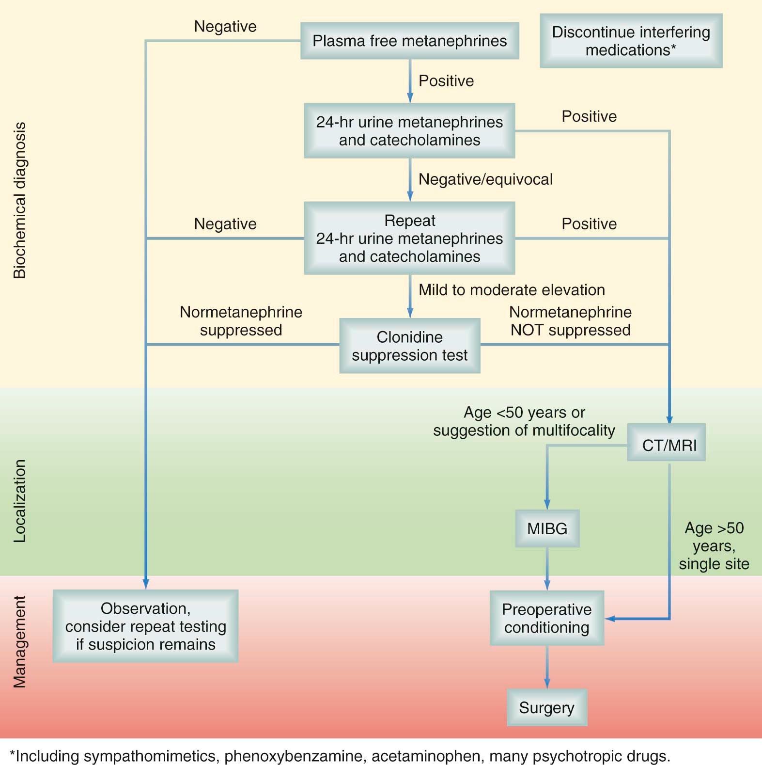
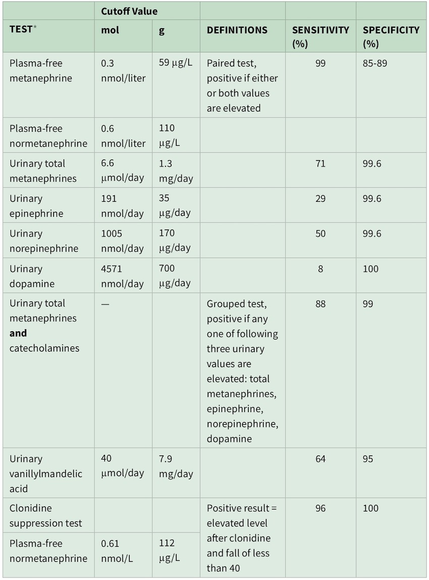
Sensitivities and specificities of these biochemical tests are dependent on the cutoff value used (greater than twofold to fourfold of the upper limit of normal) and whether the patient has a hereditary predisposition for pheochromocytoma.
Plasma metanephrine should be drawn in a supine position and after an overnight fast to reduce the likelihood of false-positive results from increased levels resulting from upright posture or caffeine and nicotine use
Acetaminophen, labetalol, sotalol, alpha-methyldopa, tricyclic antidepressants, buspirone, phenoxybenzamine, monoamine oxidase (MAO) inhibitors, sympathomimetics, cocaine, sulfasalazine, and levodopa, that can cause falsely elevated levels of plasma and urinary metanephrine and normetanephrine.
Tests performed during episodes of acute pain, critical illness, or urgent hospitalization may be misleading
Plasma-free metanephrine testing carries an extremely high sensitivity, approaching 99%, and, being a one-time blood test, is more convenient than 24-hour urine testing. Specificity however is only 89% at best.
The primary usefulness of plasma-free metanephrine testing is to exclude pheochromocytoma when the test is negative
When serum metanephrines are positive, confirmatory testing with 24-hour urine levels of catecholamines and their metabolites is required
A urine collection may be considered positive if total metanephrines or any single catecholamine fraction (e.g., epinephrine, norepinephrine, dopamine) is elevated above its cutoff value
V
*
Clonidine suppression testing for equivocal cases
Two 24-hour urine collections for catecholamines and their metabolites are sufficient to make (or exclude) the diagnosis of pheochromocytoma in almost all cases. Clonidine suppression testing, the measurement of plasma-free normetanephrine levels after the oral administration of 0.3 mg clonidine, may help clarify equivocal test results
V
*
Cutoff values for biochemical Dx of pheochromocytoma
Screen Shot 2020-01-21 at 14.50.58 copy
*
Urine vanillylmandelic acid is no longer used for screening for pheochromocytoma because of the lower specificity of this test
>
Imaging
V
*
CT neck/chest/abdomen/pelvis: heterogeneous & have higher attenuation
The tumors may appear homogeneous or heterogeneous, necrotic with some calcification, solid, or cystic
CT scans of the neck, chest, abdomen, and/or pelvis, with and without contrast, should be performed to exclude extra-adrenal paragangliomas, using a section thickness of 2 to 2.5 mm in the neck and 5 mm through the chest, abdomen, and pelvis, especially in young patients (<40 years) and in those who have germline SDHx mutations or a family history of pheochromocytomas and paragangliomas
*
MRI (more sensitive) preferred in pediatrics & pregnancy: may have pathognomonic high signal intensity on T2-weighted images
>
Functional (nuclear) scans are indicated for age < 50Y & for suspicion of metastasis or multifocal disease:
*
123I-Metaiodobenzylguanidine (MIBG)
*
FDG PET/CT
*
FDOPA
*
Ga-DOTA peptide PET/CT
*
Metastatic work up: FDG PET/CT & Ga-DOTA peptide PET/CT are most accurate
>
Management
*
Patient selection: all pheochromocytomas require resection
>
Preoperative management
*
Cardiac evaluation for all
V
>
PreOp anti-HTN to reduce HD instability intraOp:
Goal BP ≤ 130/80 mmHg while sitting
Goal HR ~ 70
V
*
α-antagonists (phenoxybenzamine) are the first choice once pheochromocytoma is Dx (even if normotensive) for a period at least 2w preOp
Phenoxybenzamine (10-20 mg BID), start at least 7-14 days preOp to normalize the BP & HR
Other options: doxazosin (2 mg QD), prazosin (2 mg BID)
The period of preoperative conditioning should last at least 2 weeks to allow for adequate reversal of α-adrenergic receptor downregulation. This restores sensitivity to vasopressor agents, which can then be used to treat the patient postoperatively
*
High-salt diet 3 days after initiation of α-antagonist
V
*
β-blocker is started 2-3 days after initiation of α-antagonist if tachycardia is persistent
The use of a beta-blocker without alpha blockers will exacerbate vasoconstriction by blocking its vasodilator component.
The administration of a β-blocker before an α-blocker to a hypertensive patient with a pheochromocytoma may precipitate a hypertensive crisis and acute cardiac decompensation and profound heart failure
*
If BP is still high: CCB or metyrosine therapy is initiated.
*
1-2 L of NS the evening before surgery to reduce risk of ↓BP after tumor removal
>
Considerations:
*
“Laparoscopic adrenalectomy is a relative contraindication for lesions > 6-8 cm” — SCORE module
“laparoscopic approach to the adrenal gland is the procedure of choice for patients with small, solitary intraadrenal pheochromocytomas that have no malignant radiologic features” — UTD
*
Invasive BP monitoring intraOp
*
Nitroprusside is the first-line antihypertensive intraoperatively for pheochromocytoma.
Other options include phentolamine, nicardipine, labetalol, and esmolol
*
After dividing the adrenal vein, sudden ↓BP may develop. Ensure euvolemia and start vasopressors if needed
*
Pheochromocytomatosis (peritoneal implants with local-regional recurrence) may occur with fracturing of the tumor
>
Postoperative
V
*
24h ICU monitoring of BP & glucose levels
Patients may have hypotension resulting from the continued effects of the alpha adrenergic blockade or withdrawal of catecholamine and metabolites (adrenal insufficiency)
There have been reports of hypoglycemia resulting from rebound secretion of insulin after removal of a pheochromocytoma, and therefore blood sugars should be monitored
>
Follow Up:
>
Sr.Metanephrine should be evaluated at:
*
6 weeks and at 6 months to confirm the patient’s biochemical response
*
Annually, to rule out recurrence (malignant pheochromocytoma may present as recurrence)
*
Recurrence of pheochromocytoma is managed with resection
>
Malignant pheochromocytomas
>
Features associated with malignant pheochromocytoma
*
Tumor > 5 cm
*
Presence of necrosis & hemorrhage
*
↑Mitotic index
*
SDHB gene mutation have higher risk than SDHD, VHL, and RET mutations
*
No histopathologic criteria for determining malignancy have demonstrated the ability to predict the clinical course accurately
>
Estimated to be between 5-40% of cases depending on:
*
Evidence of metastatic disease at nonchromaffin sites distant from the tumor (usual axial skeleton, LN, liver, lung, kidney)
*
Evidence of gross local invasion
*
Evidence of regional LN metastasis
*
Malignant disease (recurrence or metastasis) may manifest decades after initial resection of pheochromocytoma
*
The most common sites of metastasis are the axial skeleton, lymph nodes, liver, lung, and kidney
>
Management
*
Recurrence warrants resection if R0 or R1 is acheivable
*
Widely metastatic disease may benefit from cytoreductive surgery to palliate symptoms and control cardiovascular events
V
*
Unresectable disease may benefit from: cyclophosphamide, vincristine, dacarbazine, or 131I-MIBG
Unfortunately, only two thirds of metastases are avid on MIBG scanning, which can limit the efficacy of this therapy.
>
Hyperaldosteronism
*
Caused by autonomous overproduction of aldosterone, not suppressed by Na loading
*
Lesions tend to be small (< 2 cm)
>
Etiology:
*
Adenoma (aldosteronoma)
*
Unilateral/bilateral adrenal hyperplasia (idiopathic hyperaldosteronism)
*
Inherited disorder of glucocorticoid-remediable aldosteronism
>
Clinically
*
Most patients are asymptomatic
*
Classically present with Conn’s syndrome: HTN, polyuria-polydipsia, ↓K
V
>
Screening
Screening for hyperaldosteronism is done only if the patient is hypertensive or hypokalemic and consists simply of measuring plasma aldosterone and renin levels
>
Indications for screening
*
Incidentaloma
*
All patients with HTN and unexplained ↓K and in HTN resistant to Rx
V
V
Screening tests
This test should be performed after discontinuation of interfering medications, such as spironolactone, ACE-inhibitors, diuretics, and β-adrenergic blockers
Screen Shot 2020-01-18 at 12.07.33 PM
*
Aldosterone:Renin ratio >20-25, and
*
Sr. Aldosterone > 15 mg/dL, and
*
Sr. Renin < 0.5
V
Dx must be verified with another test:
*
Oral Na loading followed by 24h Ur. aldosterone measurement (>12 microgram/dL)
*
IV Saline loading followed by 24h Ur. aldosterone measurement (>12 microgram/dL)
*
Fludrocortisone suppression with Na loading
*
Captopril challenge
*
Patients who test positive and are < 30 years should be genetically screened for glucocorticoid-remediable aldosteronism (familial hyperaldosteronism type I), especially if they have a family history of early-onset hypertension
>
CT findings may show
*
Normal glands
*
Unilateral/bilateral nodules
*
Unilateral/bilateral hyperplasia
*
Unilateral adrenal limb thickening
>
Adrenal vein sampling is the gold standard to localize aldosterone hypersercretion
*
Helps differentiate idiopathic hyperaldosteronism from bilateral cortical hyperplasia
>
Indications
*
Bilateral adrenal nodularity (patients may have a concurrent contralateral nonfunctioning adenoma)
*
Unilateral nodule < 1 cm
*
Normal appearing glands
*
Age > 40-45 years (because the incidence of nonfunctional incidentalomas increase with age)
*
Technique: blood samples are obtained from each adrenal vein, the inferior vena cava, and a peripheral vein.
*
Confirmation of the adequate positioning of the catheter during sampling is obtained by a ratio of cortisol 5:1 when comparing adrenal vein to IVC
>
Results
*
A side-to-side ratio of at least 4:1 for the cortisolcorrected aldosterone concentrations is accepted widely as indicative of unilateral hypersecretion
*
A ratio of 3 : 1 or less suggests bilateral hypersecretion (and thus disease that should be managed medically)
>
Management
V
Laparoscopic adrenalectomy is indicated for unilateral disease (adenoma or hyperplasia)
*
Overall cure rates range from 75% to 95% at subspecialty centers
V
PostOp care
*
Hold all anti-HTN Rx; ween down those that will cause rebound hypertension (beta blockers & clonidine)
*
High salt diet can help blunt the rebound ↑K
*
Follow up in 1w for measurement of vitals & electrolytes
*
Nonsurgical candidates are managed with potassium-sparing diuretics: spironolactone or eplerenone
V
Hypercortisolism: Cushing’s syndrome
*
Etiology of Cushing: iatrogenic > pituitary ACTH-dependent > benign/malignant adrenal adenoma
V
Clinical features to distinguish Cushing’s syndrome from obesity:
*
Easy bruising
*
Muscle weakness
*
HTN
*
Plethora
*
Hirsutism
V
V
Biochemical testing:
Screen Shot 2020-01-18 at 12.40.19 PM
V
*
Dexamethasone suppression test (must be verified with another test)
1 mg of Dexamethasone given at 11 PM
Plasma cortisol level is collected at 8 AM
Normal AM cortisol level < 3 microgram/dL
Morning cortisol level will be insufficiently suppressed by the exogenous dexamethasone
Morning serum cortisol of 5 μg/dL or more is an acceptable cut-off, suggestive of hypercortisolism with a sensitivity of 85% and specificity of 95%
V
*
Late-night salivary cortisol measurement (performed twice)
A high cutoff value of 550 ng/mL has a sensitivity of 93% and specificity of 100%.
*
24h urine free cortisol levels (performed twice)
*
Serum ACTH also should be checked, and a level of less than 10 pg/mL excludes an extra-adrenal cause of Cushing’s
*
Additional imaging that may help localize the source of an ectopic-ACTH secreting tumor: OctreoScan and DOTATATE scan
V
Management
*
Cortisol-secreting adenomas are better treated by adrenalectomy over medical management
*
Normalization of cortisol level is recommended for patients with overt Cushing’s: ketoconazole, metyrapone, mifepristone, or mitotane
*
Endocrine Society guidelines currently recommend against treatment to reduce cortisol levels or action, if there is not an established diagnosis of CS. However, several small studies, recently meta-analyzed by Iacobone and colleagues, have shown improvement in body weight, hypertension, and blood glucose levels of SCS patients treated with adrenalectomy
*
A subnormal morning cortisol level on POD1 or 2 is indicative of cure. Glucocorticoid supplementation is then resumed until the HPA axis recovers, usually for at least 6 months
V
*
If the patient has an Ectopic ACTH-dependent Cushing syndrome, patients will benefit from bilateral adrenalectomy
Don’t wait too long for surgery. These patients can deteriorate quickly
V
Virilizing & feminizing tumors
*
Rare tumors that have a high risk of malignancy
>
Clinical & biochemical manifestations
>
Estrogen-secreting tumors:
*
Precocious puberty
*
Postmenopausal vaginal bleeding
*
Feminization in men
*
↑Estrogen (estradiol, estrone)
*
↓Gonadotropins (FSH, LH)
>
Androgen-secreting tumors:
*
Hirsutism
*
Menstrual irregularities
*
↑ Testosterone, dehydro-epi-androsterone sulfate (DHEA-S), androstenedione
*
Surgical resection is the treatment for both these adenomas and adrenocortical carcinomas
V
*
Nonfunctional cortical adenomas — see incidentalomas
Hormonal conversion or tumor growth of more than 1 cm should prompt consideration of excision
*
Myelolipoma — see incidentalomas
*
Angiomyolipoma
*
Oncocytic neoplasm
>
Indications for surgery
*
Symptoms from local growth
*
Rapid growth
*
Size > 4 cm
*
Adrenal hemorrhage (may occur because of underlying mass)
>
Malignant
V
Adrenocortical carcinoma
*
Rare, with very poor prognosis; local recurrence or metastasis develops in 85%
*
Presentation & Dx are delayed; 40% have metastasis at the time of Dx
*
Any adrenal mass with associated androgen or estrogen excess should raise the suspicion of adrenocortical carcinoma
>
Most cases are sporadic, but can occur as part of familial syndromes
*
Li-Fraumeni syndrome
*
MEN 1
*
Beckwith-Wiedemann syndrome
*
50% are hormonally active; Cushing is most commonly seen, followed by virilization
V
Dx & work up
*
CT/MRI is the cornerstone of Dx & surgical planning: irregular borders and higher attenuation values on unenhanced CT
*
Typically large (> 8 cm)
V
*
Staging: CT chest/abdomen/pelvis
T1: < 5 cm
T2: > 5 cm
T3: any size with local invasion, not invading surrounding organs
T4: invasion of surrounding organs (liver, spleen, pancreas, vessels, diaphragm)
N0, N1
M0, M1
*
FNA is discouraged
V
Management
*
Open surgery is indicated. Choice of several incisions including: midline or Makuuci
*
May require en-bloc resection of adjacent organs. Extension in & thrombosis of renal vein or IVC is not prohibitive of complete resection & thrombectomy should be performed
*
The need for LN dissection is unclear
V
*
Mitotane is the principle chemotherapeutic agent for the treatment of adrenocortical carcinoma
The clear indications for adjuvant mitotane include R1 resection, vascular or capsular invasion, intraoperative tumor spillage, and high-grade disease
V
Adrenal metastases
*
It is the second most common cause of adrenal masses (after benign adrenal cortical adenomas)
>
Source
*
NSCLC
*
Breast
*
RCC
*
Melanoma
*
GI tract
*
The standard biochemical workup of adrenal nodules should be performed, even if the suspicion of adrenal metastases is high
*
Initial presentation of a tumor with metastasis detected in the adrenal is highly unlikely
*
In select patients, who are poor surgical candidates, the option of stereotactic ablative body therapy of adrenal metastasis can be considered (RFA or microwave ablation)
>
Pheochromocytoma crisis
*
A purely medical condition
V
Time to operation depends on how the patient’s condition improves with medical management
*
Electively: for transient crisis (e.g after FNA Bx — which usually shouldn’t be done)
*
Urgently: in ~ 1 month
*
Emergency: wait a minimum of 7-10d, but before the patient is discharged from the hospital. Try to have the patient condition plateau
V
>
Multiple Endocrine Neoplasia (MEN) & hereditary syndromes
The associated endocrine tumors may be benign or malignant and may develop synchronously or metachronously.
Pasted_Graphic_11
Screen Shot 2020-02-22 at 00.50.21
*
Have autosomal dominant inheritance
>
MEN 1
*
Caused by tumor suppressor gene mutation on chromosome 11q13
*
MEN1 gene encodes the protein menin. Menin promotes the maintenance of transcription of critical cell cycle regulators essential for normal endocrine cell growth control. Approximately two thirds of the reported mutations in the MEN1 gene result in premature termination of translation and truncation of the C-terminal portion of the menin protein
*
When compared with sporadic endocrine tumors, the endocrine tumors arising in association with the familial MEN1 syndrome are characterized by an earlier age of onset, multifocal involvement within a target endocrine tissue, and development of concurrent neoplasms in multiple endocrine tissues.
*
Presentation is typically 2ry to ↑Ca
>
Dx
*
Clinically, MEN1 is defined as the occurrence of neoplasms in at least two target endocrine tissues in an individual
*
Hyperpararthyroidism is almost always seen in MEN1
*
Testing for MEN1 is done by testing for the menin gene
>
Manifestation
*
Multiple parathyroid tumors develops in ~99%; hypercalcemia is often mild
V
*
Duodenopancreatic NET develops in 30-80%
Pancreatic NETs that are nonfunctioning or that secrete pancreatic polypeptide are probably the most frequent NETs that occur in patients with MEN1
The most common NET are nonfunctional
The most common functional NET in patients with MEN1 is gastrinoma
In patients with MEN1, the most common functional neuroendocrine tumors are :
gastrinomas (54%),
insulinomas (18%),
glucagonomas (3%),
and VIPomas (3%).
V
*
Pituitary tumors become clinically evident in 15-50%
The most frequent pituitary tumor in patients with MEN1 is a prolactinoma
*
The principal cause of mortality in patients with MEN1 is malignant progression of duodenopancreatic neuroendocrine cancers or intrathoracic malignant carcinoids
V
>
Management
Similar recommendations apply for MEN4
IMG_0166
*
Testing for pituitary adenomas starting the age of 5Y
*
Annual screening at age 8Y: PTH & ↑Ca
>
Annual screening at age 15-20Y in presymptomatic individuals
*
Pancreatic polypeptide
*
Gastrin
*
Glucagon
*
Chromogrannin A
>
Typical surgery:
>
Parathyroids
*
Indications for parathyroidectomy in patients with MEN1 are similar to those in patients with sporadic adenomas causing primary hyperparathyroidism. Can be offered also to help control gastrinoma symptoms
V
*
3½ gland resection with cervical thymectomy is done (may harbor parathyroid glands)
Preoperative imaging tests are not necessary for patients with MEN1 undergoing initial neck exploration because appropriate treatment requires bilateral neck exploration and identification of all four glands
*
There is a significantly higher rate of recurrent or persistent hyperparathyroidism after parathyroidectomy when compared with the results for the treatment of sporadic parathyroid adenomas
*
Parathyroidectomy is usually done before addressing NET. It may help control gastrinomas
>
Pituitary adenomas are treated in the same way as sporadic pituitary adenomas
*
Bromocriptine or cabergoline is effective in controlling the hyperprolactinemia in most patients with prolactinomas
*
Growth hormone–producing tumors causing acromegaly or ACTH-producing tumors are usually treated by surgical resection
>
Duodenopancreatic NET
V
*
Active ZES as part of the MEN1 syndrome should be treated primarily by PPI
The role of duodenal-pancreatic surgery to prevent metastatic disease is uncertain and controversial but could prove beneficial
*
Surgery is indicated for patients with functional PNET (but less for gastrinomas)
>
MEN 2
V
Caused by RET proto-oncogene mutation on chromosome 10
*
The mutations responsible for MTC are missense mutations that result in single amino acid changes that cause gain of function alterations in the protein
*
The hallmark of the MEN2 syndromes is MTC, which occurs with almost complete penetrance
>
MEN 2A (55%)
*
MTC develops in 100%
*
Pheochromocytoma in 40%
*
Primary HPT in 30%
>
MEN 2B (10%)
>
MTC develops in 100%
*
MEN2B patients expressing the M918T mutation have the most aggressive forms of MTC, with evidence of disease often present in early infancy
*
Pheochromocytoma in 40%
*
Physical features: Marfanoid habits, mucosal neuromas, ganglioneuromatosis of the GI tract, megacolon
>
Familial non-MEN MTC (FMTC)
*
Familial MTC (FMTC) is characterized by the presence of a RET germline mutation in families with MTC, or an individual with MTC, who do not develop pheochromocytoma or primary hyperparathyroidism
*
FMTC patients demonstrate an indolent form of MTC that more often presents in the later decades of life
*
High index of suspicion in families with ≥ 4 members are Dx with MTC
>
Management
*
Once identified, RET mutation analysis should also be performed in 1st- and 2nd-degree family members
>
Thyroid
*
MTC is managed with total thyroidectomy. Prophylactic bilateral central LN dissection is done routinely
*
Screening & prophylactic total thyroidectomy for known RET-mutation patients but without clinically apparent disease
Screen Shot 2020-01-18 at 7.52.10 PM
>
Pheochromocytoma
*
Annual screening is also done for pheochromocytoma. Start at age 11Y
V
*
If pheochromocytoma is present, it (unilateral) should be resected first. If bilateral disease is present, bilateral adrenalectomy is done.
For patients with bilateral pheochromocytomas, bilateral adrenalectomy is necessary. It should also be considered in a patient with unilateral disease when other family members have had unusually aggressive bilateral adrenal medullary disease. For most other patients with a unilateral pheochromocytoma, we recommend unilateral adrenalectomy as the treatment of choice (Grade 1C)
*
Cortical sparing adrenalectomy can be offered on the contralateral side of the pheochromocytoma
>
Parathyroid
*
There’s no role for prophylactic parathyroidectomy
>
Head & Neck
V
>
Assessment of neck masses
Screen Shot 2020-04-03 at 5.36.53 PM
Screen Shot 2020-04-03 at 5.12.55 PM
>
Key points
*
Inquire for risk factors: tobacco, EtOH, marijuana, HPV, EBV, ill fitting dentures
*
Inquire about Hx of: TB, HIV, foreign travel, pets, insect bites
*
Inquire about PSHx or F.Hx of cancer or paraganglioma
*
Inquire for pain/trismus
*
Inquire for B-symptoms
*
Inquire about change in voice, hearing loss, otalgia, or pharyngitis
*
Assess for skin changes
*
Assess for masses: size, duration, fixation
*
Unilateral hearing loss in Asian patient need to rule out nasopharyngeal cancer
>
Examination
*
Skin
*
Oral cavity & oropharynx
*
Larynx & hypopharynx
*
Nasal cavity & nasopharynx
*
Ear & external auditory canal
>
Gland examination
*
Thyroid
*
Parotid
*
Submandibular
*
Sublingual
*
Minor salivary glands
*
CN examination
V
>
LN assessment
Screen Shot 2020-04-03 at 2.25.08 PM
*
Posterior triangle LN are a frequent metastatic site for nasopharynx malignancy
*
Submental triangle LN are a frequent metastatic site of lip malignancy
*
Tip of the tongue drains into the submental nodes
*
The posterior 2/3 of the tongue drain into the submandibular nodes
*
Hepatosplenomegaly
>
Work up
>
Neoplasm suspected
*
Pulsatile mass: CT neck with contrast
V
*
Non-pulsatile mass: FNA
Screen Shot 2020-04-03 at 2.54.01 PM
*
Open Bx indicated for ‘persistently non-diagnostic FNA’ but suspicious clinical characteristics
>
Inflammatory or infectious process suspected
*
Empiric trial of Abx
>
Failure to improve on Abx:
*
CXR & tuberculin test
*
± CT with contrast & FNA
>
DDx
>
DDx likelihood stratified by age
*
Children: congenital > inflammatory > malignant
*
Young adult: inflammatory > congenital > malignant
*
Adult > 50Y: malignant > inflammatory > congenital
>
DDx based on etiology (VINDiCATE)
*
Vascular: carotid body tumor, hemangioma
*
Inflammatory: lymphadenitis, thyroiditis
*
Neoplastic: primary malignancy, metastatic LN
*
Degenerative: atherosclerosis, MNG
*
Congenital: cystic hygroma, branchial cyst
*
Autoimmune: sarcoidosis, Sjogren’s syndrome
*
Traumatic: fracture, hematoma
*
Endocrine: thyroid nodule, thyroglossal cyst
>
DDx based on location
V
Anterior triangle
*
Thyroid/thyroglossal cyst
*
Plunging ranula
*
Carcinoma larynx
*
Carotid body tumor
*
Branchial cleft cyst
*
Salivary gland tumors
>
Posterior triangle
*
Metastatic node (most common in adults)
*
Lymphoma
*
Soft tissue tumor
*
Cystic hygroma
*
Vascular/lymphatic tumor
*
Neurogenic tumor
>
Conditions
>
Carotid body tumors
*
Demonstrate splaying of the ICA & ECA (Lyre’s sign)
*
MRI shows salt & pepper pattern
*
Treatment is excision with proximal and distal control ± bypass
>
Vagal paraganglioma
*
Managed with active surveillance ± radiation to arrest growth
*
Surgery results in CN deficit resulting in change in voice and aspiration
>
Vagal schwannoma
*
Dx with FNA showing spindle cell neoplasia
*
Manage as with vagal paraganglioma
>
Lymphangioma
*
Soft compressible mass
*
Satellite lesions
*
Surgery indicated for well localized lesions
>
Branchial cleft cyst/sinus
*
Need to rule out SCC
*
Excision is warranted with excision of the tract
>
Thyroglossal duct cysts (TGDC)
>
It’s the epithelial remnant of the thyroglossal tract & presents characteristically as a midline neck mass at the level of the thyrohyoid membrane
*
The cyst may occur anywhere along the thyroglossal duct tract from the foramen cecum at the base of the tongue to the level of the suprasternal notch
*
Usually causes no symptoms but may be slightly tender
*
Moves up with swallowing or protrusion of the tongue
*
The incidence of primary carcinoma of the thyroglossal duct is < 1% in all age groups, the majority of which are PTC
>
Patients with a TGDC often have ectopic thyroid glands
*
Ectopic thyroid tissue can be confused with a TGDC
*
All cases of thyroid ectopia should have thyroid function tests, ultrasonography, and a thyroid scan performed to locate additional functioning thyroid tissue
>
Most TGDCs have some degree of infection or inflammation at presentation
*
It is essential to avoid surgery during the phase of acute inflammation as this can lead to recurrence
V
*
An infected TGDC should be managed with Abx: 1st Gen. cephalosporin, Clavulin, or clindamycin
The cysts are usually infected by oropharyngeal flora; hence, antibiotics should be directed toward the most common oral organisms, which include various streptococcal species and oral anaerobes
*
I&D is indicated only for failure to respond to Abx
*
Once the infection clears completely, an elective Sistrunk procedure is performed
V
>
Sistrunk procedure
Screen Shot 2020-04-03 at 1.50.32 PM
*
Performed for all TGDC unless the patient is not a surgical candidate
*
If a TGDC is not removed, ~½ become infected, and infection before surgery is a well-described cause of recurrence
V
*
Technique: resection of the cyst and the mid portion of the hyoid bone in continuity & resection of a core of tissue from the hyoid upwards toward the foramen cecum
General Surgery Review Course: also approximate supra- and infra-hyoid muscles
*
Poor surgical candidates are managed with percutaneous ethanol injection after the presence of malignancy is excluded
*
Injury to the spinal accessory nerve results in inability to raise the arm above the shoulder
>
Operative considerations
>
Classification of neck dissection
V
Comprehensive neck dissection: level 1-5
*
Radical neck dissection: standard basic procedure
>
Modified radical neck dissection preserves a non-lymphatic structure
*
Type I: CN XI is preserved
*
Type II: CN XI & IJV are preserved
*
Type III: CN XI, IJV, & SCM are preserved
*
Selective neck dissection: preservation of ≥ 1 LN group
*
Extended radical neck dissection removes additional LN groups or non-lymphatic structures
V
*
Frey syndrome
UTD:
Frey syndrome, also known as auriculotemporal syndrome or gustatory sweating, is characterized by sweating and flushing of the facial skin over the parotid bed and neck during mastication. Frey syndrome is thought to be due to aberrant regeneration of cut parasympathetic fibers between the otic ganglion and salivary tissue, which leads to innervation of sweat glands and subcutaneous vessels.
Frey syndrome is reported by approximately 10 percent of patients, although rigorous testing may detect gustatory sweating in as many as 95 percent of patients after parotidectomy. Frey syndrome may occur from two weeks to two years after surgery due to a latency in the regeneration of parasympathetic nerve fibers; the area of involved skin may increase over time
>
If CN VII is transected during surgery
*
Neurorrhaphy (direct approximation)
*
Interpositional graft (gap > 1 cm) using the greater auricular nerve or sural nerve
*
Nerve transfer using the CN XI, CN XII or phrenic nerve
>
Adjuvant radiation indications
*
T3-4 (tumors with soft tissue infiltration)
*
High grade tumors
*
Extensive nerve/bone invasion
*
LVI or PNI⊕
*
Close or positive resection margin
*
Recurrent/residual tumors
*
Extranodal extension
*
Deep lobe tumors
>
Salivary glands
V
*
Parotid anatomy
Screen Shot 2020-04-03 at 5.10.14 PM
>
General
V
>
Neoplasms incidence: parotid (70%) > submandibular (22%) > sublingual & minor glands (8%)
“As the gland gets smaller, the risk of malignancy is higher”
*
The majority of parotid gland neoplasms are benign
*
The majority of minor salivary gland neoplasms are malignant
V
*
Submandibular gland neoplasms are 50% malignant
Large tumors may require mandibulotomy with resection of hypoglossal and lingual N
*
Ratio of benign to malignant tumors: parotid (80:20)> submandibular & sublingual (50:50)
*
When the presenting symptoms is of unilateral facial paralysis, Bell’s palsy may be misdiagnosed as the cause.
*
FNA has high sensitivity, specificity, & accuracy
>
Conditions
>
Sialadenitis
>
Acute
*
Commonly affects parotid & submandibular glands
*
Caused by bacterial (Staph. Aureus) or viral (mumps)
*
Subacute
>
Chronic
*
Associated with sarcoidosis, actinomycosis, TB, cat scratch disease
*
Sialolithiasis
>
Benign lymphoepithelial lesions (non-neoplastic glandular enlargement)
*
Associated with autoimmune diseases such as Sjogren’s syndrome
V
>
Benign tumors
Treatment of choice is surgical excision with a margin of normal tissue
The facial nerve should not be sacrificed when removing a benign lesion
>
Pleomorphic adenoma
*
Contains groups of epithelial and myoepithelial cells in varying states of organization
*
Accounts for 40-70% of all tumors of the salivary glands
*
Usually occur in the tail of the parotid
*
Although usually well encapsulated, simple enucleation is associated with local recurrence
V
>
Malignant potential (carcinoma ex pleomorphic adenoma):
Frequent local recurrences and distant metastases mark the clinical course of invasive carcinoma ex pleomorphic adenoma. Carcinoma ex pleomorphic adenoma should be considered a high-grade malignancy requiring aggressive treatment at the initial diagnosis, as subsequent salvage options are limited
>
Incidence
*
1.5% within the first 5 years
*
Increases to 9.5% once the benign tumor has been present for ≥ 15 years
*
May present with sudden growth
V
Monomorphic adenomas
>
Warthin’s tumor (papillary cystadenoma lymphomatous)
*
The second most common benign parotid tumor
*
Often occurs in older white ♂
*
Lights up on Technetium-99m scan
*
If FNA suggests a slow-growing Warthin tumor with confirmatory technetium scan in a patient with contraindication to surgery, the tumor may be closely monitored because it has no malignant potential
*
Oncocytoma
*
Basal cell adenomas
*
Ductal papilloma
*
Capillary hemangioma
>
Malignant tumors
>
Mucoepidermoid carcinoma
*
The most common malignant tumor of the parotid
V
*
High grade vs low grade
High-grade lesions have a propensity for both regional and distant metastases and corresponding shorter survival rates than low-grade mucoepidermoid carcinomas
V
*
Adenoid cystic carcinoma (the second most common malignant salivary gland tumor)
An indolent growth pattern and a relentless propensity for perineural invasion characterize adenoid cystic carcinoma
*
Carcinoma ex pleomorphic adenoma (See pleomorphic adenoma)
*
Malignant transformation of pleomorphic adenoma
V
*
Lymphomas (usually NHL)
Risk for malignant lymphoma in patients with Sjogren’s syndrome is 44-fold higher than in normal population
>
Management of malignant tumors
>
PostOp radiation is administered for high-grade malignancies demonstrating (any):
*
Extraglandular disease
*
Perineural invasion
*
Direct invasion of surrounding tissue
*
Regional metastases
*
Gross tumor should not be left in situ but if the facial nerve can be preserved by peeling the tumor off it, the nerve should be preserved & radiation used for microscopic residual disease
*
Elective neck dissections are performed for high-grade malignancies
V
Thyroid & parathyroid
V
V
Anatomy, physiology and embryology
Screen Shot 2020-04-03 at 5.07.28 PM
>
Parathyroid
*
Chief cells of the parathyroid gland secrete PTH
*
Parafollicular cells of the thyroid secrete calcitonin
*
PTH t½ is 4m
V
*
PTH function
Screen Shot 2020-01-10 at 10.57.05 AM
0e54a3cc418d64bcdd4ea0183903da48
*
The inferior parathyroids originate from the third branchial pouch, whereas the superior parathyroids descend from the fourth branchial pouch
*
Supernumerary parathyroid glands occur in 7-10% of the general population
V
>
Inferior thyroid artery
dca771a97571404b8cc4e95524d1856a
*
Supplies all parathyroid glands (20% superior parathyroid glands receive their blood supply from elsewhere)
*
The superior parathyroid glands is located superior to it
*
The inferior parathyroid gland is located inferior to it
*
If the main trunk of the inferior thyroid artery is sacrificed for dissection, both parathyroids on that side become devascularized because there is usually no collateral blood supply to maintain viability
>
Thyroid
*
Parafollicular (C cells) arise from the fourth pharyngeal pouch and produce calcitonin. These C cells are the only component of the adult gland that is not of endodermal origin
V
*
The superior edge of the isthmus sits just below the cricoid cartilage
reic2_c026f003
*
A pyramidal lobe is present in 30% of patients & represent the most distal portion of the thyroglossal duct
*
TSH is secreted from the anterior pituitary in a pulsatile fashion
*
Peripheral conversion of T4 to T3 can be impaired in many clinical circumstances, such as overwhelming sepsis and malnutrition, thionamide (propylthiouracil) use, high-dose corticosteroids, beta blockers, iodinated contrast agents, and amiodarone use
*
t½ of T3 is 8-12h; t½ of T4 is ~ 7 days
>
Vessels
>
Arteries
>
Superior thyroid artery
*
It is the first branch of the external carotid after the common carotid bifurcation
>
Inferior thyroid artery originates from the thyrocervical trunk from the subclavian artery
*
The RLN is usually directly adjacent (in an anterior or posterior position) to the inferior thyroid artery, within 1 cm of its entrance into the larynx.
*
Almost always supplies all 4 parathyroid glands
>
Thyroid ima artery is present in < 5% and arises from the innominate artery or the aorta
*
If the main trunk of the inferior thyroid artery is sacrificed for dissection, both parathyroids on that side become devascularized because there is usually no collateral blood supply to maintain viability
>
Veins
*
Superior thyroid vein
*
Middle thyroid vein exists in > 50% & courses immediately lateral into the internal jugular vein
*
2-3 Inferior thyroid veins drain into the innominate and brachiocephalic veins
>
Nerves
>
SLN branches off from the vagus at the base of the skull
V
*
SLN descends toward the superior pole of the thyroid along the internal carotid artery.
Screen Shot 2020-01-11 at 20.00.26
>
Branches
*
External branch innervates the cricothyroid muscle, which is the only tensor of the vocal cords
*
Internal branch provides general sensation, including pain, touch, and temperature for the tissue superior to the vocal folds
>
RLN branches off from the vagus
*
Supplies four intrinsic laryngeal muscles (Lateral cricoarytenoid, posterior cricorytenoid, transverse and oblique interarytenoid and thyroarytenoid) but not the cricothyroid muscle
*
The interarytenoid muscle, the only unpaired muscle of the larynx, receives innervation from both RLNs
V
>
RLN in relation to vessels
Anatomy_Recurrent_Laryngeal_2GG
*
The right nerve loops around the subclavian artery
*
The left nerve loops around the aortic arch, lateral to the ligament arteriosum
V
*
The RLN is found immediately anterior or posterior to the main arterial trunk of the inferior thyroid artery in the tracheoesophageal groove
A329068_1_En_3_Fig1_HTML
*
Incomplete RLN injury results in inability to abduct the vocal cords while adduction remains functional. Complete RLN injury affects abduction and adduction
V
*
RLN injury
Photo Mar 16, 20 30 30
V
V
RLN in relation to parathyroid glands:
Screen Shot 2020-01-09 at 22.08.56
*
Superior parathyroid gland is lateral and posterior to RLN
*
Inferior parathyroid gland is medial and anterior to RLN
V
>
LN stations
Pasted_Graphic_19
>
Central nodes (level VI) boundaries:
*
Laterally: carotid arteries
*
Medially: midline
*
Superiorly: hyoid bone
*
Inferiorly: sternal notch / brachiocephalic vessels
*
Superior third of the thyroid may drain directly into the lateral compartment
>
Inhibition of thyroid synthesis
V
Drugs
V
Thionamides (propylthiouracil (PTU), methimazole)
*
Inhibit organification & oxidation of inorganic iodine
*
Inhibit linkage of MIT & DIT to form T3 & T4
*
PTU inhibits peripheral conversion of T4 to T3
*
Methimazole requires single daily dosing, and is the preferred agent for the nonpregnant
*
Agranulocytosis is rare but serious side effect that needs to be monitored
V
Steroids
*
Suppress the pituitary-thyroid axis
*
Inhibit peripheral conversion of T4 to T3
*
β-blockers do not inhibit hormone synthesis but are valuable in controlling peripheral sensitivity to catecholamines by blocking their effects
V
Iodine
V
*
Given in large doses after the administration of antithyroid medication can inhibit thyroid hormone release (Wolff-Chaikoff effect) — a transient but effective strategy, especially prior to surgery
Wolff-Chaikoff effect:
- Increasing doses of iodine increase hormone synthesis initially
- Higher doses cause cessation of hormone formation
- This effect is countered by iodide leak from normal thyroid tissue
- Pateints with autoimmune thyroiditis may fail to adapt and become hypothyroid
V
Parathyroids
>
Hyperparathyroidism (HPT)
>
Primary (most common) (PHPT)
>
Etiology
V
*
80% are a result of clonal expansion of Chief cells within a single gland
♀ 3:1 ♂, mostly middle-aged
*
10-15% are due to 4-gland hyperplasia
*
4% are due to adenomatous expansion in 2-3 glands
>
1% is due to parathyroid cancer
*
Most patients with carcinomas have marked hypercalcemia (>14 mg/dL) and are more likely to have associated bone and renal disease than those with adenomas
*
Suspicion also is raised by an extremely high iPTH level, a palpable neck mass on physical examination, significant uptake on sestamibi scan, or ultrasound evidence of invasion with loss of planes between the parathyroid and thyroid, occasionally with lymphadenopathy.
*
Parathyroid biopsy should be avoided because of the risk of seeding tumor cells into the surrounding tissue and the limited utility of frozen section in clarifying the diagnosis
*
Although fibrosis and mitotic activity are common, they are not specific for malignancy. The diagnosis of carcinoma is restricted to tumors that show invasion of blood vessels, perineural spaces, soft tissues, thyroid gland, or other adjacent structures, or to tumors with documented metastases
>
The mainstay of treatment for parathyroid carcinoma is surgical resection. The technique shown to have the best outcome is en bloc resection. All structures that the tumor is invading are resected with gross negative margins.
*
En-bloc resection could include the ipsilateral thyroid lobe, paratracheal alveolar and lymphatic tissue, the thymus or some of the neck muscles, and in some instances, the recurrent laryngeal nerve. Some, centers recommend ipsilateral lymph node dissection
*
Distant metastases generally develop in the lungs, liver, and bone; they can occasionally be treated by resection of individual tumor deposits.
>
Family Hx
*
MEN 1 — PHPT penetrance 100%
*
MEN 2A — PHPT penetrance 20-30%
V
*
Jaw tumor syndrome — PHPT penetrance 80%
HPT is the most common feature and is associated with a high incidence of severe hypercalcemia and a risk for parathyroid carcinoma
>
DDx of ↑Ca
*
Parathyroid
Primary hyperparathyroidism: Sporadic, Familial
*
Nonparathyroid Endocrine
Thyrotoxicosis
Pheochromocytoma
Acute adrenal insufficiency
Vasointestinal polypeptide hormone–producing tumor (VIPoma)
*
Malignancy
Solid tumors
Lytic bone metastases
Lymphoma and leukemia
Parathyroid hormone–related peptide
Excess production of 1,25(OH)2D3
Other factors (cytokines, growth factors)
*
Granulomatous Diseases
Sarcoidosis
Tuberculosis
Histoplasmosis
Coccidiomycosis
Leprosy
*
Medications
Calcium supplementation
Thiazide diuretics
Lithium
Estrogens, antiestrogens, testosterone in breast cancer
Vitamin A or D intoxication
*
Other
Benign familial hypocalciuric hypercalcemia
Milk-alkali syndrome
Immobilization
Paget’s disease
Acute and chronic renal insufficiency
Aluminum excess
Parenteral nutrition
>
Dx & localization
V
*
Dx is entirely biochemical: ↑Ca + ↑PTH + normal Vit-D
Thus blood vitamin D levels should be normalized using vitamin D supplementation before assigning a diagnosis of PHPT.
In up to 15% of patients, serum PTH levels fall within the upper normal range, but these levels are inappropriate relative to the elevated serum calcium levels
V
*
Consider FHH with 24h urine calcium < 100 mg. Hypercalciuria is present in 80% of PHPT
Biochemical testing for familial hypocalciuric hypercalcemia (FHH), an autosomal dominant syndrome resulting from loss-of-function mutation(s) in the calcium sensing receptor gene, is necessary before diagnosing PHPT. This condition is characterized by mild elevations in blood-intact parathyroid hormone level, with associated relative hypercalcemia, and is diagnosed by 24-hour urine calcium level testing. A value of less than 100 mg in 24 hours is diagnostic of FHH and rules out PHPT.
FHH is not corrected by parathyroidectomy
V
*
Imaging plays no role in Dx, but does in localization. Start with neck US + sestamibi-SPECT
Combined (US & sestamibi-SPECT) true-positive rate of 90%
V
>
Localization options
There is general consensus that the single best study is sestamibi, especially when combined with SPECT, and it is now the most common nuclear medicine study performed
A subset of patients who require reexploration will have negative, discordant, or nonconvincing noninvasive localization studies. Current guidelines recommend that these patients undergo invasive localization in the form of selective arteriography in conjunction with venous sampling for PTH
Screen Shot 2020-01-09 at 2.56.40 PM
*
US
V
*
Technetium-99m sestamibi scan ± single-photon emission CT (SPECT)
Can also localize ectopic disease, which is present in 20% of cases.
The sensitivity of sestamibi is limited in multiglandular disease.
SPECT, which allows localization of structures in the anteroposterior plane, is particularly helpful in detecting smaller lesions and adenomas located behind the thyroid.
The study works by mitochondrial uptake of 99mTc-sestamibi, and parathyroid cells typically have a large number of mitochondria. Sestamibi, a monovalent lipophilic cation, diffuses passively across cell membranes and concentrates in mitochondria.
A significant limitation of sestamibi scans is related to the coexistence of thyroid pathology or other metabolically active tissue (e.g., lymph nodes or thyroid cancer) that can mimic parathyroid adenomas by causing false-positive results on sestamibi scans
V
*
4D CT: useful once sestamibi-SPECT & US fail to localize the disease (provides anatomic & functional information)
Four-dimensional CT (4D-CT), a novel imaging modality similar to CT angiography, is derived from three-dimensional (3D)-CT scanning with an added dimension from the changes in perfusion of contrast over time
This technique involves fine-cut (1- to 3-mm) imaging of the neck and upper chest using noncontrast, arterial contrast, and venous contrast phases
Sensitivity of 4D-CT is 88%
*
MRI
V
*
Venography with venous sampling / Selective Venous Sampling
Selective venous sampling (SVS), which involves catheterization of the internal jugular vein for lateralization and if necessary multiple veins in the neck and mediastinum, including the superior, middle, and inferior thyroid veins as well as the subclavian vein. A limitation of this approach is that the source of PTH overproduction often is only roughly regionalized. Furthermore, the venous drainage pattern for each parathyroid gland often is altered by previous neck operations, making exact localization of the gland challenging
*
Patients with PHPT for whom preoperative localization fails often have multigland disease and should undergo bilateral neck/four gland exploration
V
*
Ectopic parathyroid glands
Screen Shot 2020-01-09 at 18.26.09
V
>
Indications for surgery
The benefits of parathyroidectomy for PHPT patients who meet criteria for surgery are well established and include resolution of preoperative symptoms and improvement in renal function and bone mineral density. Surgical cure is achieved in greater than 95% of cases when bilateral exploration is required, with comparable results among patients undergoing MIP (cure rates as high as 98%).
Patients with elevated PTH levels and consistently normal serum calcium levels, in whom secondary causes of hyperparathyroidism have been excluded, may represent the earliest presentation of primary HPT. It is believed that during this early phase, termed normocalcemic hyperparathyroidism, elevated serum PTH levels cause a reduction in cortical bone density
*
Symptomatic PHPT (“stones, bones, abdominal groans, & psychiatric moans”)
>
Asymptomatic with:
*
Age < 50Y
*
Serum Ca 1.0 mg/dL (0.25 mmol/L) above normal
*
24h urine Ca > 400 mg/d (100 mmol/d)
V
*
Cr clearance < 60 ml/m or nephrolithiasis
15% of patients with renal stones will have PHPT
*
Bone density T-score < -2.5 or documented vertebral fracture
>
Management in patients that don’t meet criteria for surgery
*
Bisphosphonates: etidronate, alendronate, pamidronate
*
± Selective estrogen receptor inhibitors: tamoxifen, raloxifene
*
Calcimimetic agenst: cinacalcet
*
Q1Y assessment for symptoms & blood tests
*
Q1-2Y bone mineral density
>
IntraOp adjuncts
V
*
IntraOp blood parathyroid hormone level testing improves cure rates and may be used for all patients going for PHPT surgery
Cameron: ”Because of potential involvement of hyperfunctional supernumerary parathyroid glands, we employ intraoperative blood parathyroid hormone level testing whenever performing parathyroidectomy for PHPT”
Sabiston: In patients with multigland disease in particular, intraoperative PTH testing has been shown to be essential
Miami criteria:
- PTH checked pre-incision, pre-excision of the gland, and at 5, and 10m after excision
- A drop by 50% of PTH level suggests > 90% success rate
*
RLN monitoring
*
Gamma probe-mediated IntraOp parathyroid adenoma localization using technetium-99m
>
Surgery
>
“Minimally Invasive” Parathyroidectomy
*
Baseline venous blood parathyroid hormone level is checked immediately preOp
*
Procedure performed under GA/region/local anesthesia
*
Patient positioned in semi-Fowler’s position (semi-sitting position)
*
Transverse incision 2-fingerbreadths above sternal notch
*
Platysma is divided and subplatysmal flaps raised: superiorly to the cricoid cartilage, inferiorly to the level of the clavicles
*
Midline raphe between strap muscles is divided
*
Thyroid gland is rotated medially to expose parathyroid
*
Dissection carried out at the level of the parathyroid capsule
*
Upon gland excision, frozen section analysis is done
V
*
PTH level is assessed after excision & then 5, 10, and 20 mins later
The probability of cure is estimated on the basis of the Miami criteria, which stipulate a drop in blood-intact parathyroid hormone level of 50%, or more, relative to either the preoperative baseline value obtained on the day of surgery or to the highest value obtained intraoperatively before gland excision. A 97% cure rate has been reported when blood-intact parathyroid hormone levels fulfill the Miami criteria, and we therefore cease operative exploration under this circumstance.
Blood samples acquired via aspiration from a peripheral intravenous line may be diluted by saline within the associated tubing, leading to false decreases in blood-intact parathyroid hormone levels
*
Any normal parathyroid glands identified during exploration are inspected for viability, and the anatomic integrity of the recurrent laryngeal nerve(s), if exposed, is verified.
*
Patients with PHPT for whom preoperative localization fails often have multigland disease and should undergo bilateral neck/four gland exploration
>
Bilateral neck exploration
*
Remains the gold standard, with > 95% success rate
>
Indications for bilateral neck exploration
*
Failure to localize source of PHPT preOp
*
Failure of PTH levels to decrease appropriately after excision of preoperatively localized enlarged gland
*
Suspected 4-gland disease (MEN 1 & MEN 2A)
*
Ectopic localization
*
Bilateral neck exploration is continued until PTH level decreases by 50% on gland excision or subtotal/total parathyroidectomy with autotransplantation is performed
>
Technique as for parathyroidectomy but with:
*
4-glands are explored and kept in situ. Questionable glands are biopsied
*
In the setting of two or three enlarged parathyroid glands, all diseased glands should be excised
V
*
When four gland enlargement is present, a subtotal (3½ -gland) or total (4-gland) parathyroidectomy with autotransplantation is performed
“When performing a total parathyroidectomy with autotransplantation, we transplant into the nondominant brachioradialis muscle rather than the ipsilateral sternocleidomastoid muscle because the former practice allows avoidance of potential complications associated with reoperative neck surgery should the transplanted tissue subsequently grow to produce recurrent disease”
V
*
If a large, gray-white, locally invasive parathyroid carcinoma is suspected on exploration, an initial aggressive surgical approach involving en bloc tumor resection, ipsilateral thyroid lobectomy, and resection of adjacent soft tissues is performed because this is the only potentially curative treatment.
A frozen section biopsy is not performed before resection because it could lead to capsular rupture and potentially spread tumor cells within the neck
>
Complications (< 4%)
>
Nerve injury 1%
*
RLN injury
*
SLN injury → subtle voice change
*
Hypoparathyroidism
*
Hematoma
*
Wound infection
*
2% of patients develop recurrent HPTH
>
Secondary
*
4-gland hyperplasia accounts for all cases of secondary HPT (preoperative imaging before initial parathyroidectomy for secondary HPT is not indicated because bilateral neck exploration is required)
>
Etiology
V
*
ESRD (most common) → ↑PO4 + ↓Vit-D conversion → ↓ intestinal Ca absorption → ↑PTH
↑PO4 & ↓Vit-D also directly stimulate production of PTH
V
*
Vit-D deficiency
Current recommendations from the Institute of Medicine for adequate daily intake of vitamin D are 200 IU for adults up to age 50 years, 400 IU for adults ages 51 to 70 years, and 600 IU for adults age 71 years and older. Patients with vitamin D deficiency can take vitamin D 2 (50,000 IU weekly for 8 weeks, or 3000 IU daily) or vitamin D 3 (1000 IU daily); both have been shown to be effective and cost-conscious methods of supplementation
*
Malabsorption
*
Metabolic disorders
*
Rx: Lithium
*
Characterized by low to normal serum Ca levels and ↑PTH
*
Patients with severe secondary HPT should not undergo renal transplantation until their secondary HPT has been treated
*
Localization studies are not required because the pathology is hyperplasia rather than a single adenoma
>
Management
>
Initial management is nonoperative
>
Dietary
*
↓PO4
*
↑Ca-based phosphate binders
*
↓Mg
V
*
Vitamin-D analogues / calcimimetics (cinacalcet)
Cinacalcet reaches peak plasma levels within 2 to 3 hours of oral administration, and it can lower circulating PTH levels within this same period
>
Indications for parathyroidectomy
>
Symptomatic
*
Hypercalcemia and refractory hyperphosphatemia are the most common reasons for parathyroidectomy to treat refractory hyperparathyroidism
V
*
Renal osteodystrophy: osteomalacia, adynamic bone disease, osteitis fibrosa
Established on bone Bx, measurements of alkaline phosphatase, PTH, serum aluminum, and bone scintigraphy
V
*
Calciphylaxis: calcification of the media of small to medium-sized arteries
It results in ischemic damage in dermal and epidermal structures. Calcification can lead to nonhealing ulcers, gangrene, sepsis, and death
Definitive diagnosis of calciphylaxis requires a skin biopsy, preferably a punch biopsy, which allows for differentiation from other similar skin conditions
*
Parathyroidectomy effectively treats hyperparathyroidism-related bone pain, pruritus, and myopathy when retractable
*
Asymptomatic with PTH > 1000 pg/ml refractory to Rx
>
Surgery
*
Optimal time for dialysis is the day before surgery (with reassessment in the immediate postOp period)
*
Cervical thymectomy should be performed in all patients undergoing surgery for secondary HPT because supernumerary, intrathymic parathyroid glands are a common cause of persistent or recurrent disease
>
Total parathyroidectomy with autotransplantation
*
All glands are removed
*
Autotransplantation should be performed with a 40- to 50-mg remnant of the most normal appearing parathyroid gland
*
1 gland is minced into 1-mm pieces & 12-18 pieces are embedded into well-vascularized muscle
*
Clip is used to mark the site of autotransplantation
>
Subtotal parathyroidectomy (may be preferred)
*
Removal of 3 glands (more if supernumerary glands are identified)
*
50-75% of the last gland is removed “with preservation of a viable, histologically confirmed remnant”
*
The remnant is marked with a clip to facilitate identification in case of reoperation
>
Tertiary
*
Occurs in the setting of prolonged ESRD in which autonomous hyperfunction develops and the parathyroids no longer respond to calcium feedback inhibition
*
Patients with symptomatic bone disease or other serious sequelae of uremic HPT may benefit from surgery
*
Localization studies are not required because the pathology is hyperplasia rather than a single adenoma
>
Surgical is reserved for
*
Failure of resolution of symptoms
*
Hormonal and chemical abnormalities (Ca > 12 mg/dL X1Y post-transplantation)
*
Acute ↑Ca > 12.5 mg/dL in the immediate post-transplant period
*
Subtotal parathyroidectomy is the preferred approach
>
Persistent & recurrent HPT
*
Recurrent = successful operation + normalization of PTH & Ca + ↑PTH after 6 months of cure
*
Persistent = failure of normalization of PTH for 6 months postOp
>
Etiology
V
*
Missed glands — most common cause of persistent HPT
Screen Shot 2020-01-10 at 10.14.11 AM
*
Multigland disease wrongly diagnosed as a single adenoma
*
Incomplete resection of a single gland
*
Implantation of disease from a ruptured parathyroid cyst
V
>
Workup
Screen Shot 2020-01-10 at 10.17.45 AM
*
Repeat workup exactly as per PHPT
*
For reoperative parathyroidectomy, a minimum of two concordant imaging studies are required for proper operative planning
*
FNA biopsy of a suspicious lesion can be obtained
>
Management
*
Lack of confirmation of the Dx = an absolute contraindication to reoperative surgery
*
Blind explorations should not be performed
*
Inconclusive localization studies are a relative contraindication, and the decision to reoperate should be made on an individual basis
>
Indications for re-operation are stricter than than initial parathyroidectomy
*
Worrisome hypercalcemia
*
Ongoing nephrolithiasis
*
Worsening bone disease, as evidenced by bone mineral density scores
*
Worsening renal function
*
Associated psychiatric symptoms
*
Associated neuromuscular symptoms
>
“Missing gland Maneuvers”
*
Open the capsule of the thyroid
*
Identify, retract, and remove the thymus
*
Ligate middle thyroid vein, inspect entire length of tracheoesophageal groove & retroesophageal space
*
Take down upper pole of thyroid
*
Open carotid sheath
*
Consider thyroid lobectomy
>
Approach
*
The reoperative surgical approach selected is guided by two factors: preoperative localization studies and the initial operation. If there is adequate localization and the surgeon is experienced, a unilateral focused approach is possible
>
The lateral incision approach is an excellent option in reoperative cases to avoid scar tissue in the central compartment of the neck if the initial operation used a central approach.
*
The lateral technique approaches the gland between the strap muscles (retracted medially) and the sternocleidomastoid muscle (with the medial edge of this muscle retracted laterally)
*
The lateral approach is particularly useful for missed superior glands in the retroesophageal space or in the carotid sheath
*
A central incision is ideal for glands in the thymus
*
In rare cases, removal of a mediastinal parathyroid gland requires a partial or total sternotomy or a thoracoscopic approach
>
Complications
*
Risk for nerve injury is 2-15%
*
Videostroboscopy is done preOp to assess vocal cord status (unilateral paralysis may already be present yet unnoticed)
*
Hypothyroidism may be permanent in 10-16%
>
Inherited parathyroid disease
>
MEN 1
>
Consists of
*
Primary HPT from parathyroid hyperplasia (the most common and first glandular manifestation in MEN 1)
*
Pituitary lesion
*
Pancreas lesion
*
Occurs in 3rd-5th decades of life
*
There is high incidence of supernumerary glands (20%) & asymmetrical enlargement
>
Management
*
Parathyroid surgery is thought of as debunking or palliative (recurrence is inevitable)
*
Timing of surgery is controversial
*
Procedure: subtotal parathyroidectomy or parathyroidectomy with heterotrophic autotransplantation
*
Transcervical thymectomy is performed at the initial surgery
*
Reoperative debulking surgery is done when necessary
>
MEN 2A
>
Consists of
*
Medullary thyroid cancer
*
Pheochromocytoma
*
Primary HPT from a single adenoma ± hyperplasia (the least common manifestation; occurs in 20-30%)
*
Indications for parathyroidectomy and diagnostic criteria are more similar to those of sporadic primary HPT than with MEN 1
*
Compared to MEN 1, MEN 2A tends to be milder and more often asymptomatic
*
Enlarged parathyroids encountered during thyroidectomy for medullary thyroid cancer in a normocalcemic patient are resected
*
Most but not all endocrine surgeons leave normal-appearing parathyroids in situ, although total parathyroidectomy with autotransplantation to the forearm has been advocated by some.
>
Reimplantation options for total parathyroidectomy
*
SCM
*
Brachioradialis
*
Pectoralis muscle below the clavicle
*
The gland may be cryopreserved also for later implantation, but the success is lower than immediate autotransplantation
>
Hypercalcemic crisis management
>
Risks of ↑Ca in pregnancy
*
Preterm labor
*
Sudden fetal demise
*
Early abortion
*
Bisphosphonates, loop diuretics, and calcitonin are not of proven safety in pregnancy
>
Management
*
1. Stop Rx associated with or adversely affected by ↑Ca, specifically digoxin
V
*
2.1 Resuscitation with NS: 200-300 ml/h ± loop diuretics
In the absence of renal failure or heart failure, loop diuretic therapy to directly increase calcium excretion is not recommended, because of potential complications and the availability of drugs that inhibit bone resorption, which is primarily responsible for the hypercalcemia.
*
2.2 IHD is needed instead of aggressive resuscitation for renal failure
V
*
3.1 Hydrocortisone 200-400 mg/d IV QDay X 3-5d
Glucocorticoids are particularly effective in the setting of hypercalcemia secondary to granulomatous disease, in which the hypercalcemia stems from vitamin D toxicity.
Glucocorticoids are ineffective in most cases of hypercalcemia associated with malignancy.
Schwartz specifies that it is useful for hematologic malignancies.
*
3.2 Calcitonin (acts within 24-48h) is more effective when used in combination with glucocorticoids
*
3.3 Concurrent administration of zoledronic acid or pamidronate
*
4. Urgent localization & urgent parathyroidecotmy after patient stabilizes and Ca levels to acceptable levels for induction of anesthesia
>
Management of hypercalcemia in malignancy
*
Surgery
*
Chemotherapy
*
Radiation
V
*
Gallium nitrate or pamidronate
Gallium nitrate to inhibit osteoclast resorption & ↓Ca levels: 200mg/m2 QDay X5d
>
Hypoparathyroidism
*
The nadir for hypocalcemia typically occurs 24-48h after surgery
>
Etiology
*
Almost always iatrogenic
*
It can occur as part of a multiglandular endocrine deficiency syndrome (type 1), usually characterized by hypoparathyroidism, adrenal insufficiency, and mucocutaneous candidiasis.
*
Idiopathic hypoparathyroidism also occurs sporadically in adults and is associated with antiparathyroid antibodies
*
Parathyroid gland function can be impaired by infiltrative involvement of the glands in diseases such as hemochromatosis, Wilson’s disease, sarcoidosis, tuberculosis, or amyloidosis
*
Exposure to external radiation or very large doses of 131I for Graves’ disease or well-differentiated thyroid cancer has rarely been associated with hypocalcemia
*
Abnormalities in Mg are associated with a reversible abnormality of PTH secretion.
>
IV replacement:
*
Either 10% Ca-gluconate (90 mg of elemental calcium per 10 mL) or 10% Ca-chloride (270 mg of elemental calcium per 10 mL) can be used to prepare the infusion solution. Calcium gluconate is usually preferred because it is less likely to cause tissue necrosis if extravasated
*
Infusion is done over 10 to 20 minutes to avoid the risk of serious cardiac dysfunction, including systolic arrest
>
Thyroid
>
Perioperative considerations
*
Near-total thyroidectomy = leaving < 1g of tissue adjacent to the RLN at the ligament of Berry on one side
*
Standard of care now is that every patient should have laryngoscopy prior to OR
V
*
Central neck dissection: station VI (between the medial border of the carotid sheaths, sternal notch, and hyoid bone)
570447D5-D27E-4CE2-856F-29588481801E_1_105_c
V
>
Modified radical neck dissection
Screen Shot 2020-01-12 at 18.10.31
V
*
Modified radical neck dissection preserves the sternocleidomastoid muscle, internal jugular vein, and spinal accessory nerve
Some centers advocate dissection of only those levels of the neck documented to contain biopsy-proven disease or at highest risk of containing disease (i.e., levels III–IV or levels II–IV)
*
It is safe to say that this operation is most widely performed in patients with documented disease in whom obvious and palpable lymphadenopathy lateral to the carotid sheath exists at the time of the original diagnosis or occurs after preceding thyroid surgery
*
Cervical incision is used, extended laterally & superiorly along the border of the SCM
*
Operative field is deep to the SCM, anterior to the carotid sheath, above the clavicle
*
The phrenic nerve is identified laterally and preserved
*
On the left side, the phrenic nerve is immediately adjacent to the thoracic duct at the level of the junction of the internal jugular and subclavian veins
*
The goal of dissection is removal of all tissue between the superficial and prevertebral fascia, except for the carotid artery, jugular vein, vagus, and phrenic and spinal accessory nerves
*
As the dissection proceeds in a more cephalad direction, the hypoglossal nerve is encountered
*
If one chooses to ligate the internal jugular vein, care must be taken not to injure the hypoglossal nerve as it crosses in this area.
V
*
Medially, the surgeon must take care not to injure the cervical sympathetic chain, which lies deep to the carotid sheath, just anterior to the prevertebral fascia
Injury to the sympathetic chain in this area results in Horner’s syndrome, which includes ptosis, miosis, anhidrosis, and increased skin temperature on the involved side.
*
It is not usually necessary to extend dissection into the suprahyoid area unless there is extensive lymph node involvement
*
Dysphagia, dyspnoea, neck pain, hoarseness → preOp vocal cord assessment is mandatory
*
Nodulectomy is never appropriate
>
Nodulectomy or leaving >1 gram of thyroid tissue in a subtotal thyroidectomy is an inappropriate surgical option for thyroid malignancy
*
More aggressive resection facilitates radioiodine therapy/ablation, T4 suppression, & surveillance
>
Benign thyroid diseases
>
Hypothyroidism
>
Iodine deficiency
*
In underdeveloped countries, iodine deficiency “results in a large proportion of hypothyroid conditions”
*
In developed countries, most cases are caused by Hashimoto’s thyroiditis, radioactive iodine therapy, or surgical removal.
V
*
Iodine deficiency can lead to endemic goiter and cretinism
Cretinism: neurological impairment, stunted growth, mental deficiency, and overt hypothyroidism caused by profound iodine deficiency in utero
>
Postradiation
*
Occurs after treatment of Grave’s disease & toxic multi nodular goiter
*
External beam mediastinal radiation for lymphoma or for head and neck cancer is associated with subclinical hypothyroidism (except in patients who have had previous thyroid resection)
*
Postsurgical
>
Pharmacological
*
Antithyroid medication
V
>
Amiodarone
Amiodarone induced thyrotoxicosis occurs more frequently in populations that are iodine-depletes at baseline, whereas in the United States, an area of higher iodine intake, hypothyroidism predominates
*
Type 1 develops in patients with pre-existing goiter
*
Type 2 (more common) occurs without pre-existing thyroid disease from a chemical induced thyroiditis and resultant release of hormones
*
Amiodarone associated thyrotoxicosis is refractory to medical management
*
Surgery may be necessary
*
Lithium
*
Cytokines: IFN-α and IL-2 —reversible with discontinuation
>
Thyroiditis
>
Hashimoto’s thyroiditis
*
It’s the major cause of hypothyroidism in the adult population
*
Autoimmune destruction of thyrocytes → infiltration of lymphocytes and resultant fibrosis
*
Sabiston: Thyroid peroxidase antibodies (anti-TPO Abs) are produced that are key mediators in the initial complement fixation process
*
Cameron: Almost all patients have elevated antimicrosomal antibodies (90%) and less often antithyroglobulin antibodies (20% to 50%) and TSH receptor blocking antibodies (10%)
*
Nontender, diffusely firm, bumpy, or bosselated thyroid gland
*
A rapidly enlarging goiter in a patient with Hashimoto’s thyroiditis should raise suspicion for lymphoma
*
The diagnosis of Hashimoto’s thyroiditis also can be made by FNA biopsy, which reveals a predominance of lymphocytes with histiocytes, plasma cells, and Hürthle cells
*
Hashimoto’s thyroiditis is usually asymptomatic and requires no treatment
*
In patients with hypothyroidism, treatment consists of thyroid hormone replacement
>
Indications for surgery
*
Compressive symptoms
*
Suspicion of malignancy
>
Postpartum thyroiditis
*
Self limited, destruction induced thyroiditis
V
*
Characterized by a thyrotoxic phase followed by a euthyroid phase, a hypothyroid phase, and a recovery phase
The hypothyroid phase May last up to 1 year
*
The thyrotoxic phase of silent or postpartum thyroiditis can be treated with a beta-blocker agent when necessary.
>
Acute suppurative thyroiditis
*
Extremely rare, potentially life threatening
*
A result of severe pyogenic URTI
*
Staphylococcus aureus and Streptococcus pyogenes are the most common causative organisms
*
Requires Abx and abscess drainage
>
Subacute thyroiditis AKA subacute granulomatous thyroiditis and de Quervain’s thyroiditis
*
The exact cause is not known, but believed to have a viral or autoimmune
*
May develop diffuse swelling in the cervical area & sudden increase in pain
*
⅔ of patients develop fever, weight loss, and severe fatigue. Possibly transient thyrotoxicosis
*
FNA can be diagnostic if it demonstrates giant cells of an epithelioid foreign body type
*
Steroids & NSAIDS (even steroids) may relieve symptoms. The disease is usually self limited with resolution in 8w
>
Riedel’s struma
*
Rare
*
Inflammation may extend into adjacent structures and cause airway obstruction or dysphagia
V
*
Surgical pathology reveals dense fibrous tissue & almost total obliteration of normal follicular architecture
FNA biopsy of the mass reveals a paucity of follicular epithelial cells and extensive fibrotic change, findings that cannot be distinguished from the fibrotic change that occurs in patients with poorly differentiated or anaplastic thyroid cancer
*
Treatment with thyroid hormone replacement, steroids, tamoxifen
*
Surgery is reserved for palliation, resecting only the portion of the thyroid that is causing constriction
>
Myxedema coma
*
Hallmarks: decreased mental status and hypothermia, but hypotension, bradycardia, hyponatremia, hypoglycemia, and hypoventilation are often present as well
>
Management
*
Thyroid hormone
*
Glucocorticoids (until the possibility of coexisting adrenal insufficiency has been excluded)
*
Supportive measures
*
Appropriate management of coexisting problems (eg, infection)
V
>
Hyperthyroidism
Radioactive iodine uptake is elevated in Graves’ disease and may be either elevated or normal in nodular goiter with hyperthyroidism. In contrast, the uptake is low or undetectable in thyroiditis or amiodarone-associated thyrotoxicosis. A thyroid scan may be helpful to distinguish Graves’ disease—diffuse uptake—from toxic multinodular goiter—focal areas of increased uptake with intervening suppressed uptake.
Screen Shot 2020-01-13 at 8.40.02 PM
>
Grave’s disease
*
The most common cause of hyperthyroidism
*
♀ aged 20-40Y are most affected
*
Caused by stimulatory antibodies to TSH-R
>
Classic triad:
*
Thyrotoxicosis signs & symptoms
*
Visibly enlarged neck mass
*
Exophthalmos: optic nerve damage and blindness can be a long-term consequence if the underlying condition is not corrected.
*
If nodules are present in conjunction with Graves’ disease, they should be evaluated in the usual fashion
>
Toxic nodular goiter (Plummer’s disease) & toxic adenoma
*
Toxic nodular goiter: nodule contained in an otherwise goitrous gland that has autonomous function
*
Patients have a milder course and are older than those with Grave’s
*
Antithyroid antibodies are not detected
*
131I radionuclide scan is performed to localize one or two autonomous areas of function while the rest of the gland is suppressed
*
In toxic multi nodular goiter: cardiac symptoms such as tachycardia, heart failure, or arrhythmia and atrial fibrillation are most frequent.
>
Management
*
Thionamides (but nodules rarely resolve with thionamides alone)
V
*
Radioiodine therapy
Radioiodine is widely used for patients with toxic adenomas, although it is not as effective as for those with Graves’ disease
V
>
Surgery
Surgical approach is lobectomy or near-total thyroidectomy, particularly when clinical symptoms are pronounced. In the case of a single hyperfunctioning adenoma, lobectomy is generally curative.
V
>
Indications
UTD:
Indications — Surgery is used more commonly for the treatment of patients with a toxic adenoma or MNG than it is for Graves' hyperthyroidism. It is indicated for patients with:
Obstructive goiters or very large goiters (>80 g)
Coexisting malignancy or primary hyperparathyroidism
Need rapid and definitive correction of hyperthyroidism
*
Substernal goiter
*
Compressive symptoms
*
Suspected malignancy
*
Nodule > 4 cm
*
Because of the size of MNG, the surgical risks are often higher than with bilateral thyroidectomy for either cancer or Graves’ disease.
>
Management of hyperthyroidism
V
*
PTU or methimazole
Antithyroid medication is effective for gaining rapid control of thyrotoxicosis, but the relapse rate after discontinuation of medication approaches 50% within 12 to 18 months after cessation
Drugs are seldom favored for definitive treatment but commonly utilized for preoperative preparation or for temporary management of pregnant patients with Graves’ disease
*
β-blockers to lessen the adrenergic symptoms
V
*
Steroids
Corticosteroids in combination with beta blockers can help gain rapid control of the hypermetabolic effects of increased peripheral T4 and T3
>
Radioactive iodine ablation therapy
*
Some patients have significant fear of any radioactivity, even though the frequencies of major genetic or carcinogenic effects are not increased in more than 25 years of follow-up experience with adults.
*
Radioiodide ablation with 131I is the therapy of choice in the United States for Graves’ disease
*
It is also a good option for the treatment of toxic adenoma and toxic multinodular goiter
*
It ablates the thyroid within 6 to 18 weeks
*
Overall cure rate is 90%
*
Hypothyroidism will develop in cured individuals
>
Contraindications to radioactive iodine:
*
Pregnant women
*
Lactating women
*
Presence of suspicious nodule
>
Disadvantages:
*
Exacerbation of cardiac arrhythmias
*
Possible fetal damage in pregnant women
*
Worsening ophthalmic problems
*
Thyroid storm
>
Surgical ablation
V
*
Preoperative preparation is absolutely required (see medications above)
The administration of iodine in the form of potassium iodide (SSKI) or Lugol’s solution frequently has been used as the sole means of preoperative preparation in the past. It rapidly but temporarily restores normal thyroid function and reduces thyroid gland vascularity.
>
Primary indications
*
Obstructive goiter
*
Fear of radioactivity
*
Noncomplicance
*
AE to thionamides
*
Concern for concomitant malignancy
*
Pregnancy
>
The amount of residual tissue is a subject of debate
*
Patients with ophthalmopathy are better stabilized by total thyroidectomy
*
Near-total thyroidectomy or subtotal thyroidectomy are other options
>
Thyroid storm
*
Occurs after thyroid resection in an uncontrolled hyperthyroid patient
>
Manifested by
*
Severe ↑HR
*
Fever
*
Confusion
*
Vomiting to the point of dehydration
*
Adrenergic overstimulation → mania & coma
>
Treatment
*
Resuscitation
*
Antithyroid Rx
*
β-blockers
*
Iodine solutions
*
Steroids
*
If life-threatening, hemodialysis may be effective in lowering T4 & T3 levels
>
Nonfunctioning goiter
>
Multinodular goiter
*
Diffusely heterogenous thyroid gland
*
The cause is usually iodine deficiency. Other causes include genetic factors, smoking, autoimmune thyroid disease, malignancy, and infiltrative diseases
*
Initially the mass is euthyroid, but with increasing size, ↑T3 & T4 levels and gradually progress to hyperthyroidism
>
Indications for resection
*
Local composite symptoms
*
Nodule > 4 cm
*
Suspected or proven malignancy
*
Substernal goiter
*
Total thyroidectomy is warranted for bilateral goiter, patients with family history if thyroid disease/cancer. Otherwise lobectomy is appropriate
*
Substernal goiter
>
Solitary thyroid nodule
*
Thyroid nodules are present in ~70% of ♀ above 40Y — Dr. Mitmaker
*
50% of thyroid nodules are malignant in children under 14 years
*
Palpable thyroid nodules are present in 1-5% of population
V
>
Most solitary thyroid nodules are nonfunctioning benign lesions, but up to 5-15% are cancers
Multiple nodules or diffuse nodularity are more associated with a benign diagnosis, whereas a firm solitary nodule, particularly in older men, is more suggestive of malignancy
>
Risk factors for malignancy in solitary nodule:
*
Children
*
Males
*
Adultx < 30Y or > 60Y
*
Exposure to radiation
*
The finding of a purely cystic lesion may be reassuring, but such lesions represent a small minority of thyroid nodules (1% to 5%)
V
*
All nodules require thyroid profile testing
↓TSH correlated with lower likelihood of malignancy
*
Thyroglobulin is not checked with the initial evaluation of a thyroid nodule
*
If TSH is low (hyperthyroidism), radioisotope scan is indicated
>
Imaging
>
US
*
All nonthyrotoxic nodules should be evaluated with diagnostic US
>
Aim of US
>
Assess primary tumor
*
Location
*
Size
*
Cystic vs solid
V
>
Findings suspicious for malignancy:
Screen Shot 2020-01-11 at 4.11.32 PM
*
Microcalcifications
*
Hypervascularity
*
Infiltrative/irregular margins
*
Hypoechoicity
*
Shape that is taller than its width in transverse view
>
Identify abnormal LN
*
Central neck
*
Lateral Neck
V
>
TIRADS reporting
04abb98b24678bf8f71b8f91053fe9_gallery
*
TIRADS < 3 = no need to biopsy
>
Features with risk of malignancy
*
Fine stippled calcification
*
Enlarged regional LN
*
Module that is taller than it is wide
*
Irregular borders
*
Hypoechoic
V
*
Nodule < 1 cm + no high risk features + no suspicious lymphadenopathy: require no further evaluation
Nodules smaller than 1 cm in patients with previous radiation exposure, family history of thyroid cancer, or other high-risk clinical findings, such as hoarseness or rapid growth, may warrant biopsy.
>
Thyroid scanning (123 Iodine or 99mTc)
>
Recommended for >1 cm nodule + suppressed TSH
*
In euthyroid or hypothyroid patients with thyroid nodules, radioisotope scanning is not routinely indicated
*
If done, and scan shows the nodule is hyperfunctioning, pathologic examination is not required (very low risk of cancer) — (Fischer’s)
*
131Iodine is optimal for imaging of thyroid cancer & is the screening modality of choice for the evaluation of distant metastasis
*
Malignancy has been shown to occur in 15-20% of cold nodules and in 5-9% of warm or hot nodules
*
Scans are contraindicated in pregnancy
>
Other indications for radioscintigraphy:
*
Thyroid remnant survey after surgery
*
Detection of functioning thyroid cancer metastasis
*
Evaluation of focal functional thyroid abnormalities
>
CT & MRI
*
Considered for large, fixed, or substernal lesions
*
Appropriate for suspicious mass with palpable cervical LN
*
CT or MRI is advisable in preoperative planning for larger thyroid masses that show significant tracheal deviation suggestive of a substernal goiter on chest radiographs
*
Although the use of IV contrast improves anatomic definition, the large iodine load may interfere with subsequent plans for radioactive iodine imaging or therapy
>
FNA
*
The single most important test in evaluating thyroid masses
*
Sensitivity 86% & specificity 91%
*
“Tissue diagnosis remains as the best predictor for surgical intervention”. The ‘next step’ after detecting a thyroid nodule is FNA, rather than US, if palpable
*
Performed ± US-guidance
*
If bloody aspirate is obtained, position patient upright & repeat biopsy with a finer needle (25-30 gauge)
>
Will not be able to differentiate benign nodules from:
*
FTC (requires demonstration of invasion into capsule or vessels)
*
Lymphoma (requires a core Bx)
V
>
When to biopsy?
Sabiston: Essentially all dominant nonfunctioning thyroid nodules that are 1 cm or larger should be evaluated by FNA biopsy if a decision for operative intervention has not already been made
V
>
American Thyroid Association & UTD guideline (2016)
Screen Shot 2020-01-11 at 5.27.46 PM
>
Memory tip:
*
Purely cystic = benign → no biopsy
*
Spongiform or partially cystic nodule = very low suspicion → FNA if > 2 cm
*
Isoechoic or hyperechoic solid nodule = low suspicion → FNA if > 1.5 cm
*
Hypoechoic solid nodule = intermediate suspicion → FNA if > 1 cm
*
Calcifications or irregular margins = high suspicion → FNA if > 1 cm
>
UTD: FNA should be performed in any nodule (regardless of size) with the following suspicious sonographic features:
*
Subcapsular locations adjacent to the recurrent laryngeal nerve or trachea
*
Extrathyroidal extension
*
Extrusion through rim calcifications
*
Associated with sonographically abnormal cervical lymph nodes
*
Cameron: patient with ↑risk of thyroid cancer (Hx of radiation or family Hx of thyroid cancer)
>
Results
V
*
Bethesda reporting
Repeat biopsy is warranted if result is “Atypia of undetermined significance = Bethesda 3”
Screen Shot 2020-01-13 at 9.05.31 PM
*
FTC or Hürthle cell carcinoma is diagnosed by documenting vascular or capsular invasion; thus surgery usually is performed for FNA findings of follicular neoplasm, Hürthle cell neoplasm, or follicular lesion of undetermined significance
V
>
65% Benign
The presence of colloid and macrophages suggests a benign lesion
*
Adenomatous nodule
>
Cyst
>
75% resolve with aspiration
*
Sabiston: recurrent cysts are considered surgical candidates
>
Risk of malignancy in
*
> 4 cm cyst
>
Complex cyst with solid & cystic component
*
During FNA, the solid portion should be sampled
*
PTC occasionally manifest as a cystic lesion. Cysts that have a residual palpable mass after aspiration or that recur should be considered for resection
>
Colloid nodule
>
Management: observe with serial US & Tg levels
*
Repeat FNA if enlarges
>
If Hx of radiation or Hx of thyroid Ca
*
Total or near-total thyroidectomy
*
< 3% Risk of malignancy
*
5-10% False negatives
>
For benign nodules repeat US in 6-18 months
*
If stable, repeat Q3-5 years
*
If > 50% change in volume or > 20% in 2 dimensions repeat FNA
>
20% Suspicious / indeterminate
*
>50% Risk of malignancy (most are FTC)
*
Perform RadioActive Iodine scan
*
Sabiston: If FNA demonstrates suspicion of papillary carcinoma or Hurthle cell neoplasm, no radionuclide scan is needed and resection should be planned
*
Hemithyroidectomy or total thyroidectomy is recommended
*
5% Malignant
V
>
10% Non-diagnostic — Management options:
Lesions in which FNA is persistently nondiagnostic are known to have a significant rate of malignancy and therefore must continue to be followed closely or excised
*
Close follow up
*
Repeat FNA (yield after 2nd FNA ~65%)
*
Perform lobectomy (SCORE Thyroid module)
V
>
The diagnostic accuracy of FNA biopsy can be improved by the use of molecular markers
Screen Shot 2020-01-21 at 5.22.17 PM
*
BRAF V600E mutation is the most commonly used marker. It is present in 40% to 70% of PTCs and, if identified on a thyroid nodule FNA biopsy, confers a nearly 100% risk of PTC
V
>
Management
Algoevalthyroidnodule
*
Solitary nodule + ↓TSH + hot nodule on scan: surgery vs 131Iodine
*
Solitary nodule + normal TSH → US ± FNA
*
A histopathology report on a lobectomy showing NonInvasive Follicular Thyroid neoplasm with Papillary-like nuclear features (NIFTP) indicates a benign or an indolent thyroid tumor. Further surgical management is not warranted at this time
>
Malignancy
>
Thyroid cancer incidence: 40/million
*
75% occur in ; the 6th MC malignancy in
*
The rising incidence of thyroid cancer in the United States is largely attributable to the rising incidence of papillary thyroid carcinoma (PTC), the most common endocrine malignancy.
>
Incidence of thyrotoxicosis with thyroid malignancy = 2%
*
↑↑↑ T4 suspect metastatic cancer
>
Recurrence risk is 40%
*
Most are detected within 5Y of follow up
*
Firm solitary nodule is suggestive of malignancy
*
Multiple nodules/diffuse modularity are more associated with benign Dx
>
Symptoms to illicit
*
Pain
*
Dysphagia
*
Dyspnoea
*
Chocking
*
Hoarseness may be 2ry to RLN involvement
*
All thyroid cancers should be evaluated with lateral neck US to examine LN status
*
Fixed, bulky, or substernal lesions warrant cross-sectional imaging
*
Metastatic disease may warrant thyroidectomy to allow radioactive iodine therapy
>
Risk factors
>
Genetic alterations related to thyroid neoplasia
>
Mutations in proto-oncogenes
*
RET encodes for a membrane receptor tyrosine kinase — the most frequent genetic alteration in PTC
*
Activating mutations of BRAF induce the MAPK pathway and initiate malignant transformation
>
Loss of function in tumor suppression genes
*
Loss of function of the p53 tumor suppressor gene is one of the most common genetic alterations seen across all human cancers and is associated with radiation exposure
V
>
External beam radiation
Sabiston: Radiation is the only well-established environmental risk factor for thyroid malignancy.
*
Risk is greater in those exposed during childhood
>
Risk increases linearly from 6.5-2000 cGy
>
> 2000 cGy results in
*
Incidence of cancer
*
Thyroid tissue destruction
*
Thyroid nodule + Hx of radiation = 40% risk of cancer
*
Ionizing radiation is associate with PTC > FTC
>
Nonmedullary thyroid cancer occurs in association with
>
Cowden’s syndrome
*
PTEN gene
*
FTC > PTC
>
Werner’s syndrome
*
AKA adult progeroid syndrome
>
FAP
*
Risk of PTC
V
*
Staging of differentiated thyroid cancer & anaplastic cancer
Screen Shot 2020-01-20 at 19.32.23
Screen Shot 2020-02-22 at 00.32.45
>
Differentiated thyroid cancers
>
Papillary Thyroid Cancer (PTC)
>
General
*
5-10% of PTC are thought to be familial
*
Accounts for 80% of all thyroid malignancies in I-deficient areas
*
Incidence almost 200% in between 1973 to 2003
*
It is the predominant thyroid cancer in children & individuals with external radiation
*
2:1 ♂; Mean age: 30-50Y
*
Presence of thyroid cancer in a FDR is associated with PTC
>
Pathology (FNA) & diagnosis
>
Histologically
*
Papillary projections
*
Mixed pattern of papillary & follicular structures
*
10% have a “pure follicular pattern” (follicular variant of PTC)
>
Other variants with worse prognosis:
*
Columnar
*
Tall cell
*
Insular
*
Diffuse sclerosing
*
Clear cell
*
Trabecular
*
Poorly differentiated
>
Dx is established by characteristic nuclear cellular features
*
Orphan Annie nuclei = crowded nuclei demonstrate “grooving” & intranuclear cytoplasmic inclusions
*
± Psammoma bodies (diagnostic) = microscopic calcified deposits representing clumps of sloughed papillary projections
>
Microscopic multifocality is present in 85% of cases
*
May represent intraglandular metastasis or multiple primaries
*
Associated with risk of cervical LN metastasis
>
Rarely invade adjacent structures
*
RLN
*
Trachea
*
Esophagus
V
>
Once diagnosed, complete neck US to evaluate
May appear partially cystic on US
*
Ipsilateral & contralateral thyroid lobes
>
LN metastasis in
*
Central neck compartment
*
Lateral neck compartment
*
90% Have microscopic LN
>
At Dx, LN involvement is common, especially in children
*
50% Have clinically detectable nodal disease
*
“Lateral aberrant thyroid” denotes a cervical LN invaded by cancer
>
CT scan is indicated for
*
Hoarseness with vocal cord paralysis/paresis
*
Progressive dysphagia
*
Mass fixation to surrounding structures
*
Respiratory symptoms, hemoptysis, stridor, or positional dyspnea
*
Large size or mediastinal extension
*
Rapid progression
*
US suspicion for extra thyroid invasion
*
Bulky, posterior LN
*
US not available
>
Distant metastasis
*
in < 5% at initial presentation
>
Ultimately develop in 20% of patients
*
Lung > bone > liver, & brain
*
Have poor prognosis
>
Surgical treatment
>
Total thyroidectomy indication (NCCN)
*
Metastatic disease
*
Extrathyroidal extension
*
Tumor > 4 cm
*
Cervical LN ⊕
*
Macroscopic multifocal disease > 1 cm
*
Poorly differentiated
*
Consider total thyroidectomy for prior radiation or bilateral nodularity
>
Unilateral lobectomy + isthmusectomy is appropriate for
*
Hx of neck radiation
*
Tumor < 4 cm
*
LN
*
⊖ Metastasis
*
Tumor removed then found to be PTC + angioinvasion + multifocality + < 1 cm + resection margin + ⊖ poorly differentiated — otherwise completion thyroidectomy is warranted.
*
For tumors 1-4 cm with no clear indication for total thyroidectomy: completion thyroidectomy vs active surveillance with levothyroxine therapy can be offered
>
LN
*
With no evidence of radiologic or clinical LN involvement, we rarely do LN dissection nowadays - Dr. Mitmaker
>
Central LN
*
Clinically ⊕ central LN → central neck dissection
>
Some recommend routine (prophylactic) bilateral central neck dissection
*
“Improved rates of recurrence & survival”
*
Arguing that T3-4 tumors should have central LN dissection
*
This should be balanced with risk of hypoparathyroidism
*
“There is a recent trend towards a more conservative approach nowadays” - Dr Mitmaker
>
Lateral LN
*
Bx proven ⊕ lateral LN → therapeutic lateral neck dissection
*
Prophylactic lateral LN dissection is not necessary. Micrometastases often can be ablated with RAI therapy
>
In pregnancy
*
If PTC remains stable by mid-gestation or is diagnosed in the second half of pregnancy defer surgery until patient is postpartum (doesn’t affect oncologic outcome) while giving thyroid suppression therapy
*
UTD: we suggest thyroid hormone suppressive therapy with a goal of maintaining the TSH in the range of 0.3 to 2.0 mU/L
*
Locoregional recurrence is managed with completion thyroidectomy & regional lymphadenectomy
*
See PostOp management of differentiated thyroid cancer for RAI management
*
CNS metastasis not amenable to RAI therapy → resection vs stereotactic radiosurgery. Consider radiation therapy for multiple lesions
>
Prognosis
*
>95% 10Y survival rate
>
LN involvement
*
LN metastasis + <45 YO = no effect on the excellent over survival
*
LN metastasis + >45 YO = risk of death by 46%
V
>
Scoring systems
Screen Shot 2020-01-12 at 9.30.33 AM
V
*
Age at Dx is the most important prognostic factor in well-differentiated thyroid cancer
Diagnosis at an age younger than 40 years is associated with excellent survival. In women, this age benefit is extended to 50 years
>
AGES
>
Age (> or < 40Y)
*
The most important factor in well-differentiated thyroid cancer
*
Grade (histologic)
*
Extrathyroid invasion/metastasis
*
Size of tumor (<4 cm)
>
MACIS
>
Criteria
*
Metastasis status
*
Age (> or < 40Y)
*
Completeness of original resection
*
Extrathyroid Invasion
*
Size of lesion
*
Classifies patients into 4 risk groups
>
TNM
>
Differs for those older or younger than 45Y
*
<45Y + LN = “no effect on excellent overall survival”
*
> 45Y + LN = risk of death by ~ 50%
*
Thyroid cancer has the only TNM staging that is affected by age
>
TNM < 45:
*
Stage I: Any + M0
*
Stage II: Any + M1
>
T
*
T1: ≤2 cm + limited to thyroid
*
T2: >2 cm + <4 cm + limited to thyroid
*
T3: >4 cm + limited to thyroid, or any tumor with minimal extrathyroid invasion
*
T4a: Extends beyond capsule
*
T4b: Invades prevertebral fascia, or encases carotid artery or mediastinal vessels
>
N
*
N1: Regional LN metastasis
*
N1a: Metastasis to level VI
*
N1b: Metastasis to unilateral, bilateral, or contralateral cervical or superior mediastinal LN
>
Up to 50% develop recurrence & die from their disease
*
Recurrence rate + survival in patients undergoing total or near-total thyroidectomy
>
Follicular Thyroid Cancer (FTC)
>
General
*
Account for 10% of thyroid cancers
*
Occur more commonly in iodine-deficient areas
*
3:1
*
Mean age 50Y (older than PTC)
>
Presentation
*
Solitary nodule
*
± Rapid growth rate
*
± Long standing goiter
*
Pain is uncommon, unless hemorrhage has occurred
*
LN involvement is unusual in FTC and occurs in < 10% of cases
V
>
Distant metastases may be present
Unlike papillary cancer, follicular thyroid cancer typically spreads via hematogenous routes, which occurs in 10% to 15% of cases. The most common sites for metastatic deposits are lytic bone lesions and lung
*
Lytic bone lesions
*
Lung
*
Solid organs
*
<1% are hyperfunctioning
*
Follicular nodules > 4 cm in older men are likely malignant
>
Pathology
*
Usually solitary
*
Majority are surrounded by a capsule
>
Histologically
*
Microscopic changes: anywhere from almost normal follicular architecture and function to severely altered cellular architecture
*
Follicles, but lumen may be devoid of colloid
*
Malignancy is defined as presence of vascular, lymphatic or capsular invasion (not on cytology alone)
*
Loss of Heterozygosity (LOH) near von Hippel-Lindau locus of 3p25-26 is reported to be a strong discriminant of being from malignant follicular lesions on FNA
*
Gene expression classifier (Afirma and miRInform) may be helpful for follicular nodule or atypical of undetermined significance, thereby possibly eliminating the need for 'diagnostic hemithyroidectomy'
V
>
After FNA shows follicular lesion:
The diagnosis of the carcinoma cannot be determined by preoperative FNA or intraoperative frozen section diagnosis of a follicular lesion. The surgeon is left to select the most efficacious treatment of a follicular lesion, which, lacking the obvious gross characteristics of malignancy and widely invasive follicular cancer, is most likely a benign lesion
>
80% will have benign disease
*
FNA may be able to reliably identify benign follicular lesions. It’s those with indeterminate significance that you cant know are malignant on FNA - Mitmaker
*
Start with lobecotmy +isthmusthectomy
*
Lesion < 2 cm → consider lobectomy & isthmusectomy
*
Lesion > 2 cm → “Surgeon may well proceed with total thyroidectomy”- Sabiston
*
Sabiston: In general, the recommendations for surgical management of follicular cancer mirror those of papillary cancer
>
IntraOp
>
Frozen-section
*
Generally not helpful
*
Should be considered if there capsular/vascular invasion or lymphadenopathy
*
Prophylactic nodal dissection is unwarranted (LN involvement is infrequent)
>
Completion thyroidectomy
>
Indications
*
Frank invasive carcinoma
*
FTC with angioinvasion ± capsular invasion
*
May not be necessary if there is minimal capsular invasion and no angioinvasion
*
Is followed by 131Iodine to detect & ablate metastases
>
Prognosis
*
Age is the most important predictor of survival
*
Age < 40Y have 95% 5-10Y survival but overall, FTC is worse than PTC
*
Predictors of poor long-term prognosis are similar to PTC
>
Hurthle Cell Carcinoma
*
Considered a subtype of FTC
*
Tumors contain sheets of eosinophilic cells packed with mitochondria — derived from oxyphilic cells of thyroid gland
>
Differences from FTC
*
Multifocal & bilateral in 30%
*
Usually do not take up RAI
>
More likely to metastasize to LN (25%) & distal sites
*
Unlike PTC, LN is associated with 70% mortality
*
Worse prognosis: 20% at 10 years
*
Recurrence rate: Hurthle > FTC
*
Management as with FTC
*
Non-Invasive Follicular Thyroid Neoplasm with Papillary-like nuclear features (NIFTP) is a new entity on pathology that is benign. No suppressive therapy or RAI is required after the diagnostic hemithyroidectomy
>
Medullary Thyroid Cancer (MTC)
>
General
*
Accounts for 5% of thyroid malignancies
*
Arise from parafollicular cells (C-cells) (concentrated superolaterally in the thyroid lobes)
>
Genetics
*
Unilateral in 80% of sporadic cases
>
Familial
>
25% of MTC occur within the spectrum of inherited syndromes — all are a result of germline mutations in the RET proto-oncogene
*
Familial MTC
*
MEN 2A
*
MEN 2B
*
Multicentric & bilateral in 90% of familial cases
V
*
Associated with ‘C-cell hyperplasia’, a premalignant lesion
Is of no malignant potential in patients without RET mutations
*
In MEN 2B kindreds, RET testing should be performed shortly after birth and before age 5 years in Familial MTC and MEN 2A kindreds
>
Secrete
>
Calcitonin & CEA
>
Monitor annually to detect persistent/recurrent MTC
*
CEA is a better predictor of prognosis
*
Calcitonin is a more sensitive tumor marker
*
The most sensitive test for tumor recurrence is the pentagastrin-stimulated peak plasma calcitonin level
*
PGE2, PGF2
*
Serotonin
>
Presentation
*
Neck mass + palpable LN in 20%
*
Pain is common
*
Distant bone metastases occur later in disease to liver & bone
*
1.5:1
*
Mean age 50-60Y; but familial disease presents earlier
>
Dx
*
Immunohistochemistry for calcitonin (± CEA) is commonly used as a diagnostic tumor marker
V
*
Mass + calcitonin = diagnostic for MTC
However, the finding of an elevated basal calcitonin level in the absence of a thyroid mass might require further workup, including repeat basal calcitonin measurement and a calcium-stimulated or gastrin-stimulated test
*
Amyloid is diagnostic
*
NCCN: consider contrast‑enhanced CT of chest and liver MRI or 3‑phase CT of liver for MTC on FNA
>
All new patients with MTC should be screened for
*
RET mutations (100% Penetrance for MTC in RET mutations)
*
Hyperparathyroidism
*
Pheochromocytoma (if present, needs to be operated on before MTC)
V
*
ATA guidelines recommend additional imaging to detect distant metastatic disease if the patient has extensive neck disease, signs or symptoms of distant metastasis, or a serum calcitonin level greater than 500 pg/mL.
Evaluation should include chest CT to evaluate for metastases to the lung and mediastinal lymph nodes, three-phase contrastenhanced multidetector liver CT or contrast-enhanced magnetic resonance imaging (MRI) to evaluate for liver metastases, and both bone scintigraphy and axial MRI to detect bone metastases
V
*
Staging
Screen Shot 2020-01-20 at 19.34.34
V
>
Management
*
Surgical treatment of local-regional MTC is often more aggressive than the surgical management of differentiated thyroid cancer because no other truly effective therapy exists for MTC
V
*
For known or suspected MTC, “at least” total thyroidectomy usually with bilateral central LN dissection
Rationale being: the high incidence of multi centricity, and that iodine therapy is not effective
V
*
If MTC is diagnosed only on postOp pathology, completion thyroidectomy & appropriate LN dissection is carried out
An exception to this are patients with an incidental finding of MTC in a thyroid lobectomy in which the MTC is sporadic and unifocal, there is no C cell hyperplasia, and an otherwise normal ultrasound of the neck, negative surgical margin, and normal serum calcitonin level are all confirmed
>
LN
*
Bilateral central neck dissection is routinely performed
*
Prophylactic lateral neck dissection (stations 2-4) is controversial
*
Clinically or radiologically ⊕ lateral group LN warrants lateral compartment dissection
>
Indications for adjuvant radiation
*
Microscopic or macroscopic residula MTC
*
Extrathyroidal extension
*
Extenseive LN metastases
*
Those at risk of airway obstruction
*
Tyrosine Kinase Inhibitors have more role than chemotherapy
>
Regarding parathyroids
*
For patients without hyperparathyroidism, the parathyroids are preserved
>
For patients with primary hyperparathyroidism
*
If single adenoma, it is excised
*
If multiglandular disease, 4-gland excision with implantation or preservation of the equivalent mass of a normal parathyroid gland
V
>
PostOp
*
PostOp levothyroxine to normalize TSH
*
At 2-3m — NCCN: Calcitonin ≥ 150 pg/mL warrants whole body imaging with PET FDG or Ga-68 DOTATATE. Bone scan is considered also
*
Follow up with calcitonin and CEA Q6-12m
*
Locoregional recurrence is best managed with resection
*
Distant or persistent disease is managed with Vandetanib or Cabozantinib
>
Anaplastic thyroid cancer
>
General
*
Accounts for 1% of all thyroid malignancies
*
>
*
Fast growing and the most aggressive form of thyroid cancer
*
Patients frequently have a Hx of prior or coexisting differentiated thyroid cancer
>
Presentation & Dx
*
Typical manifestation: older patient with dysphagia, cervical tenderness, and rapidly enlarging neck mass. SVC syndrome may be present, as is tracheal obstruction
*
Tumor is larger (average of 7 cm) & may be fixed to surrounding structures
*
LN usually at presentation
*
Workup includes CT head/neck/chest/abdomen/pelvis or FDG PET
*
Distant metastasis in 90% at Dx (most commonly to lung)
*
FNA is accurate in 90% of cases
>
Pathology
*
Grossly: Firm & whitish, locally invasive ± ulcerated with areas of necrosis
*
Microscopically: giant cells with intranuclear cytoplasmic invaginations
V
*
Speculations: anapestic tumors arise from well-differentiated tumors
Occasionally, squamous cell elements or islands of more recognizable differentiated thyroid carcinoma, such as papillary carcinoma, can be identified within the locus of the tumor
>
Management
*
The majority of patients are not candidates for surgery; the mainstays of therapy are chemotherapy and radiation
>
Total thyroidectomy with therapeutic LN dissection is performed if the disease is resectable with R0-R1 resection
*
Prophylactic LN dissection is not warranted. Only do dissection if there is evidence of disease
*
In general, there is no survival benefit with surgical debulking unless complete microscopic tumor resection can be achieved
>
Palliation
*
Urgent radiation may help relieve acute symptoms (even respiratory compromise)
*
Isthumsthectomy ± tracheostomy may be needed to alleviate tracheal obstruction
*
Prognosis: few patients survive > 6 months after Dx
>
Lymphoma
*
Account for <1% of thyroid malignancies
*
Most are NHL of B-cell type, otherwise MALT may occur
*
May develop in patients with chronic lymphocytic thyroiditis (Hashimoto’s)
*
Pain usually present with symptoms similar to anaplastic cancer
*
US may demonstrate a classic pseudocystic pattern
>
Dx
V
*
Usually suggested by FNA
May be diagnostic using flow cytometry for monoclonality
*
Needle-core or open biopsy may be necessary
>
Management
V
>
CHOP followed by radiation
Thyroid lymphoma often has excellent responses to chemotherapy alone unless significant compressive symptoms are present. For diffuse large B-cell lymphoma, the therapy is CHOP (Cytoxan®, hydroxy doxorubicin, Oncovin®, Prednisone) followed by radiation.
*
Cyclophosphamide
*
Doxorubicin
*
Vincrestine
*
Prednisone
*
Consider thyroidectomy & nodal resection to alleviate airway obstruction if not resolving quickly
>
Prognosis
*
50% 5Y OS
*
Extrathyroid disease ‘marked lower survival’
>
Metastasis to the thyroid
*
Most commonly from renal cell carcinoma and lung cancer
*
In oligometastatic disease, hemithyroidectomy may be warranted
>
Postoperative management of differentiated thyroid cancer
>
Follow up
*
Hx, physical examination, TSH, Tg level, antithyroglobulin antibody at 6w, 12w, 6m, 12m then q12m
*
US to be done ‘periodically’
*
Assessment for RAI ablation using 123 Iodine whole body diagnostic imaging with TSH-stimulation is done at 6-12w post-thyroidectomy for the population who’s indicated to get RAI therapy
>
Tg (thyroglobulin)
*
If a patient has undergone complete thyroid ablation, Tg levels should be undetectable
*
Thyroglobulin is not a reliable marker to Dx cancer. There is no role for measuring it preoperatively
*
> 2 ng/mL is highly suggestive of metastatic disease or persistent normal thyroid tissue
*
Measure Tg & Tg-antibody levels Q6months until patient is clinically disease free
*
Recurrent disease with stimulated Tg 1-10 ng/mL + non-resectable tumor + non-RAI responsive → TSH-suppression with levothyroxine
>
RAI Therapy / radioablation
>
Screening sensitivity for metastases: RAI > CXR or CT
*
Tg measurement is more sensitivie than RAI, except Hurthle cell tumors
*
Consider TSH‑stimulated radioiodine whole body imaging in high‑risk patients, patients with previous RAI‑avid metastases, or patients with abnormal Tg levels
>
Can detect and treat 75% of metastatic differentiated thyroid cancers
*
Treats >70% of lung micrometastases
*
Treats <10% of lung macrometastases
*
Postoperatively, patients are usually withheld from thyroid replacement therapy so that TSH levels may become elevated, rendering the thyroid iodine avid and thus maximizing the effect of 131I
>
NCCN indications for RAI ablation
*
No/minor thyroid bed uptake + unstimulated Tg < 1 ng/mL → follow without RAI ablation
>
Recommended for PTC & FTC/Hurthle with
*
Gross extrathyroidal extension
*
Tumor > 4 cm
*
Extensive vascular invasion in FTC/Hurthle cell
*
PostOp unstimulated Tg > 5-10 ng/mL
*
Bulky or > 5 ⊕ LN
*
PTC unresectable or gross residual disease with RAI uptake
*
Metastatic disease amenable to RAI
>
Considered RAI ablation for PTC & FTC/Hurthle with
*
Tumor 2-4 cm
*
High-risk histology
*
LVI ⊕
*
LN ⊕
*
Macroscopic multifocality (> 1 cm)
*
R1 resection
>
Scanning considerations
>
6 Weeks prior to 131Iodine
*
Switch Rx from T4 to T3
>
2 Weeks prior to treatment
*
Stop T3
*
Recommend a low iodine diet
>
Screening dose: 1-3 mCi is administered and then uptake measured 24h later
*
Value should be <1% after total thyroidectomy
*
Hot spot after initial screening represents residual normal tissue in thyroid bed
>
If there is significant uptake therapeutic dose of
*
30-100 mCi for low-risk patients
*
100-200 mCi for high-risk patients
*
Max one-time dose without dosimetry = 200 mCI; with a cumulative dose of 1,000-1,500 mCi
*
Up to 500 mCi can be given with proper pretreatment dosimetry
*
Contrast given preoperatively (example: CT scan) will delay postoperative treatment with RAI
>
Complications
>
Acute
*
Swelling/tenderness
*
Thyroiditis, if remnant present
*
Hemorrhage (brain metastases)
>
Chronic
*
BM suppression (>500 mCi)
*
Leukemia (>1,000 mCi)
*
Infertility
*
Pulmonary fibrosis
*
Hypoparathyroidism
>
Cancers
*
Breast; lung; HCC; gastric
>
External Beam Radiation & Chemotherapy
>
Indication
*
Control unresectable, locally invasive or recurrent disease
*
Treat metastases in support bones ( fracture risk)
*
No role for routine chemotherapy in disseminated disease
*
Doxorubicin & paclitaxel are the most frequently used agents
>
T4 (TSH-suppressive therapy)
>
Function
*
Replacement therapy
*
TSH growth stimulus for possible residual cancer cells
>
Considerations
>
Administered to ensure
*
Euthyroid
>
Target TSH level
*
0.5-2 uU/L in low-risk patients
*
<0.1 uU/L in high-risk patients
>
Balance risk of recurrence with those of prolonged TSH suppression
*
Osteopenia
*
Cardiac problems
*
Lenvatinib is an inhibitor of VEGFR, RET, and fibroblast growth factor receptor kinases 1 to 4. Approved by the FDA for the treatment of locally recurrent or metastatic, progressive DTC that no longer responds to radioactive iodine treatment.
>
Preparation for oral exam:
>
Key family Hx questions for thyroid patients
>
For PTC
*
Any family Hx of thyroid cancer?
*
Any Hx of goiter?
*
Any family Hx of early aging, excessive skin wrinkling or gray hair by the age of 20Y?
*
Any Hx of osteosarcoma?
*
Any Hx of endometrial cancer?
*
Any Hx of kidney cancer?
*
Any Hx of breast cancer?
*
Any Hx of enlarged head circumference?
>
For MTC
*
Any Hx of pheochromocytoma?
*
Any Hx of hyperparathyroidism?
>
For benign goiter
*
Any Hx of hearing loss at birth?
>
Key family Hx questions for parathyroid patients
*
Any Hx of pituitary tumors?
*
Any Hx of PNET?
*
Any Hx of pheochromocytoma?
*
Any Hx of jaw tumors?
*
Any Hx of uterine cancer?
*
Any Hx of kidney cancer?
*
Any family Hx of hyperparathyroidism?
>
Key family Hx questions for adrenal patients
*
Any Hx of HTN at young age or difficult to control HTN in middle age?
*
Any Hx of neurofibromas or cafe-au-lait spots?
*
Any Hx of early childhood tumors?
*
Any Hx of cleft palate or abdominal wall defects?
*
Any Hx of colorectal cancer?
*
Any Hx of ovarian cancer?
*
Any Hx of endometrial cancer?
>
Breast
>
Anatomy
*
Screen Shot 2020-01-05 at 12.38.48 PM
*
Anatomy
*
Histology: about 12 large branching ducts lead down from the nipple ending in glands or acini arranged into lobules (terminal duct lobular unit)
>
Normal & Abnormal Development
>
Normal Development
*
Prepubertal gynecomastia: symmetrical enlargement and projection of the breast bud in a young girl before the average age of 12 years, unaccompanied by the other changes of puberty
*
With pregnancy, there is diminishment of the fibrous stroma and the formation of new acini or lobules (Adenosis of Pregnancy)
V
In the premenopausal phase, there is accumulation of fluid & interlobar edema. This edema can produce pain and breast engorgement
*
Ill-defined masses in premenopausal women are generally observed through the course of the menstrual cycle prior to intervention
*
Menopause results in a general decrease in the epithelial elements of the resting breast. The changes include: ↑ fat deposition, ↓ connective tissue, & the disappearance of lobular units
V
>
Fibrocystic disease
UTD: While cyclical breast pain has traditionally been attributed to fibrocystic changes, chronic cystic mastitis, and mammary dysplasia, breast pain and nodularity are so common that the term fibrocystic "disease" has become obsolete, and we suggest that it no longer be used.
*
Occurs during 4th-5th decade of life, lasts until menopause
*
Characterized by breast pain, & tenderness nodularity
*
Cyclical mastalgia is a result of normal ovarian hormonal influence
*
Patients may present with a palpable mass
V
Classification of fibrocystic changes:
*
Nonproliferative (normal breast tissue)
*
Proliferative without atypia (normal breast tissue)
*
Proliferative with atypia
>
Abnormal development
*
Flattening or inversion of the nipple can be caused by fibrosis in certain benign conditions, especially subareolar duct ectasia. In these cases, the finding is frequently bilateral and the history confirms that the condition has been present for many years
*
Accessory nipples are usually removed only for cosmetic reasons
>
Accessory mammary tissue
*
Is usually located above the breast, in the axilla
*
Is surgically removed if it is large or cosmetically deforming or to prevent enlargement with pregnancy
>
Breast hypertrophy / virginal breast hypertrophy / gestational breast hypertrophy
*
Rare condition
*
Breasts becomes extremely large
*
Enlargement may be unilateral or bilateral
*
Gigantomastia occurs when breast tissue is > 2.5kg per breast
*
Virginal breast hypertrophy occurs at puberty
V
>
Gynecomastia
Screen Shot 2020-04-05 at 6.56.09 PM
*
Definition: benign proliferation of the glandular tissue of the male breast and clinically by the presence of a rubbery or firm mass extending concentrically from the nipple
*
Caused by an increased ratio of estrogens/androgens (not necessarily pathologic)
>
Features
*
50-55% bilateral
*
Smooth, firm, & symmetrically distributed beneath the areola
*
Frequently tender
*
If a dominant mass is suspected, it should be biopsied
*
Pseudogynecomastia is the deposition of fatty tissue in the breast and is seen in obese patients
>
Causes of gynecomastia
>
Physiologic (50%)
*
Neonatal — resolves within 1-2 months
*
Pubertal — may be bilateral or unilateral
*
Senescent hypertrophy is Dx is men > 50Y — frequently unilateral
V
>
Pharmacologic
Cardiovascular medications are the most common cause
*
Spironolactone
*
Thiazides
*
Antidepressants
*
Digoxin
*
Estrogens
*
Flagyl
>
Pathologic
*
Pituitary adenoma → ↑ prolactin
*
↑ Estrogen caused by: testicular tumors, adrenocortical tumors, obesity, Klinefelter syndrome
*
Cirrhosis
*
ESRD
*
Malnutrition
*
Chronic disease: DM, CHF
*
Hyperthyroidism
>
Assessment & work up
>
Clinical examination: breast, abdomen, testicles
*
Tends to be smooth, firm, and symmetrically distributed beneath the areola
*
Frequently tender
*
Suspicious findings include: eccentric location, hard consistency, unilateral, irregular mass, nipple retraction, skin changes
V
*
Physiologic gynecomastia < 5 cm does not require further evaluation
General Surgery Review Course
*
Tender lumps require investigation
*
Labs: LFT, TSH, renal panel, testosterone, ± (LH, FSH, prolactin, β-hCG)
>
Management
*
Bx is done for suspicious lesions or a dominant mass
*
The approach to unilateral gynecomastia is the same as for bilateral gynecomastia
V
*
Reassurance and observation: it generally regresses within 18 months, and persistence is uncommon in men older than 17 years
Cameron: Both pubertal and senescent gynecomastia may be managed nonoperatively and can be fully characterized with ultrasonography. There is little confusion with carcinoma occurring in the male breast
*
Discontinue offending Rx or treat underlying condition if present
*
Pharmacological management: tamoxifen, anastrozole, danazol
>
Considerations for surgical excision:
*
UTD: Surgical therapy should be considered in men whose gynecomastia does not regress spontaneously, is causing considerable discomfort or psychological distress, or is longstanding (greater than 12 months) and the fibrotic stage has been reached
>
Cameron:
*
Unilateral gynecomastia
*
Fails to regress
*
Cosmetically unacceptable
>
UTD ‘potential indications’:
*
> 4 cm
*
Pain
*
Embarrassment
*
Persistence of pubertal gynecomastia into late adolescence or early adulthood
V
Surgical procedure:
*
Subdermal mastectomy ± liposuction
*
US-assisted liposuction
V
Galactocele
*
It is a round, well circumscribed, milk-filled cyst, & easily movable
*
Occurs after the cessation of lactation (may occur up to 6-10 months after cessation) or when feeding has curtailed significantly
V
Dx & management is with fluid aspiration
*
Imaging demonstrates no diagnostic features unless a fat-fluid level is seen on MMG
*
Needle aspiration produces: thick creamy material tinged dark green or brown (Although it appears purulent, the fluid is sterile)
*
Excision is reserved for cysts that cannot be aspirated or those that become infected
>
Genetics
V
>
Mutations
Pasted_Graphic_9
Screen Shot 2020-04-12 at 3.53.11 PM
>
BRCA
*
Autosomal dominant transmission
*
Function as tumor suppressor genes (loss of both alleles is required for the initiation of cancer)
*
5-10% of breast cancers are caused by BRCA1 & BRCA2 mutations
>
BRCA1
*
Location: Chromosome arm 17q
*
Some BRCA1 mutations occur at a 10-fold higher frequency in Ashkenazi Jewish population
V
Risk in BRCA1 mutation carriers:
*
55-70% lifetime risk for breast cancer
*
40-65% risk for contralateral breast cancer
*
40% lifetime risk for ovarian cancer
V
*
Other associated cancers: prostate, pancreas, fallopian tube
Memory cue: P.P.p → Prostate, Pancreas, fallopian tube
>
BRCA2
*
Location: Chromosome arm 13q
>
Risk in BRCA2 mutation ♀ carriers:
*
40-70% lifetime risk for breast cancer
*
40-65% risk for contralateral breast cancer
*
15% lifetime risk for ovarian cancer
*
Risk in BRCA2 mutation ♂ carriers: 6% risk of breast cancer
>
Features associated with BRCA breast cancer
*
Invasive ductal carcinoma
>
Histology
*
BRCA1: Poorly differentiated
*
BRCA2: Well differentiated
V
ER/PR hormonal status
*
BRCA1: ER/PR ⊖
*
BRCA2: More likely to express ER/PR than BRCA1
*
Higher prevalence of bilateral breast cancer
*
Early age of onset
V
*
Screening (NCCN)
Clinical breast exam, every 6–12 mo, b starting at age 25 y.
Breast screening
Age 25–29 y, annual breast MRI e screening with contrast f (or mammogram with consideration of tomosynthesis, only if MRI is unavailable) or individualized based on family history if a breast cancer diagnosis before age 30 is present.
Age 30–75 y, annual mammogram with consideration of tomosynthesis and breast MRI e screening with contrast.
Age >75 y, management should be considered on an individual basis.
For women with a BRCA pathogenic/likely pathogenic variant who are treated for breast cancer and have not had a bilateral mastectomy, screening with annual mammogram with consideration of tomosynthesis and breast MRI should continue as described above.
>
Management of ♀ BRCA carriers who do not have cancer:
V
*
Risk-reducing BSO once childbearing is complete & between the age of 35-40Y
May be delayed to 40-45Y in BRCA2 as they tend to develop cancer later than BRCA1
UTD: rrBSO not only decreases the risk of ovarian cancer in BRCA mutation carriers, but also decreases mortality.
V
*
Prophylactic bilateral mastectomy decreases the incidence of breast cancer by ≥ 90% ~ offering it at the age of 20-25Y is reasonable
This may not affect all-cause mortality rate (as opposed to rrBSO)
*
Regarding tamoxifen: “For female BRCA2 carriers who opt against mastectomies, we offer tamoxifen or aromatase inhibitors for risk reduction for women. However, based on the patient's age and general health, the option of risk-reducing mastectomy should be rediscussed periodically with patients, as medical chemoprevention is less effective than surgery”
>
Cowden Hamartoma
*
PTEN mutation
*
85% lifetime risk of breast cancer
>
Other tumors:
*
Papillomas of the lips & mucous membranes
*
Endometrial cancer
*
Thyroid cancer
*
Colon cancer
>
Memory queues:
*
Cow”den” → P”TEN”: Thyroid + (breast Ca 85% lifetime risk)
*
COwden → colon Ca
*
cowdEN → Endometrial Ca
>
Li-Fraumeni syndrome
*
Autosomal dominant transmission
*
Associated with P53 tumor suppressor gene mutations
*
100% lifetime risk for breast cancer that manifests at a very early age
V
Other tumors:
*
Soft tissue sarcoma; osteosarcoma
*
Brain tumor
*
Leukemia; adrenocortical malignancies
*
Lung cancer
*
Li-Fraumeni patients are not candidates for breast conserving surgery as the rate of sarcoma development post-radiation is high
*
CDH1 Hereditary diffuse gastric cancer is associated with 40-60% lifetime risk for lobular breast cancer
*
Peutz-Jeghers Syndrome (STK11 gene)
V
*
PALB2
General Surgery Review Course:
“• Partner and localizer of BRCA2
• Loss of function mutation in PALB2 gene
• Up to 58% risk breast cancer
• Other cancers risks: Pancreatic, ovarian
• Require high risk screening for breast”
>
Ataxia-Talengiectasia mutated gene
*
~50% lifetime risk of breast cancer
>
Radiation sensitive
*
Whole breast radiation is relatively contraindicated
*
Yearly MMG is relatively contraindicated
>
Other associated malignancies
*
Pancreas
*
Prostate
*
Screening is done with MRI
*
Patients likely need mastectomy to avoid radiation
*
CHEK2
>
Criteria for genetic evaluation:
*
Breast cancer < 50Y
*
Triple negative breast cancer
*
≥ 2 Primary cancers
*
Male breast cancer
*
Ovarian or tubal or primary peritoneal cancer
*
HBOC (Hereditary Breast or Ovarian Cancer) associated tumors
V
*
3 FDR with breast cancer over 2 generations
Source: SCORE
>
Genetic testing involves:
*
Proband = affected individual
*
Pre-test counseling
*
Pedigree evaluation
*
Testing
*
Once a positive results is identified in an affected individual the remaining family members are eligible for testing
*
A negative result is only relevant in reference to a pertinent positive; otherwise the test is non-informative
V
*
Post-test counseling
Variation in specific gene sequence with unknown risk of disease association may be detected
Consider re-testing at a later time (as BRCA testing becomes more refined)
Screen patients with variance of unknown significance in a high-risk clinic & don’t offer them prophylactic surgery
>
Breast screening modalities
V
*
Breast Self-Examination (BSE)
Large randomized trials have failed to show a reduction in breast cancer–specific or all-cause mortality from regular BSE in populations at average risk
Cameron: BSE is deemphasized or absent in newer versions of screening guidelines. We share the opinion still held by many, however, that BSE has value
V
*
Clinical Breast Examination (CBE)
Because several randomized controlled screening trials included both CBE & MMG, the contribution of CBE to the early detection of breast cancer is unclear. That said, 10% to 20% of breast cancers are not visible on screening mammography, and CBE performed by trained personnel has been shown to increase breast cancer detection over mammography alone.
CBE remains an important screening tool globally in places where mammography is not available but has become less emphasized in U.S. screening guidelines
V
*
BIRADS classification & management
Pasted_Graphic_26
Source: SCORE: Architectural distortion note related to prior intervention = BIRADS 4
V
>
Mammography
Mammography is the primary imaging modality for the early detection of breast cancer among asymptomatic women because it is the only method of breast imaging that consistently has been found to decrease breast cancer-related mortality.
Diagnostic mammography (which is more expensive than screening mammography) should be the first test ordered in the cases of a palpable abnormality or if a screening mammogram is abnormal.
9 RCT showed 20% relative risk reduction for breast cancer mortality in women invited to screening as compared with controls, with the benefit most pronounced in older age groups (60 to 69 years).
>
Experienced radiologist can detect breast cancer with:
*
False-positive rate of 10%
*
False-negative rate of 7%
V
>
Abnormal screening MMG
Abnormal MMG in 40-49Y + ⊕ F.Hx of breast cancer = 3X more likely to be cancer than in ♀ without F.Hx
>
Features not appreciated on clinical exam:
V
*
Clustered microcalcifications
Fine, stripped Ca in & around a suspicious lesions = occurs in 50% of nonpalpable cancers
As the cells inside the ductal membrane grow, they have a tendency to undergo central necrosis, perhaps because the blood supply to these cells is located outside the basement membrane. The necrotic debris in the center of the duct undergoes coagulation and finally calcifies, thereby leading to the tiny, pleomorphic, and frequently linear forms of microcalcifications seen on mammograms
*
Densities
*
Architectural distortions / asymmetric thickening
*
Solid mass ± stellate features
>
Tomosynthesis (three-dimensional 3D digital mammography)
*
Advantage: Early data suggest it may ↓ false-positive tests and ↑ the cancer detection rates over 2D digital MMG; however, it is not clear whether these additional cancers detected would have become clinically significant (i.e., whether they represent over diagnosis).
>
Disadvantage:
*
Twice the amount of radiation of 2D digital MMG
*
It is generally done in addition to a 2D MMG screening test
*
Higher cost
>
Whole breast ultrasound
*
Ultrasound is not currently a routinely useful screening tool
*
For investigating a palpable breast mass, start with an US (rather than mammography)
*
US does not reliably detect lesions ≤ 1 cm
V
US findings suggestive of malignancy
*
Spiculated, irregular margins
*
Margins with acoustic enhancement
V
*
MRI
Low specificity currently limits MRI as a screening tool. See indications for MRI in high-risk populations
V
*
Molecular breast imaging
Currently there is not enough evidence to support MBI as a breast cancer screening tool
>
Stereotactic biopsy
>
Tissue core biopsy has replaced FNA biopsy because it distinguishes in situ from invasive cancer and usually provides ample material for biomarkers
*
FNA, which provides a sampling of cells rather than tissue, is inadequate to distinguish between invasive and in situ disease
*
With core biopsy: if malignancy is detected, histologic subtype, grade, & receptor status should be determined
*
It is the standard of care for initial diagnosis of breast lesions & is cost effective
V
*
Its concordance with surgical biopsy is 91-98%
The false-negative rate of stereotactic biopsy ranges from 11.8% to 28.6%
>
Factors that increase the sensitivity & accuracy of stereotactic core Bx
*
Increasing the number of cores taken to 6 or more
*
Larger needle size
>
Use of vacuum-assisted devices
*
The standard technique now is the vacuum-assisted biopsy needle device (as opposed to the spring-loaded biopsy guns)
*
The vacuum-assisted devices can obtain multiple specimens in a 360-degree fashion, require only one needle insertion, and provide larger specimens
V
*
Indications for stereostatic Bx
Pasted_Graphic_5
*
Stereotactic biopsy is not indicated in pregnant women
V
*
After the specimen cores have been obtained, a postbiopsy marker clip should be placed at the biopsy site & a postprocedure MMG performed
This marker not only facilitates mammographic surveillance but also serves as a guide if surgical excision is required
Clip migration can occur in up to 20% of cases.
V
>
Certain lesions require surgical excision in order to exclude a higher-grade lesion / malignancy
The purpose here is to decrease the risk of sampling error
Although the biopsy specimen is considered benign, these women should be offered high-risk screening with the goal of early detection of any subsequent malignancies
>
Discordance between imaging and pathology
*
When pathologic findings do not correlate with imaging, excisional biopsy is recommended
*
Example: the cores show no calcifications while the radiograph demonstrated calcifications
*
Atypical Ductal Hyperplasia
*
Atypical Lobular Hyperplasia
*
Radial scar, complex sclerosing lesion
*
Papillary lesions / Papillomas
*
Cellular fibroepithelial lesions with complex features (to rule out Phyllodes tumors)
*
LCIS
*
Mucocele-like lesions
>
Risk assessment models
>
Gail model includes the following factors:
*
1
Age
*
2
Age at menarche
*
3
Age at 1st live birth
*
4
Number of previous breast biopsies
*
5
Number of FDR with breast cancer
*
6
Race
*
7
Presence of proliferative disease with atypia
*
BRCAPro model estimates the risk of BRCA1 & BRCA2
>
Increased risk for breast cancer is defined as a 5-year calculated risk of 1.7% or higher using NCI risk calculator
>
High risk patients may be managed with:
*
Close observation: CBE, MMG, MRI
*
Chemoprevention: tamoxifen or raloxifene
V
*
Bilateral prophylactic mastectomy or oophorectomy
Reduces the risk of developing breast cancer in high-risk women by 90%
The use of risk reducing salpingo-oophorectomy reduces the incidence of ovarian cancers from 5.8% to 1.1% and the incidence of breast cancers from 19.2% to 11.4%
V
>
Breast screening guidelines
Screen_Shot_2019-06-25_at_12.52.26
V
*
Average risk — MMG Q1Y starting at age ≥ 40Y
2019 NCCN recommends to start screening for average risk at ≥ 40Y:
- Annual clinical encounter
- Annual screening MMG (consider tomosynthesis)
- Breast awareness
Summarizing ACS 2015 screening recommendations:
- Women may be offered screening between the age of 40-44 years
- Start annual MMG at age 45
- Switch to biennial screening at age 55 years (but can be offered to screened annually)
- Continue screening MMG as long as life expectancy is ≥ 10 years
- CBE is not recommended at any age
The Canadian Task Force on Preventive Health Care recommends
- Routine screening MMG every 2 to 3 years in women ages 50 to 74 years
- Recommends against screening women ages 40 to 49 years.Pasted_Graphic_27
>
High risk
>
High risk population
*
BRCA mutations
*
♀ with previous Dx of breast cancer
*
♀ with previous atypical ductal or lobular hyperplasia
*
♀ with previous LCIS
*
FDR who tested positive for a breast cancer-associated genetic mutation
*
Family Hx suggestive of familial breast and/or ovarian cancer
*
Mantle cell irradiation between the age of 10-30 years
*
Risk prediction models
V
*
Screening recommendation
In general, annual screening mammography is recommended for women of appropriately high risk beginning at 30 years, supplemental screening with breast MRI is recommended for a subset with very high risk, and screening ultrasound scan is recommended for women in whom MRI is unavailable
NCCN — BRCA PATHOGENIC/LIKELY PATHOGENIC VARIANT-POSITIVE MANAGEMENT:
• Clinical breast exam, every 6–12 mo, starting at age 25 y
• Breast screening
Age 25–29 y, annual breast MRI screening with contrast (or mammogram with consideration of tomosynthesis, only if MRI is unavailable) or individualized based on family history if a breast cancer diagnosis before age 30 is present.
Age 30–75 y, annual mammogram with consideration of tomosynthesis and breast MRI screening with contrast.
Age >75 y, management should be considered on an individual basis.
Treated for breast cancer and have not had a bilateral mastectomy, screening as for Age 30-75 y (above)
SCORE: The expert recommendations for screening in patients with known or suspected BRCA mutations include:
- Annual ovarian cancer screening (including TV ovarian ultrasounds, CA-125, pelvic exams beginning at age years or 5-10 years before the earliest age of first diagnosis of ovarian cancer in the family)
- Biannual clinical breast exam beginning at age 25
- Monthly breast self-exams/breast awareness starting at age 18
Screen_Shot_2019-04-14_at_12.16.20
>
Indications for annual screening MRI
>
2007 ACS recommends annual screening MRI for:
*
Patients with BRCA mutation, & FDR
V
*
Patients with a predicted lifetime risk of ≥ 20-25% according to risk modeling
Consider for LCIS
>
NCCN also recommends annual MRI screening for:
*
Radiated chest between the age 10-30 years
*
Li-Fraumeni sydnrome, & FDR relatives
*
Cowden syndrome, & FDR relatives
*
Bannayan-Riley-Ruvalcaba syndrome, & FDR relatives
*
The FDA recommends MRI screening for silent rupture 3 years after silicone breast implant placement and Q2Y after that
>
Benign
>
Masses
>
Mixed connective & epithelial tumors
>
Fibroadenoma
*
It is the most frequently encountered solid benign mass, especially in younger women (<30 years of age)
*
10-15% will have multiple (synchronous or metachronous) fibroadenomas
*
They may increase in size over several months
*
‘Giant fibroadenoma’ ( > 5 cm) tend to have rapid growth and need excision
*
Complex fibroadenomas are ones that present with a second pathology, such as a cyst
V
*
May be mildly symptomatic: pain (worse at menstruation) — become less symptomatic with age
Fibroadenomas are believed to be hormonally sensitive and can increase in size or become more symptomatic at the time of menstruation or with hormonal changes associated with pregnancy or the use of oral contraception
*
Often calcify in postmenopausal women
*
On exam: mobile with rubbery consistency, lobulated (not fixed)
*
MMG may not be able to discriminate between a cysts & fibroadenoma
*
US can distinguish a fibroadenoma from a cyst
>
Features that increase the risk of malignancy transformation in fibroadenoma
*
Family Hx of breast cancer
*
Complex fibroadenoma features
*
Associated with proliferative lesion
>
Management
V
*
Observation with F/U imaging Q6m X 2 years
Source: General Surgery Review Course
V
*
A well-defined solid mass with benign features is managed with core-needle Bx or short-term reassessment (US & CBE)
Source: UTD
*
Core-needle Bx cannot distinguish fibroadenoma from Phyllodes tutor
>
Indications for excision:
*
Size > 3 cm
*
Increasing in size
*
Pain associated with the lesion
*
Atypia on Bx
*
Inability to rule out Phyllodes
*
Patient axiety
*
Cryoablation is safe & effective primary therapy for selected fibroadenomas
>
Phyllodes tumor
*
On MMG: indistinguishable from fibroadenoma
*
Core needle biopsy cannot distinguish fibroadenoma from Phyllodes
*
Cytologic analysis is unreliable in differentiating low-grade phyllodes from fibroadenoma
V
*
The Dx is suggested by the larger size, history of rapid growth, and occurrence in older patients
Thus, the final diagnosis is best made by excisional biopsy, followed by careful pathologic review
*
Can be locally aggressive & recurrent
V
*
Metastasis occurs from hematogenous spread. Usual sites: lung, bone, abdominal viscera, & mediastinum
Minimal success is seen with systemic therapeutic agents in metastatic disease
V
>
Classified to benign, borderline, or malignant based on:
Source: UTD
Benign = ↑ stromal cellularity with mild-moderate atypia, circumscribed tumor, < 4 mitoses/10HPF, & lack of stromal overgrowth
Borderline = greater atypia, 4-9 mitoses/10HPF, microscopic infiltrative borders, & lack of stromal overgrowth
Malignant = Marked atypia, infiltrative margins, > 10mitoses/10HPF, presence of stromal overgrowth
*
Degree of stromal cellular atypia
*
Mitotic activity
*
Infiltrative or circumscribed tumor margin
V
*
Presence or absence of stromal overgrowth (i.e presence of pure stroma devoid of epithelium)
This feature is the most one most consistently associated with aggressive/metastatic behavior
>
Management
V
Surgical
*
Benign: wide local excision if negative margins are obtainable
*
In general positive margins require re-excision
*
Borderline: wide local excision with 1 cm margin to prevent local recurrence
V
*
Malignant: treated similar to soft tissue sarcomas: complete excision with a margin of normal tissue is advised
Similar to other soft tissue sarcomas, regional lymph node dissection is not required for staging or locoregional control.
Malignant phyllodes tumors rarely metastasize to the axillary/lymph nodes and staging is not indicated with or without sentinel lymph node biopsy. Core biopsy is an accurate diagnostic procedure for this diagnosis. Wide excision with negative margins is appropriate; genetic testing is not indicated for this diagnosis
*
Surgical axillary staging is not necessary unless examination is abnormal
*
Adjuvant radiation is offered to borderline and malignant phyllodes after resection
*
Adjuvant chemotherapy is reserved for highly selected patients with large, high-risk, or recurrent phyllodes
*
No hormonal therapy is indicated
>
Recurrence:
*
Recurrence usually occurs within the first 2 years after excision
*
Include CT chest to assess for metastases
*
Local recurrence that is resectable: wide re-excision followed by radiation
*
Resectable pulmonary metastasis should be resected
*
Unresectable recurrence: palliative radiation
*
Fibrocystic changes now mostly classified under mastalgia
>
Hamartoma
*
They’re indistinguishable from fibroadenoma on exam, MMG, & gross inspection
*
They may increase in size during pregnancy & lactation
*
FNA and core-needle Bx are not sufficient to establish the Dx
*
UTD: As coexisting malignancy can occur, excision is recommended
>
Adenoma
*
They are pure epithelial neoplasms of the breast (sparse stromal element distinguish them from fibroadenomas)
*
Lactating adenomas occur commonly in pregnancy
*
They have no malignant potential
*
They may require excision because of their size
>
Cyst
*
50% of cysts are multiple or recurrent
*
There is no evidence of increased risk for breast cancer associated with cyst formation.
*
Cysts are influenced by ovarian hormones — incidence steadily increases until menopause then sharply declines afterwards
*
Cysts are anechoic on US
*
Simple cyst = well circumscribed + thin walled + without septations or solid intracystic component + without debris
*
Complicated cyst = have debris + no septation + no solid component + no thick wall
V
*
Complex cysts = irregular wall + internal septation + has solid & cystic component
Risk of malignancy may be up to 20%
>
Detected and aspirated by US
*
Serous aspirate with complete collapse → no Bx is needed
V
Indications for Bx
*
Incomplete cyst collapse after aspiration
*
Complex (solid) component in the cyst
>
Management
V
Simple cyst
V
*
Asymptomatic: reassurance (70% disappear after 5 years)
Only caveat is that cysts > 5 cm may obscure imaging on MMG. This may be aspirated
V
>
Complicated cysts
Screen Shot 2020-04-05 at 6.29.51 PM
*
Aspiration is indicated for complicated cysts only. If benign, F/U Q6m X 1-2Y
*
If aspirate is blood, it needs to be sent for cytopathology to rule out malignancy
*
If the cyst does not collapse fully after aspiration, image guided core-Bx should be performed
*
If cyst recurs after 2 aspirations, send the fluids aspirate for cytology & evaluate for a solid component for Bx
>
Complex cyst (with solid component)
*
Core-needle Bx targeting the solid component
*
Tissue Bx with leaving a clip in place in the event that an excisional Bx is warranted later
*
If benign: imaging F/U Q6m X 2 years
*
Excisional Bx is warranted for any discordance
*
Lipoma
>
Fat necrosis
V
*
Etiology: Breast reduction surgery, breast irradiation, and post-operative or post-traumatic breast hematoma may all result in fat necrosis.
Lactation, unless complicated by severe mastitis or abscess, seldom results in fat necrosis.
*
Frequently mistaken for cancer
*
Has no malignant potential
V
*
Radiologic finding: radiolucent well-defined cysts, lucent-centered calcifications, and fat-fluid level layering within a cyst are all classic imaging findings of fat necrosis.
Source: SCORE
Pathognomonic sign: central sparing
>
Diabetic mastopathy (lymphocytic mastitis)
*
Most common in premenopausal ♀ with DM1
*
Present with suspicious lump with dense pattern on MMG
*
Core-Bx shows keloid-like fibrosis & periductal lymphocytic reaction
*
Excision is not required
*
Has no increased risk of breast cancer
>
Pseudoangiomatous stromal hyperplasia (PASH)
V
*
Benign stroll proliferation that simulates a vascular lesion
May be confused with mammary angiosarcoma
*
Has no increased risk of subsequent breast cancer
*
May present as a mass or thickening on physical examination
*
US: solid, well-defined, non-calcified mass
*
Histologically: slit-like spaces in the stroma between glandular units
*
If there are no suspicious features on imaging and core-Bx shows PASH → surgical excision is not necessary
*
If there are any suspicious features on imaging, the Dx of PASH on a core-Bx should not be accepted as a final Dx → warrants excisional Bx
>
Breast infection
*
Tends to occur in premenopausal women and lactating women
V
Causative organisms
V
Mastitis in lactating females is often caused by skin flora (Staph. infection)
*
Lactating women should be encouraged to continue breastfeeding & ensure the breast is emptied with each feeding
>
In non-lactating women, mastitis is caused by bacteria introduced through the nipple: usually aerobic & anaerobic skin flora
*
Infections are usually retroareolar
>
Risk factors:
*
Nipple piercing (highest odds ratio)
*
Smoking
*
DM
*
Obseity
*
Zuska's disease is a rare and recurrent disorder of abscess around the nipple, epithelial squamous metaplasia causing plugging and obstruction of the duct
*
A 1-week course of Abx is a reasonable first step. Failure to improve should prompt a punch biopsy
*
UTD: In patients who have recently undergone chemotherapy or are neutropenic, coverage should be broadened to include gram-negative rods such as Pseudomonas aeruginosa
>
Managing
V
*
Non-lactating women must be advised to stop smoking or the recurrence rate is as high as 100%
As per the General Surgery Review course
*
If presents early with an abscess, prior to developing loculations, may be managed with US-guided aspiration/drainage (and lavage under ultrasound - as per the Review Course)
*
Need documentation of the resolution of the abscess cavity
>
Indications for surgical drainage
*
Thin or necrotic skin
*
Recurrent abscesses may be caused by undrained pockets or retained debris that may require formal debridement to prevent recurrence
>
Fistula management:
V
*
Radial duct excision
This is the better option
- Probe the fistula with a lacrimal probe
- Perform an elliptical incision removing the skin and nipple over the fistula
- Remove the fistula tract, surrounding inflammatory tissue, and granulation
- Primary closure to recreate the nipple
*
Fistulectomy
>
DDx
>
Chronic Granulomatous Mastitis
*
A rare chronic inflammatory breast disease with unclear cause
*
Patients have a painful mass often associated with fistulas, abscesses, and inflammatory changes
*
Typically caused by Cornybacterium
*
Clinical presentation and radiologic findings mimic breast cancer (peau d’orange)
*
Diagnostic workup with Bx is often warranted: shows granulomas
*
Test Bx specimen to rule out tuberculosis & sarcoidosis
V
>
Management
With all treatment options, the time to resolution is usually several months, and recurrences are common
*
Antibiotics
*
Steroids ± methotrexate
*
Prolactin-lowering medication
*
Observation with reassurance
V
*
Never excise the mass, even if you feel one on clinical examination — they have high rate of failure to heal
Source: General Surgery Review Course
V
>
Inflammatory breast cancer needs to be considered in the DDx
Inflammatory breast cancer is the most aggressive subtype of breast cancer with significant LN & distant metastasis
*
It should always be suspected in non-lactating, postmenopausal women without any precipitating factors or systemic signs of infection
*
If inflammatory symptoms do not respond within 24-48h response to antibiotics, Bx of any underlying abnormality should pursued
*
Progression of inflammatory breast cancer usually happens over a period of weeks to 3 months
>
The Dx is both clinical & pathologic
*
Histologic finding: adenocarcinoma in the dermal lympatics within the breast & overlying skin
*
Finding carcinoma in the dermal lymphatics without the associated rapid onset of erythema and edema is not inflammatory breast cancer.
*
A negative skin biopsy does not exclude the Dx — consider MRI
>
Management
*
Survival is better in patients who receive NACTx
*
MRM (SLNBx is not accurate)
*
Post-mastectomy radiation is indicated
*
Paget's disease (see section on “Invasive/infiltrating epithelial breast cancer”)
>
Duct-ectasia
*
Blocked ducts can predispose to infection, leading to mastitis or breast abscess
*
Subareolar ducts become fibrotic and distended
*
Not associated with periareolar inflammation
*
Results in nipple inversion & creamy discharge
*
Often resolves spontaneously, sometimes with a residual subareolar nodule
*
Surgical excision may be warranted for persistent or recurrent symptoms or an associated persistent cyst
>
Periductal mastitis
*
Strongly associated with smoking
*
The glands keratinize and eventually block
*
May present with an abscess
>
Management
*
Antibiotics & drainage
V
*
Total ductal excision may be required for failed medical therapy
Need to remove 2 cm of the duct
>
Mondor disease
*
It is sclerosing thrombophlebitis of the subcutaneous veins of the anterior chest wall
*
May present with skin retraction & a tender palpable cord
*
The most common etiology is idiopathic
>
It may be triggered by:
*
Recent breast surgery
*
Vigorous exercise
*
Axillary shaving
*
Tight dressings & tight-fitting bras
*
Treatment is entirely symptomatic: Hot, wet dressings & NSAIDs
>
Mastalgia / Breast pain
>
Key questions in history (that will trigger further imaging)
V
*
Is it cyclic?
If cyclic, it tends to be bilateral and diffuse and often improves after the onset of menses
For noncyclic mastalgia, the most common causes are cysts, fibroadenoma, & costochondritis
*
Unilateral or bilateral?
*
Associated symptoms
*
Timeline
*
Aggravating & alleviating factors
V
*
Cyclinal mastalgia is bilateral, diffuse, and occurs monthly — relieved by the onset of menses
SCORE suggests that the most common location of pain for cyclical mastalgia is the upper outer quadrant
>
Conditions associated with non-cyclical mastalgia
*
Underlying bone disease & Costochondritis
*
Fibromyalgia
*
Ductal ectasia
*
Rx: antidepressants, Abx
*
Tobacco & caffeine
*
Trauma
*
NCCN: Focal (occupying less than a quadrant) pain requires workup (US ± MMG depending on age)
V
*
SCORE: Evaluation of any patient over the age of 35 should include a MMG
It’s indicated for any positive finding on clinical examination. US is appropriate for women < 30 years old.
>
Conservative measures for cyclical mastalgia
*
Well-fitted, supportive bras
*
Weight loss
*
Warm compresses
*
↓ Dietary fat intake
*
↓ Caffeine intake
*
NSAID are very useful for persistent pain
*
DynaMed: Bromocriptine (dopamine agonist) may improve symptoms of mastalgia
*
DynaMed: Danazol may improve symptoms of mastalgia, and reported to be more effective than bromocriptine or evening primrose oil (level 2 evidence)
*
Two RCTs of evening primrose oil demonstrated no difference between treatment and control groups
>
Sclerosing adenosis
*
Refers to an increased number of small terminal ductules or acini
*
May present as a mass or finding of calcium deposition
*
Sclerosing adenosis is the most common pathologic diagnosis in patients undergoing needle-directed biopsy of microcalcifications in many series
*
Has no significant malignant potential
*
Can be confused with carcinoma both grossly and histologically
*
No treatment is needed (chemoprevention is not indicated)
*
F/U imaging in 6 months
>
Radial scars (AKA Complex Sclerosing Lesion)
*
SCORE: A fibroelastic core that pulls and distorts the ducts and lobules characterizes radial scars. On low magnification, the elongated ducts resemble an asterisk.
V
*
They require excision to rule out an underlying carcinoma
SCORE: There is ~20% risk of associated malignancy; therefore, excisional biopsy is necessary. 
*
SCORE: Radial scar is associated with 1.5-2X relative risk of malignancy, which is similar to other proliferative lesions without atypia
*
As a general rule, excise all radial scars. However, more recent evidence suggests that if there is no atypia or radiologic discordance, and the lesion is < 1 cm and was completely removed with Bx, observation is considered
V
>
Nipple discharge
Screen Shot 2020-03-17 at 16.21.28
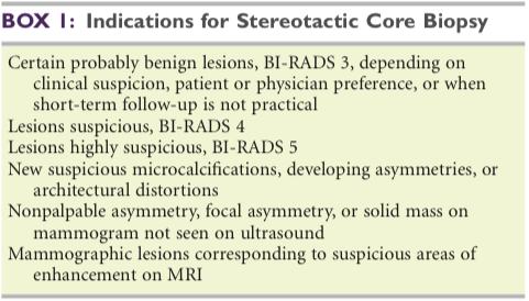
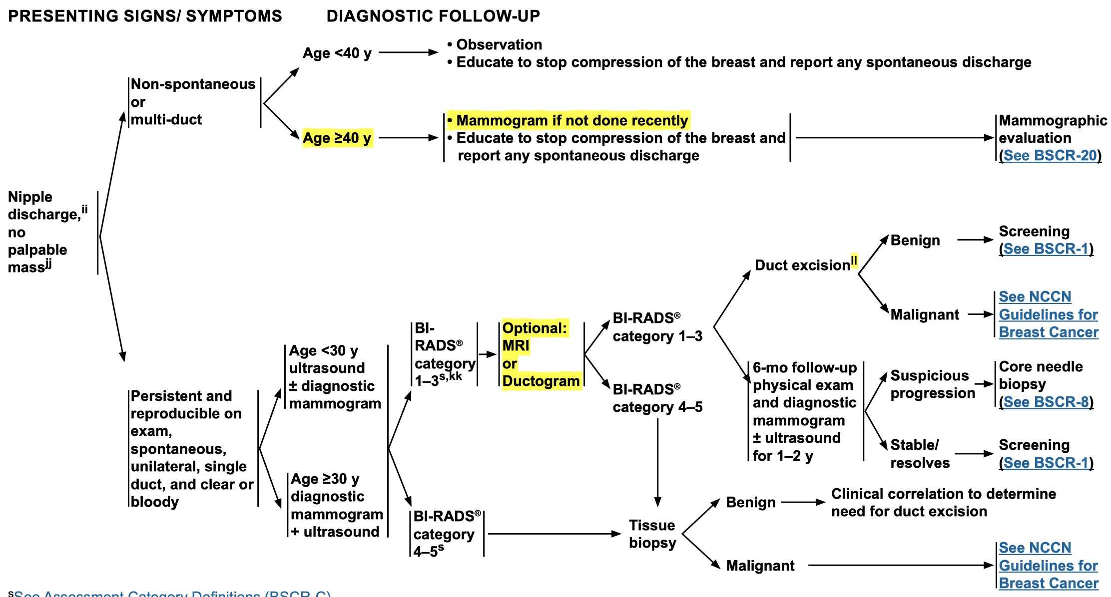
ll: Based on clinical suspicion and patient preference.
i.e With concerning clinical findings & benign workup, the investigation ladder progresses from US/MMG to MRI/ductogram to duct excision to assess for the discordance
*
It is rarely associated with an underlying carcinoma if there is no palpable mass or suspicious mammogram
>
Physiologic discharge:
*
Can be induced from several ducts by nipple manipulation
*
Does not warrant further evaluation
>
Features requiring further evaluation with US/MMG
*
Spontaneous
*
Recurrent
*
Unilateral
*
Involving a single duct
*
Bloody or clear
*
New occurrence in a woman > 50Y
V
>
Management of nipple discharge with worrisome features:
Memory tip: With concerning clinical findings & benign workup, the investigation ladder progresses from US/MMG to MRI/ductogram to duct excision to assess for the discordance
*
Bloody or clear discharge warrant cytologic assessment but it is of very low sensitivity & specificity
V
*
If a specific lesion is identified on imaging, then it warrants tissue Bx
Ductogram may be useful: the fluid-producing duct is cannulated and contrast material is injected, then a MMG shot
On ductography, cancer appears as an irregular mass or as multiple intraluminal filling defect
V
*
Ductogram/ductoscopy/MRI are indicated if US/MMG are negative
Ductoscopy is done with a 3mm scope and has no therapeutic channel. The procedure is done in the OR because of the need to dilate the nipple
V
Options if no suspicious lesion is identified:
V
V
Microductectomy: Excisional biopsy of the duct in question is warranted
Cannulation of the offending duct with a lacrimal probe is helpful
*
Excise 1-2 mm around the duct
*
Options for failed localization: abort vs perform major duct excision
V
*
Subareolar ductal exploration with duct ligation & excision
The patient may be unable to breast-feed after this procedure
V
V
Indications for nipple duct excision:
Source: SCORE
*
Spontaneous nipple discharge
*
Bloody nipple discharge
*
Subareolar mass & nipple discharge
*
Persistent subareolar infection
>
Etiology
V
Majority are benign
>
Intraductal papilloma
*
It is the most common cause of bloody nipple discharge (even in women with high risk of breast cancer)
V
*
If it is visible on imaging, it will need a biopsy to rule out atypia or carcinoma
64% will show upgrade on pathological examination
V
>
Indications for surgical excision
UTD: newer data suggest that not all papillomas diagnosed by CNB require excision.
V
Definite indications
V
*
Atypia on biopsy
Papillomas with atypia have a risk of 10-15% of concomitant malignancy.  Therefore, surgical excision is recommended.
*
Image discordance
*
High risk patients / patient preference
V
Relative indications
*
> 1 cm
*
Symptomatic
*
Age > 60
*
Nipple discharge
*
A small papilloma with no atypia may be managed with vacuum-excision
*
Surgical approach is through a circumareolar incision
*
Duct-ectasia
*
Fibrocystic breast disease
V
*
Mild inflammation
As per General Surgery Review Course
*
Galactorrhea: milk secretion can continue up to 6 months post-partum and post cessation of breast feeding
*
Malignant: DCIS, invasive cancer
V
V
Hyperprolactinemia (milky discharge)
No breast surgical intervention is required, and they are referred to endocrinologist for further evaluation
*
Primary pituitary tumor
*
Hypothyroidism
*
Medication related
>
Neoplasia & malignancy
>
General
>
Breast cancer is:
*
The most common site-specific cancer in ♀
*
The leading cause of death from cancer in ♀ between 20-60Y
*
The second leading cause of cancer-related deaths (after lung cancer)
*
Responsible for 15% of cancer-related deaths in ♀
*
Occurs at a younger age distribution in African Americans
*
Average lifetime risk = 12%
>
Multicentricity = a 2nd breast cancer outside the breast quadrant of the primary cancer (or > 4cm away)
*
Occurs in 40-80% of DCIS
*
Occurs in 60-90% of LCIS
*
Multifocality = a 2nd breast cancer within the same quadrant (or within 4 cm) of the primary cancer
>
Risk factors
V
>
Hormonal
Terminal differentiation of breast epithelium associated with full-term pregnancy is protective
>
Exposure to estrogen
*
↑ Number of menstrual cycles
*
Early menarche
*
Nulliparity or older age at 1st live birth (age > 35Y)
*
Late menopause
*
Postmenopausal HRT for 4 years → 3-4 fold risk of breast cancer + no ↓ in CAD or strokes
>
Nonhormonal
V
*
Dense breast
With use of average breast density as a reference point, the risk among women with heterogeneously dense breasts is 1.2 times as great as the average, and with extremely dense breasts, 2.1 times as great. This is equivalent to the elevated risk of breast cancer associated with having a first-degree relative with unilateral, postmenopausal breast cancer. Breast density does not appear to be associated with increased mortality from breast cancer
*
Radiation exposure (75X greater risk)
*
↑ Risk with corresponding ↑ EtOH consumption
V
>
They can be generally divided into 7 broad categories:
From Sabiston
V
*
Age
“Age is probably the most important risk factor for breast cancer development”
V
*
Family history of breast cancer
Family history of breast cancer and atypical hyperplasia increases the risk of developing breast cancer to 9X that of the general population
V
*
Hormonal factors
Including the use of OCP for premenopausal women and HRT for postmenopausal women
Women who receive combination HRT with estrogen and progesterone for 5 years have approximately a 20% increase risk of breast cancer development. Women who take estrogen-only formulations do not suffer from that increased risk
*
Proliferative breast disease
*
Irradiation of the breast or chest wall at an early age
*
Personal history of malignancy
*
Lifestyle factors
>
Mixed connective & epithelial tumors
>
Phyllodes tumor
*
On MMG: indistinguishable from fibroadenoma
*
Core needle biopsy cannot distinguish fibroadenoma from Phyllodes
*
Cytologic analysis is unreliable in differentiating low-grade phyllodes from fibroadenoma
V
*
The Dx is suggested by the larger size, history of rapid growth, and occurrence in older patients
Thus, the final diagnosis is best made by excisional biopsy, followed by careful pathologic review
*
Can be locally aggressive & recurrent
V
*
Metastasis occurs from hematogenous spread. Usual sites: lung, bone, abdominal viscera, & mediastinum
Minimal success is seen with systemic therapeutic agents in metastatic disease
V
>
Classified to benign, borderline, or malignant based on:
Source: UTD
Benign = ↑ stromal cellularity with mild-moderate atypia, circumscribed tumor, < 4 mitoses/10HPF, & lack of stromal overgrowth
Borderline = greater atypia, 4-9 mitoses/10HPF, microscopic infiltrative borders, & lack of stromal overgrowth
Malignant = Marked atypia, infiltrative margins, > 10mitoses/10HPF, presence of stromal overgrowth
*
Degree of stromal cellular atypia
*
Mitotic activity
*
Infiltrative or circumscribed tumor margin
V
*
Presence or absence of stromal overgrowth (i.e presence of pure stroma devoid of epithelium)
This feature is the most one most consistently associated with aggressive/metastatic behavior
>
Management
V
>
Surgical
Positive margins require re-excision
*
Benign: wide local excision if negative margins are obtainable
*
Borderline: wide local excision with 1 cm margin to prevent local recurrence
V
*
Malignant: treated similar to soft tissue sarcomas: complete excision with a margin of normal tissue is advised
Similar to other soft tissue sarcomas, regional lymph node dissection is not required for staging or locoregional control.
Malignant phyllodes tumors rarely metastasize to the axillary/lymph nodes and staging is not indicated with or without sentinel lymph node biopsy. Core biopsy is an accurate diagnostic procedure for this diagnosis. Wide excision with negative margins is appropriate; genetic testing is not indicated for this diagnosis
*
Adjuvant radiation is offered to borderline and malignant phyllodes after resection
*
Adjuvant chemotherapy is reserved for highly selected patients with large, high-risk, or recurrent phyllodes
*
No hormonal therapy is indicated
>
Recurrence:
*
Recurrence usually occurs within the first 2 years after excision
*
Local recurrence that is resectable: wide re-excision followed by radiation
*
Resectable pulmonary metastasis should be resected
*
Unresectable recurrence: palliative radiation
>
Angiosarcoma
*
Usually occurs after irradiation of the breast
*
May develop in the upper extremity of patients with lymphedema post-radical mastectomy
*
Clinically: reddish brown to purple raised rash within the radiation portals & on the skin of the breast
*
MMG is unrevealing in most cases
*
Histologically: composed of an anastomosing tangle of blood vessels in the dermis and superficial subcutaneous fat
V
*
Grading is based on the appearance and behavior of endothelial cells
Pleomorphic nuclei, frequent mitoses, and stacking of the endothelial cells lining neoplastic vessels are features seen in higher grade lesions
*
Resection is advised in absence of metastatic disease. Frequently requiring split-thickness skin graft or myocutaneous flaps
*
Radiation therapy is of benefit in primary angiosarcoma but not for radiation-related angiosarcoma
*
Chemotherapy is recommended in the adjuvant setting and improves outcomes
V
*
Recurrence is high
Median time to recurrence is 8 months; & the median survival is 2 years
*
Metastasis occurs hematogenously to: lung, bones, abdominal viscera, brain & the contralateral breast
*
Carcinosarcoma
*
Adenocarcinoma
V
>
Molecular markers & targets in breast cancer
SCORE: Aberrant expression of E-cadherin can be shown immunohistochemically and is used to distinguish between lobular and ductal phenotypes
V
>
Pathways and markers reported to affect breast cancer outcomes:
There are numerous pathways and molecular markers that have been reported to affect breast cancer outcomes, including steroid hormone receptor pathway (ER and PR), human epidermal growth factor receptor pathway (HER family), angiogenesis, cell cycle (e.g., cyclin-dependent kinases [CDKs]), apoptosis modulators, proteasome, cyclooxygenase-2 (COX-2), peroxisome proliferator-activated receptor-γ (PPAR-γ), insulin-like growth factors (IGF family), transforming growth factor-γ (TGF- γ), platelet-derived growth factor (PDGF), and p53. Most these markers are not routinely tested on breast cancer specimens at the time of diagnosis nor would it be feasible to do so.
*
ER: Assessment of ER is performed on all primary tumors
>
HER-2
*
Is amplified in ~ 20% of breast cancers
*
Four receptors make up the Epidermal Growth Factor Receptor family: HER1, HER2, HER3, HER4
>
The best characterized of the EGFR family is HER2
V
*
The HER2 gene, also known as, HER2/neu or ERBB2 is a proto-oncogene located on chromosome 17q21
HER-2 or erb-B2/neu protein is the product of erb-B2 gene
*
Is measured by IHC and is scored on a scale from 0 to 3+
*
FISH directly detects the quantity of HER2 gene copies (normal copy number is 2)
*
HER2 testing is a standard part of pathologic reporting on primary tumors
V
>
Molecular profiling of breast cancer
Pasted_Graphic_6
V
V
ER-Positive
Accounts for ⅔ of all breast cancers
Are generally low grade with favourable prognosis
ER expression measures ER-α levels (not ER-β) by IHC
V
*
Luminal A
They are the most common subtype
Have higher expression of ER → have the best response to endocrine therapy
Low Ki-67
V
*
Luminal B
Have worse prognosis compared to Luminal A, but superior prognosis compared to Basal-like and HER2-enriched tumors
Some Luminal B subtypes are HER2⊕
High Ki-67
V
*
HER2-Enriched
Account for 10-15% of all breast cancers
These tumors are usually HER2⊕ & ER/PR⊖
These breast cancers often are associated with high-grade, & axillary lymph node involvement.
These characteristics alone lead to increased risk of recurrence and decreased survival.
V
*
Basal-like (usually referred to as triple-negative breast cancer but are not identical)
Has a gene cluster profile similar to basal epithelial cells and is characterized by low expression of the luminal and HER2 gene clusters
ASCO/CAP guidelines used to define TNBC include lack of ER/PR (<1%) and HER2 expression (0 or 1+) by IHC and confirmation of HER2 status by fluorescent in situ hybridization (FISH) if indeterminate (2+) by IHC.
Triple negative breast cancer accounts for about 10% to 20% of all breast cancers and is biologically the most aggressive
These tumors also tend to be larger, palpable, higher grade, and more often seen as interval cancers (between mammograms).
Distant metastases often occur at visceral sites and are seen within the first 3 years after diagnosis.
*
Normal-like
V
>
Treatment according to subtype
Pasted_Graphic_7
>
ER⊕
>
Endocrine resistance
*
Is either: de novo Vs acquired during treatment
>
Methods of overcoming resistance:
*
Optimizing schedule & dose of therapeis
*
Combining different agents
*
Target crosstalk between ER & other signaling pathways (such as HER2, CDK, PI3K/AKT)
>
Addressing overtreatment
>
'Oncotype Dx' = 21-gene reverse transcriptase–PCR assay
*
Allows for individualized risk estimate or recurrence score for each tumor sample
*
NCCN breast cancer guidelines recommend the use of gene expression profiles in patients with ER-positive, node-negative breast cancers to assess the benefits of additional therapy
>
Recurrence Score range from 0 to 100
*
Low risk: < 18 → hormone therapy alone is adequate
*
Intermediate risk: 18-30 → the benefit of chemotherapy followed by hormonal therapy is being tested in the TAILORx trial
*
High risk: ≥ 31 → patients would benefit from chemotherapy followed by hormonal therapy
>
PAM50 genomic assay
*
PAM50 gives a Prosigna Score (0-100)
*
Indicated for postmenopausal ♀ with ER⊕, node-negative, breast cancer
V
*
MammaPrint = 70-gene microarray assay
It gives a low-risk or high-risk result to provide assistance with determining the benefit of adjuvant chemotherapy in selected patients with ER-positive or ER-negative disease
>
HER2⊕
*
Trastuzumab is an inhibitor of the HER-2 tyrosine kinase-linked surface receptor
*
Trastuzumab along with chemotherapy is indicated for tumors > 1 cm or N⊕ disease that is > 2 mm. It’s ‘considered’ for smaller tumors & smaller nodal-metastatic disease
*
Regimen is given for 18 months
*
Trastuzumab may increase the risk of cardiomyopathy seen with chemotherapy
*
Sabiston: The use of targeted therapies, such as trastuzumab, in combination with chemotherapy can be safely administered in the neoadjuvant setting in HER2⊕ breast cancer patients, resulting in markedly increased rates of pathologic complete response
V
Overcoming HER2 resistance
*
Resistance is: de novo Vs acquired during treatment
V
Options for HER2⊕ metastatic breast cancer:
V
*
Pertuzumab in combination with docetaxel & trastuzumab
Based on results from the NeoSphere phase II study demonstrating that pertuzumab increased pathologic complete response rate, the FDA subsequently approved the use of pertuzumab in combination with trastuzumab and chemotherapy for early-stage HER2-positive breast cancers in the neoadjuvant setting.
*
Lapatinib in combination with capecitabine is indicated for transtuzumab-refractory disease
>
Triple negative cancer
*
The standard care is: anthracycline + taxane-based combination chemotherapy
V
*
Triple-negative paradox: they are the tumors with the poorest prognosis but the most sensitive to chemotherapy
20-5% of TNBCs achieve pCR after anthracycline or anthracycline/taxane-based treatments
*
Triple negative cancers lacking functional BRCA1/BRCA2 might benefit from PARP-inhibitors (olaparib, iniparib, veliparib)
V
>
Noninvasive epithelial breast lesions
Screen Shot 2020-04-12 at 6.45.08 PM
*
CIS: do not invade the basement membrane
*
Accurate Dx necessitates analysis of multiple microscopic sections to exclude invasion
>
Classification
*
Nonproliferative changes: include mild-moderate hyperplasia within the breast ducts. They do not confer an increase risk in breast cancer development
V
*
Proliferative epithelium without hyperplasia (AKA severe hyperplasia or Usual Ductal Hyperplasia) require no treatment
Do not increase the risk of breast cancer
Doesn’t need F/U imaging
V
>
Types of proliferative lesions with atypia:
UTD: Atypical hyperplasia & LCIS are managed as risk indicators rather than precursor lesions, as the cancers that subsequently develop may occur in either breast & not necessarily at the site of atypia
Screen_Shot_2019-06-25_at_13.27.34
Screen Shot 2020-04-06 at 1.00.16 PM
V
>
Atypical Hyperplasia
AH confers a substantial increase in the risk of subsequent breast cancer (relative risk 3.7 to 5.3)
AH is associated with a generalized, bilateral increase in breast cancer risk
Patients with both ADH and ALH should be offered active surveillance and options of chemoprevention.
SCORE: There are two types of atypical hyperplasia, atypical ductal hyperplasia, and atypical lobular hyperplasia. Both occur with equal frequency, and both predispose to similar risk of invasive breast cancer.
>
Atypical Ductal Hyperplasia
*
Histology: shares the cytologic & architectural features of DCIS but is of limited extent (< 2mm)
*
After wide local excision of ADH, there is 20% rate of upstaging to DCIS or invasive disease
>
Management
*
After core biopsy: Wire-localization breast biopsy is the standard of care
*
After surgical excision: no additional surgery is indicated; re-excision is not indicated when ADH is present at the margin
>
Indications for re-excision when ADH is present at the margin:
*
If the Dx is bordering on reaching criteria for DCIS at the margin
*
Concern that the imaging target was not completely excised
>
Atypical Lobular Hyperplasia
*
It is usually an incidental finding on breast Bx performed for other reasons
*
Histology: ALH shares cytologic & architectural features of LCIS but is quantitatively lesser in extent
>
Management
>
After core biopsy: incidental radiologic-pathologically concordant ALH diagnosed no longer require excision, provided excision is not indicated for the target lesion
*
Any discordant lesions do require surgical excision
*
After surgical excision: no additional surgeries indicated, even if ALH is present at the margin
>
Flat Epithelial Atypia (FEA) AKA ‘columnar cell changes with atypia’
*
Atypia which does not meet criteria for ADH or DCIS
*
FEA is present in < 2% of core-needle Bx specimens done for micro-calcifications
*
FEA seems to represent a non-obligate precursor lesion to low-grade DCIS and tubular carcinoma
>
Management
*
Radiologic average-risk surveillance is sufficient for radiologic-pathologic concordant cases in absence of residual MMG calcifications
*
When diagnosed after surgical excision: no further management is necessary
*
No chemoprevention needed
>
Carcinoma In Situ
>
LCIS
*
Occurs 12X more frequently in White than African American
*
85% occur in premenopausal ♀
*
LCIS cells are strongly ER ⊕
*
It is recognized by its conformity to the outline of the normal lobule, with expanded & filled acini
*
Originates from the terminal duct lobular units (develops only in ♀ breast)
*
Cytoplasmic mucoid globules are a distinctive cellular feature
*
Occurs bilaterally in 50-70%
>
Risk for invasive breast cancer:
*
LCIS is regarded as a marker of ↑ risk of invasive BC rather than as an anatomic precursor. The magnitude of risk associated with LCIS is much greater than with ALH
*
Invasive breast cancer develops in 30% of ♀ with LCIS
*
Invasive cancer occurs synchronously with LCIS in 5%
*
LCIS is usually diagnosed incidentally on a breast biopsy performed for some other reason
>
Histologic classification (alters management):
*
Classic LCIS
*
Pleomorphic LCIS
*
Florid LCIS with comedo necrosis
>
Management
V
After core Bx:
*
Surgical excision is recommended for any nonclassical LCIS, any LCIS with imaging-pathologic discordance, or extensive/multifocal LCIS
>
Classical LCIS without imaging-pathology discordance can be managed in either of 3 ways:
*
Close observation
V
*
Chemoprevention with tamoxifen or raloxifene
Provides 56% reduction in breast cancer risk
V
*
Bilateral mastectomy
Is the recommended procedure for patients who elect for surgery
Most experts now consider prophylactic bilateral mastectomy too drastic for the moderate level of risk associated with LCIS in the absence of other contributory risk factors
>
After surgical excision:
*
Classic LCIS: no further surgery is required even if LCIS occurs at the margin, as long as it’s classic LCIS
V
*
Pleomorphic LCIS: re-excision to negative margins is recommended if present at the margin
There are no data on the optimal width of negative margin or the benefit of radiation therapy for patients with pleomorphic LCIS
*
F/U in high-risk clinic & consider breast MRI
*
Radiation therapy is not indicated
>
DCIS / Intraductal carcinoma
*
25% of breast cancer diagnosed in the US is DCIS (less common than invasive breast cancer)
*
DCIS is uncommon in women < 30 years
*
They usually present with a cluster of microcalcifications
*
Risk of development of metastases and/or death in pure DCIS is rare (<1%)
*
Characterized by proliferation of the epithelium that lines the minor ducts → papillary growths within the duct lumina → comedo growth pattern (outgrow their blood supply) → calcification at necrosis (usually without an associated palpable abnormality on examination)
*
It is recognized as discrete spaces filled with malignant cells with recognizable basal cell layer of normal myoepithelium
V
*
Carries high risk for progression to invasive BC
Risk for invasive cancer is increased 5-fold in ♀ with DCIS
*
DCIS transforms into an invasive cancer, usually recapitulating the morphology of the cells inside the duct
*
The rate of upstaging to either microinvasion or invasive cancer is ~ 15%
*
Occurs bilaterally in 10-20% of cases
>
Assessing grade and risk of recurrence
V
>
4 Pathological types:
The four morphologic categories of DCIS are rarely seen as pure lesions, but in reality are often mixed
>
Lower Grade:
*
Papillary
*
Cribriform
>
Higher Grade:
*
Solid
*
Comedo (has the worse prognosis of all DCIS)
>
Van Nuys score
*
Is sometimes used to advocate for mastectomy rather than breast conserving surgery
*
↑Score = ↑recurrence
>
Score elements
*
Size
*
Margin
*
Grade
*
EIC (extensive intraductal component) & central necrosis are indicative of high recurrence
V
>
Work up for suspicion of DCIS: diagnostic bilateral MMG with magnification views
Findings suggestive of DCIS on MMG:
- linear branching or segmental types of pleomorphic microcalcifications
- comedo type lesions
*
MMG often underestimates the extent of DCIS and the number of tumor foci in instances of multifocal disease
>
Management:
V
Excision with ≥ 2 mm negative margin (breast conserving surgery vs mastectomy)
>
Criteria for breast conserving surgery:
*
Multifocal disease is not a contraindication
*
Multicentric disease is a relative contraindication
*
Cosmetically acceptable resection is achievable
V
*
Ability to obtain negative margins (i.e lesion is not < 2 mm from the skin or muscle)
Negative margins are defined by tumor-filled ducts separated by a measurable distance from the inked surface (ie, 2 mm).
*
No contraindications to radiation therapy
>
Regarding mastectomy:
*
Women treated with mastectomy are candidates for immediate breast reconstruction
V
*
SLNBx is considered for ♀ requiring mastectomy for DCIS
Mastectomy will alter lymphatic drainage and make subsequent SLNBx unreliable. DCIS may be upgraded to invasive ductal cancer on final pathology necessitating axillary assessment.
>
If breast conserving surgery yields a questionable margin:
*
1. Postexcision MMG is obtained prior to initiation of radiation
*
2. Residual suspicious calcifications warrant image-guided re-excision
V
*
Adjuvant whole-breast radiation therapy is indicated following breast conserving surgery, except in low-risk disease
4 Randomized trials have shown that adjuvant RT significantly reduces the risk of in-breast tumor recurrence by ≥ 50% compared with excision alone
It is reasonable to omit in selected patients with advanced age, extensive comorbidities, or small foci of low-grade disease resected with wide negative margins.
2 Gy daily fractions over 5 weeks = 50 Gy
V
*
Adjuvant post-mastectomy radiation is indicated for positive margins at the chest level or skin level
Source: General Surgery Review Course
V
>
SLNBx can be avoided in most women, but should be obtained in high-risk features as excision may compromise the ability to perform SLNBx in the future
Omitting SLNB at the time of breast-conserving surgery for DCIS decreases perioperative morbidity
If invasive breast cancer is identified after a breast-conserving surgery is performed for DCIS (occurs in 10-20%), SLNB can be performed as a second procedure
>
Indications for SLNBx in DCIS:
*
DCIS requiring mastectomy
>
DCIS with high suspicion for invasive disease:
*
High-grade
*
Large (> 5 cm)
*
Palpable DCIS
V
*
Antiestrogen therapy X5 years is administer for ER/PR ⊕ after breast conserving surgery or mastectomy (but not bilateral mastectomy)
Following mastectomy it offers prevention for the contralateral breast. Antiestrogen therapy is not offered after bilateral mastectomy
Sabiston: Patients at highest risk for local recurrence, and therefore those most likely to benefit from tamoxifen, were patients with positive margins, comedo necrosis, a mass on physical examination, and age younger than 50 years
V
*
General Surgery Review Course: Following local treatment of DCIS, the decision for endocrine therapy is based the receptor status. However, tamoxifen is not given for risk reduction in the contralateral breast if mastectomy is done
General Surgery Review Course
*
There is no current indication for HER2-directed therapy in the management of DCIS
*
Ongoing trials are assessing whether any treatment is necessary for low grade DCIS
>
Surveillance (2019 NCCN DCIS):
*
Interval history and physical exam every 6–12 mo for 5 y, then annually
*
Mammogram every 12 mo (first mammogram 6–12 mo after breast conservation therapy)
*
GYN exam Q12m while on tamoxifen & uterus is present
>
Recurrences:
*
Local recurrence after optimal therapy is 10-15%
*
½ of all loco-regional recurrences are invasive & are staged & treated as newly diagnosed invasive breast cancer
V
>
Management is based on prior intervention:
Summary:
- The recurrence is excised
- Mastectomy is indicated if not candidate for radiation
- Radiation therapy is given if not previously given
- Antiesterogen is given if not previously given
*
Prior breast conserving surgery + radiation: mastectomy is usually performed because they are not candidates for further radiation therapy
>
Prior breast conserving surgery without radiation, options:
*
Repeat excision + radiation
*
Mastectomy
*
Prior mastectomy with recurrence in the mastectomy flap: wide local excision + radiation
*
Antiestrogen therapy is indicated if not given previously
V
*
UTD: We offer endocrine therapy as chemoprevention for women with high-risk breast lesions (regardless of receptor status)
UTD also mentions: chemoprevention has not been shown to confer a survival advantage to patients with high-risk lesions.
>
Invasive/infiltrating epithelial breast cancer
*
Account for 50-70% of invasive breast cancer
>
Invasive cancers are recognized by (either):
*
Lack of overall architecture
*
Infiltration of cells haphazardly into a variable amount of stroma
*
Formation of sheets of continuous monotonous cells without respect for form & function of a glandular organ
>
Classification
>
Paget’s disease of the nipple
*
Characterized by chronic, eczematous eruption of the nipple with possible ulceration
>
Usually associated with extensive DCIS or invasive cancer in the large sinuses under the nipple
*
Paget cells do not invade through the dermal basement membrane and are therefore categorized as CIS
*
> 95% have an underlying breast carcinoma
*
Biopsies are required: skin biopsy of the nipple-areolar complex ± core Bx of deeper lesion if present
*
Usually starts at the nipple and spreads outwards as opposed to benign causes of eczema that start at the skin then involve the nipple
*
A palpable mass may not be present
*
Pathognomonic feature: large, pale, vacuolated cells (Paget cells) in rite pegs of the epithelium
*
MRI is required prior to treatment as 50% are MMG-occult
*
Managed according to the extent of involvement of invasive cancer
V
>
Management
Screen_Shot_2019-06-15_at_6.27.13_PM
>
Paget’s + palpable mass or imaging abnormality:
*
The nipple-areolar complex & underlying cancer must be excised
V
*
Breast conserving surgery with radiation is not contraindicated if adequate margins are obtainable
If the breast is large, breast conserving surgery with reconstruction and contralateral breast reduction may be considered
>
Mastectomy is indicated for:
*
Many women with PDB and a palpable mass are not amenable to BCT because of the distance between the primary tumor and the nipple-areolar complex
*
Multicentric cancer or diffuse calcifications are best managed with mastectomy
V
>
Paget’s + no palpable mass or imaging abnormality:
UTD: Despite the absence of a palpable mass or mammographic abnormality, underlying carcinoma is present in most patients with PDB. The majority will have DCIS, but invasive cancer is present in one-fourth to one-third of cases
*
Obtain MRI to ensure no missed lesion
*
Central lumpectomy or complete resection of the nipple-areolar complex followed by whole breast radiation is a reasonable alternative to mastectomy as long as good cosmetic outcome and negative margins can be achieved
V
*
Paget’s without mass & clinically negative axilla: there is controversy as to the need for axillary evaluation when breast conservation is planned.
UTD: For patients with pure PDB without an associated mass and a clinically negative axilla, there is some controversy as to the need for axillary evaluation when breast conservation is planned. Given the high incidence of DCIS alone in these patients, SLN biopsy may not be necessary and can be considered as a second operation if invasive disease is discovered. In a series of 19 patients with PDB only, an invasive component was found in 27 percent and positive SLNs in 11 percent. Based on these numbers, some consider the likelihood of invasive disease high enough to recommend an SLN biopsy in this situation routinely.
>
Evaluation and treatment of the axilla in PDB are the same as for any breast cancer
*
Patients with DCIS do not require axillary investigation unless the disease is extensive enough to merit mastectomy
*
For invasive, N0 disease: SLNBx at the time of wide local excision
*
cN⊕ or suspicious LN → US FNA → ALND if pN⊕
*
NCCN guideline suggests that radiation can be omitted if SLNB is done with central lumpectomy
>
Ductal
*
Tend to grow as a cohesive mass
*
Almost always is E-cadherin ⊕
V
>
When infiltrating ductal carcinomas take on differentiated features, they are named according to the features that they display:
To qualify as a subtype, at least 90% of the cancer must contain the defining histologic features
V
>
Invasive ductal carcinoma not otherwise specified (IDC NOS) is the most common form of breast cancer
Schwartz: 80% of invasive breast cancers are described as “invasive ductal carcinoma of no special type (NST)”
*
Occurs usually in perimenopausal or postmenopausal ♀
*
Has poorly differentiated margins
*
60% present with ⊕ LN metastasis (macroscopic or microscopic)
*
They have worse prognosis than type-specific cancers
>
Low grade
V
>
Infiltrating tubular carcinoma: cells for small glands lined by a single row of bland epithelium
Accounts for 2-3% of invasive breast cancer
*
Long term survival is ~100%
V
*
Mucinous or colloid tumors: secrete copious amounts of mucin & appear to float in this material
Accounts for 2-3% of invasive breast cancer
Because of the mucinous component, cancer cells may not be evident in all microscopic sections
>
High grade
V
>
Medullary cancer
Accounts for 5% of invasive breast cancer
The borders of the tumor push into the surrounding breast rather than infiltrate or permeate the stroma
*
It is a frequent phenotype of BRCA1 breast cancer
*
Benign/hyperplastic LN enlargement may contribute to erroneous staging
V
*
Medullary variant
ER & PR negative, HER2 negative
>
In regards to resection margin
*
ASCO suggests no tumor on ink is adequate resection for invasive breast cancer
*
Excision of invasive cancer with finding of DCIS close to the margin: do not re-excise
*
Excision of invasive cancer with finding of positive margin for DCIS: re-excision is warranted
*
Posterior margin should be pectoralis fascia
*
Anterior margin is a ‘think skin flap’ — likely not candidate for re-excision even if close
>
Lobular
*
Invasive lobular carcinoma accounts for ~10% of breast cancers
V
*
Invasive lobular cancer tends to permeate the breast in a single-file nature
This explains why it remains clinically occult and often escapes detection on mammography or physical examination until the extent of the disease is large.
*
Tends to be E-cadherin ⊖ (CDH1 mutations)
*
Carries an intermediate prognosis between basal-like triple negative cancer and tubular cancer
V
*
Management of invasive lobular cancer is similar to that of invasive ductal cancer (the incidence of contralateral cancer with invasive lobular cancer does not differ greatly from that of patients with invasive ductal carcinoma; therefore, prophylactic contralateral mastectomy is not routinely indicated)
Source: SCORE
>
Other types:
*
Squamous cell
*
Apocrine
*
Adenoid cystic
V
>
Papillary
Advice below from the General Surgery Review Course
*
Treat as non-invasive (DCIS)
*
No need to stage the axilla
>
Medullary
*
May be associated with BRCA1
*
Often triple negative
V
>
Inflammatory breast cancer
Inflammatory breast cancer is the most aggressive subtype of breast cancer with significant LN & distant metastasis
*
It should always be suspected in non-lactating, postmenopausal women without any precipitating factors or systemic signs of infection
*
If inflammatory signs do not respond within 24-48h response to antibiotics, Bx of any underlying abnormality should pursued
*
Progression of inflammatory breast cancer usually happens over a period of weeks to 3 months
>
The Dx is both clinical & pathologic
*
Histologic finding: adenocarcinoma in the dermal lympatics within the breast & overlying skin
*
Finding carcinoma in the dermal lymphatics without the associated rapid onset of erythema and edema is not inflammatory breast cancer.
*
A negative skin biopsy does not exclude the Dx and is usually due to sampling error — consider MRI
*
Often the Dx is made without a palpable underlying tumor mass
>
AJCC Diagnostic criteria for IBC must include all:
*
Rapid onset erythema, edema/peau d’orange, warm breast with or without an underlying mass
*
Duration of history < 6 months
*
Erythema involving ≥ ⅓ of the breast
*
Histologic confirmation of invasive cancer
*
Patients require metastatic workup (see below)
V
>
Management
Screen_Shot_2019-06-15_at_6.37.21_PMScreen_Shot_2019-06-15_at_6.37.29_PM
*
NACT → MRM → radiation
*
SLNB is not accurate in inflammatory breast cancer
*
Failure to respond to NACT warrants alternative regimens ± radiation, not surgery
>
Male breast cancer
*
Given the absence of terminal lobules in the normal male breast, lobular carcinoma, both invasive and in situ, is rarely seen
V
>
Risk factors:
Gynecomastia is not a risk factor
*
BRCA gene mutations, especially BRCA2
*
Age
*
Radiation exposure
*
Estrogen/androgen imbalance (including cirrhosis)
*
Klinefelter’s syndrome (47, XXY karyotype)
*
Family History
>
Tumor biology:
*
90% are invasive ductal carcinomas
*
80% are ER ⊕
*
35% overexpress HER2
*
Treatment & staging follows same guidelines for female breast cancer
*
SCORE: While men may present later with breast cancer than women, stage for stage, their prognosis is the same
V
*
Adjuvant hormonal therapy (tamoxifen or aromatase inhibitors) is indicated for N⊕ and high-risk N⊖ disease
Options for adjuvant endocrine therapy for men with breast cancer include tamoxifen for 5–10 years or, if tamoxifen is contraindicated, a GnRH analog plus an aromatase inhibitor. In men, single-agent adjuvant treatment with an aromatase inhibitor has been associated with inferior outcomes compared to tamoxifen alone, likely due to inadequate estradiol suppression, and is not recommended.
>
Implant related lymphoma
*
Have a high index of suspicion for patients with ‘abrupt seroma’ development after surgery
*
Dx: send capsular fluid for lymphoma protocol
*
Treatment: removal of implant and capsule
>
Work up & Staging
>
Clinical staging:
*
Examination of bilateral breasts & nodal basins (axillary, supraclavicular, infraclavicular, & cervical)
*
If not otherwise indicated (with a negative clinical examination), radiologic staging of the axilla is not necessary (but most surgeons will obtain it anyway)
*
Thorough imaging of the ipsilateral and contralateral breast is performed to look for additional areas of concern other than the index lesion)
*
If there is no palpable disease in the axilla, performing US for the axilla is not a necessity
V
*
If NACTx is indicated, axillary sonography is done with Bx of LN with thickened cortices (> 3 mm)
For patients who will be receiving neoadjuvant chemotherapy, axillary sonography and sonoguided needle biopsy of lymph nodes with thickened cortices (>3 mm) is useful because it establishes initial nodal stage and also provides valuable prognostic information when compared with post-chemotherapy nodal status.
*
Investigations for all: CBC, LFT, CXR
>
CT chest/abdomen/pelvis, bone scans, & other imaging studies (PET scan optional) are generally reserved for:
*
Patients with abnormalities on blood chemistry
*
Abnormal CXR
V
*
Locally advanced breast cancer
Locally advanced breast cancer = Stages IIB, IIIA , IIIB = T2N1 or T3+ or N2+
*
Inflammatory breast cancer
*
Brain/spine MRI are indicated as per clinical suspicion
>
TNM - AJCC 8th Edition 2017
V
*
T
Pasted_Graphic_29
V
*
cN
Quick guide:
N1: palpable level I/II
N2: fixed level I/II or IM⊕ alone
N3: (⊕ level III infraclav.), ( ⊕ IM & level I/II), or (⊕ supraclavicular)
Screen Shot 2020-04-06 at 3.53.55 PM
V
*
pN
ITC = isolated tumor cell clusters = < 0.2 mm
Micrometastases = > 0.2 mm, but < 2.0 mm
Spread to ipsilateral supraclavicular node is N3c, but spread to contralateral supraclavicular node is M1
Screen Shot 2020-04-06 at 3.57.13 PM
Screen Shot 2020-04-06 at 3.57.57 PM
*
Ipsilateral axillary metastasis predicts outcome after surgical treatment more strongly than tumor size
V
*
M: most commonly to the liver, lungs, & bone
Screen Shot 2020-04-06 at 4.08.28 PM
V
*
Stage
Stage I:
- T1, N1mi or less
Stage III:
- any ≥ N2
- T3 with N⊕
- any T4
Screen Shot 2020-04-06 at 4.08.58 PM
V
*
Locally advanced breast cancer = Stages IIB, IIIA , IIIB = T2N1 or T3+ or N2+
Source: Sabiston
>
Work up of occult breast cancer presenting with axillary metastasis
>
US-Core biopsy of the LN with pathologic assessment for:
*
ER/PR
*
HER2
*
Cytokeratins 7 & 20
*
Mammaglobin
*
CEA
*
CA125
*
CT: Chest/Abdomen/Pelvis
*
Bone scan or FDG-PET
*
Bilateral breast MRI with biopsy of any suspicious lesion
V
Management for axillary disease from the an occult breast:
>
The breast
*
A lot of controversy exists in recommending mastectomy vs radiation vs close observation
>
The axilla
*
ALND if the axilla is operable
*
Chemotherapy if the axilla is not operable
>
Prognostic factors
*
1. Molecular/gene expression subtype
*
2. Stage (TNM)
*
3. Grade
>
Management
>
Overview of the sequence of therapy for patients planned for neoadjuvant therapy
>
1. Prechemotherapy
*
Assess complete extent of disease
*
Place clips at the site of tumor and LN
*
Categorize patients according to their molecular markers
V
*
2. Chemotherapy with ongoing communication with the oncologist to ensure there is no tumor progression
In the context of potentially operable disease, if there is confirmed progression at any time, mastectomy with surgical staging is performed
*
3. Repeat staging with imaging
*
4. Perform surgery 4-6 weeks after last chemotherapy
*
5. Radiation is delivered (6-8w) within 12w from the surgery date unless preceded by chemotherapy
*
6. Chemotherapy (~3-6m) can be given with Trastuzumab
*
7. Endocrine therapy (5-10Y) is given following chemotherapy
>
Breast resection (& reconstruction)
V
*
Cancer-specific & overall survival are the same for breast conservation and mastectomy
Ipsilateral breast recurrence rates were higher in patients undergoing breast-conserving surgery, but local recurrences could be salvaged by mastectomy at the time of recurrence, with no significant detriment in survival rates.
BCS
*
Patients unwilling to go for radiation therapy or surveillance should undergo mastectomy rather than breast conserving surgery
>
Breast conservation
*
Requirements for breast conserving surgery = ability to resect with adequate margins + acceptable cosmesis
V
>
It’s always safe to plan the incision directly over the tumor
mint_c027f001
*
Curved incision superior to the 9 o’clock to 3 o’clock horizon
*
Radial incision is used inferior to the 9 o’clock to 3 o’clock horizon
V
*
‘No ink on tumor’ is an acceptable negative margin but some margin distance is associated with ↓ recurrence
This is based on a meta-analysis that showed similar local recurrence rates for 2-mm or 5-mm margins as compared with 1-mm margins. However, local recurrence was statistically significantly greater for margins classified as “close” compared with those classified as “negative,” suggesting that there is some margin distance that would be associated with lower local recurrence risk than “no ink on tumor.”
*
A recommended resection margin is 1 cm (1.5-2 cm from the site of the clip is more appropriate for patients who have complete pathologic response)
>
Contraindications to breast conserving surgery
*
Active connective tissue disease involving the skin: scleroderma, lupus
*
Persistently positive margins after multiple surgical attempts
*
History of prior breast irradiation
*
Diffuse malignant appearing microcalcifications
>
Oncoplastic surgery:
*
Immediate reconstruction of the partial mastectomy defect is almost always preferred to a delayed approach
>
Initial surgical procedures: tissue advancement & local tissue rearrangement
*
They are not associated with a delay in delivery of adjuvant systemic therapy or radiation
*
Tissue expanders and implants are not recommended to fill partial mastectomy defects because radiation may end to capsular contracture, distortion, and infection
*
If cosmetic defect occurs following breast conservation & radiation, reconstruction is not recommended for up to 1-2 years after radiation is completed
>
Mastectomy
*
♀ < 50Y with triple-negative disease may have a survival benefit if they opt for bilateral mastectomy
*
Mastectomy is often required for tumors involving the skin or nipple
*
Positive margins are better managed with resection when possible (close margin < 1 mm is an indication for radiation)
>
Nipple-sparing mastectomy
*
Nipple-sparing mastectomy removes all of the glandular tissue while retaining all of the breast skin
*
Local recurrence rate is similar to skin-sparing mastectomy
V
*
Two-staged reconstruction that starts with sub-pectoral expanders yield good results
Immediate implant or autologous tissue reconstruction is associated with greater risk of skin or nipple necrosis especially when thin flaps are created.
V
>
Criteria for nipple-sparing mastectomy
It may be suitable for BRCA patients undergoing prophylactic mastectomy
Contraindications:
- Inflammatory breast cancer
- Paget’s disease of the nipple
- Clinically suspicious LN
- Imaging evidence of nipple-areola complex involvement
- Intraoperative frozen section positive subareolar histopathology
*
Tumor does not involve the nipple or skin
*
Tumor can be removed with negative margins
*
Incision & reconstruction choices can bring the nipple to an esthetically desirable level
>
Results are acceptable for:
*
Large or small breasts
*
Irradiated breasts
*
Breasts that will require radiation
*
Breasts previously undergone mastopexy or reduction
V
>
Risk factors for poor outcomes:
As per Dr. Tremblay
*
History of radiation: 27% infection; 10% skin necrosis; 5% nipple loss
*
Planned postoperative radiation
*
Smoking
*
BMI > 30
V
*
Large ptotic breast: higher risk of skin (10%) and nipple necrosis (<5%) — A & B cup-size are the ideal patients
There is no increased rate of complications with history of mastopexy, augmentation, or reduction
V
>
Technique
Screen_Shot_2019-04-26_at_14.46.04
*
A lateral inframammary incision provides excellent access to the entire breast and axilla with a relatively well-hidden scar
*
Subareolar breast tissue is sampled and submitted separately
*
Extension of DCIS to this level should prompt excision of the nipple
*
Skin-sparing mastectomy is performed through a total circum-areolar incision, which retains the nipple-areolar complex with the specimen
>
Breast reconstruction after mastectomy:
*
Immediate reconstruction does not have a detrimental effect on long-term survival, local recurrence rates, or detection of local recurrence
*
Reconstruction is usually delayed in patients with locally advanced cancer
>
Reconstruction options:
V
*
Tissue expanders & implants
SCORE: In the setting of planned post-mastectomy radiation therapy, a tissue expander is advisable in this situation, as the success and cosmesis of an autologous reconstruction will be compromised with radiation. 
V
*
TRAM flaps
SCORE: When compared with nonsmokers, patients who smoke are at increased risk for necrosis of the mastectomy skin flaps, the abdominal flap, and for the development of an abdominal hernia after breast reconstruction using a TRAM flap. Patients who have a smoking history >10 pack-years are at particularly high risk for the development of these complications. However, the risk can be managed and decreased significantly if a delay procedure (ligation of inferior epigastric artery) is performed, or if the patient stops smoking for at least 4 weeks before the surgical procedure. TRAM flap reconstruction has not been associated with an increased risk of wound dehiscence, bleeding, vessel thrombosis, or wound infection in patients who smoke.
h9991448_002
V
*
Latissimus dorsi flaps
h9991448_003
V
*
Deep Inferior Epigastric Perforator flap
DIEP Flap
*
VAC with consultation to plastic surgery
>
Options for skin necrosis
*
Skin graft
*
Latissmus dorsi flap
*
VAC dressing
>
The axilla
V
>
SLNBx
Published studies have demonstrated an average of 2-3 sentinel LN per patients
>
Lymphatic mapping
V
*
99m Tc sulfur colloid, isosulfan blue dye, or both can be used
A meta-analysis of 11 published studies was performed with 912 patients and compared injection techniques with lymphoscintigraphy or blue dye, or a combination of both, in patients who had SLNB followed by ALND for in situ and invasive cancer. This study reported a significantly higher rate of identification if radiocolloid and blue dye were used together or if radiocolloid was used alone than if blue dye was used alone.
Previous work has shown that for most surgeons, especially when first using the procedure, the combination of blue dye and radiocolloid has the highest success rate in SLN identification and the lowest false-negative rate. However, many experienced surgeons are comfortable with and highly successful at using a single agent, with some of the highest reported success rates.
>
Application:
*
Radioisotope or blue dye: subareolar, subdermal, or peritumoral
*
Intradermal injection is appropriate for radiocolloid but not blue dye (it may tattoo the skin)
*
Only radiocolloid has been shown to be safe in pregnancy
V
*
Radiocolloid preoperative 0.5 ml of 0.5 milliCurie of filtered 99m Tc sulfur colloid
Sabiston: A dose of 2.5 mCi of technetium-labeled sulfur colloid can be injected on the day prior to surgery for the preoperative lymphoscintigraphy
*
Blue dye: 3-5 ml, and then the area massaged for 5 minutes
V
>
Reasons for failure of uptake
Decision to proceed to completion axillary decision should reflect clinical suspicion of underestimated disease after considering above factors
>
Patient considerations
*
↑ Age: fatty tissue replaces breast tissue resulting in poor uptake
*
Previous axillary surgery
>
Tumor considerations (tumor may block lymphatics, thus limiting light up of a sentinel LN)
*
Size
*
Grade
*
ER/PR/HER
>
If uptake is found at the internal mammary
V
*
Do not go after it surgically
General Surgery Review Course: “Don’t Bx, don’t go after it”
*
Will receive radiation if node is clinically indicated
>
Indication for SLNBx:
>
cN0, T1-T2 invasive breast cancer
*
Patients with palpable axillary LN that are negative on FNA are candidates for SLNB
*
DCIS requiring mastectomy
>
DCIS with high suspicion for invasive disease:
*
High-grade
*
Large (> 4-5 cm)
*
Palpable DCIS
V
*
Clinically negative, Bx-positive LN, meeting ACOSOG Z0011 criteria: SLNBx + tangential whole breast irradiation
Screen_Shot_2019-04-28_at_18.02.15
>
Reasonable indications for SLNBx:
*
T3 cancers
*
Multifocal/multicentric disease
*
Prior radiation therapy
*
Prior breast or axillary surgery
V
>
Following NACTx for ⊕ LN, SLNBx (rather than ALND) is acceptable if:
Data from the ACoSOG Z1071 and SENTINA trials suggest that the false-negative rate for sentinel node biopsy in the initial clinically node-positive patient is acceptably low (~9%) if:
*
Patient achieves a complete clinical response to NACTx
*
Both 9mm Tc sulfur colloid & isosulfan blue dye are used for mapping
*
≥ 3 sentinel LN are removed
*
Specimen radiography confirms that the initially clipped positive LN has been removed
V
*
The false-negative rate of SLNBx is 5-9%
As per the B-32 study
*
SCORE: SLND performed many years after reduction mammoplasty, or even after prior SLND, appears accurate
>
Managing positive SLNB
*
If tracer uptake occurs only at the ipsilateral internal mammary group, no further staging of the axilla is needed. Radiation to the breast and axilla is offered
>
pN0, pN0(i+), pN0(mol+)
*
ACOSOG Z0010 showed that IHC-detected metastasis to the SLN had no adverse prognostic implications compared with patients without IHC-detected nodal disease
V
*
For pN0 (i+), & pN0 (mol+), pN1mi: “with modern adjuvant therapy regimens, additional surgical treatment of the axilla confers no advantage”
As per Cameron: patients in whom occult SLNB metastases (<2.0 mm) are identified do not require completion ALND and that decisions pertaining to further systemic therapy should not be based exclusively on the finding of sentinel node micrometastases or isolated tumor cell clusters.
V
>
pN1(mi+) & macrometastasis:
Micrometastases = > 0.2 mm, but < 2.0 mm
Screen Shot 2020-04-07 at 18.47.18
*
ACOSOG Z11 & IBCSG-23-01 reported for T1-2, cN0 undergoing lumpectomy: < 1% recurrence & equivalent disease-specific & overall survival for patients with 1-2 ⊕ LN who did not undergo ALND
*
If macrometastasis is identified in ≤ 2 LN, ALND is not needed
*
NCCN applies Z11 to cN1 disease also
*
Mastectomy + ⊕ macrometastasis SLNB → ALND
>
ALND
>
Indications for ‘primary ALND’
*
Inflammatory breast cancer or T4 disease
*
N3 disease
*
Palpable lymph node
*
Technical failure of SLNB localization
*
If SLN is not identified, level I & II ALND is warranted
V
>
Clinically negative, Bx-positive LN:
Screen_Shot_2019-04-28_at_18.02.15_1
*
Meeting ACOSOG Z0011 criteria: SLNBx + tangential whole breast irradiation
*
Not meeting Meeting ACOSOG Z0011 criteria: ALND
*
Bx proven clinically positive axillary LN require level I & II ALND
*
Perform a level III axillary dissection if the patient is going for upfront axillary dissection and has palpable disease in level II or III
*
ALND is the standard of care for patients who have clinically ⊕ LN (but it is not certain that this is required in patients who achieve a complete clinical response to NACTx)
>
Lymphedema
V
*
Occurs in 11% of patients treated with radiation
AMAROS trial
V
*
Occurs in 23% of patients treated with ALND
AMAROS trial
>
Management of the occult breast cancer presenting with axillary metastasis
*
If the node is clinically fixed or matted (N2) → ALND
*
Mastectomy of the ipsilateral breast may be offered but the optimal management of the ipsilateral breast & no primary breast lesion is controversial. Whole-breast irradiation has been reported
V
>
Adjuvant radiation
The EBCTCG has published a meta-analysis of the data from 7300 women who participated in randomized trials of breast-conserving surgery, with or without radiation therapy. In this analysis, radiation was found to reduce the 10-year rate of in-breast recurrence from 29% to 10% for patients with negative lymph nodes and from 47% to 13% for patients with positive lymph nodes. Importantly, this improvement in local control led to a reduction in the 15-year breast cancer mortality and overall death rate
*
Should be delivered within 12 weeks from the surgery date (unless preceded by chemotherapy)
>
Contraindication to radiation:
*
Prior chest wall irradiation
*
Active lupus or scleroderma
*
Active pregnancy (safe after delivery)
*
P53 mutation (Li-Fraumeni)
>
Whole breast radiation ± boost in the surgical bed
*
↓ risk of 1st recurrence by 50%
*
Improves cancer-specific survival by 18%
*
Indications: nearly any patient treated by partial mastectomy
*
Typical treatment course = 6-8 weeks
*
Postmenopausal + non-high-grade + ER ⊕ + Stage I: may be treated with 16 fraction course of treatment (3 weeks)
V
*
Radiation can be omitted safely in:♀ > 70Y with stage I, ER⊕ disease treated with partial mastectomy + tamoxifen
Data from the CALGB 9343 trial suggest that radiation therapy can be omitted safely for women age 70 years or older with stage I hormone receptor–positive breast cancer treated by partial mastectomy and postoperative adjuvant tamoxifen.
>
Post mastectomy:
*
In older trials, radiation was recommended after mastectomy for ♀ with 1-3 ⊕ LN, however, current local recurrence risks make it unlikely that postmastectomy radiation would be beneficial
V
*
Postmastectomy radiation is not indicated in T1N0 & T2N0 disease
Postmastectomy radiation is controversial for Stage II. It may be advocated with patients with high risk features or inadequate surgery
V
>
Postmastectomy radiation is indicated for:
Postmastectomy radiation improves outcomes of patients who have 20-40% risk of locoregional recurrence (i.e Stage III)
V
*
T3-4
Inflammatory breast cancer is an indication for post-MRM radiation
“Considered for T3” as per NCCN
V
*
N⊕
“Considered for N1” as per NCCN
*
Surgical margin ≤ 1 mm
>
Partial Breast Irradiation (PBI) may be delivered via
*
Brachytherapy
*
EBRT
*
Intense-modulated radiation
>
Nodal radiation (done with mapping of level I, II, & III and radiation targeted to that region)
*
Target: Infraclavicular region, supraclavicular area, internal mammary nodes, and any part of the axilla bed at risk
V
>
Indications for nodal radiation:
Stephany Wong: in Canada, any patient with positive axillary LN receives axillary nodal radiation
*
⊕ N2-3 disease (≥ 4 ⊕ LN)
>
⊕ N0-1 with additional adverse prognostic feature:
*
Extracapsular invasion
*
Young age
*
High tumor grade
*
Extensive LVI
*
Elderly patients who are not candidates for ALND
*
The targeted nodal areas are those of the non-dissected axilla supraclavicular nodes
*
SCORE: In a survey of patients receiving radiation therapy as a part of breast conservation therapy for breast cancer, 34% reported lactation from the irradiated breast and 24% reported successful breast feeding after radiation therapy
V
>
Chemotherapy
SCORE: Chemotherapy is given either before or after surgery.  There are currently no indications for adjuvant chemotherapy after a patient completes neoadjuvant chemotherapy
For women with advanced stage IV breast cancer, systemic therapy is given in efforts to palliate symptoms from cancer and potentially to prolong survival
Sabiston: Overall, a number of clinical trials have shown equivalence in survival for administration of therapy in the neoadjuvant setting versus the adjuvant setting
>
NACTx
*
May allow for breast conserving surgery rather than a mastectomy if the tumor shrinks
>
Ensure a marker clip is placed prior to commencement of NACTx to allow later identification and assessment of treatment response
*
40% will convert from Bx⊕ to ypN0 after NACTx
*
ER/PR ⊖ disease is more likely to respond to NACT and if they also express HER2/neu, the probability of a pathologic complete response to NACT combined with dual HER2 blockade (trastuzumab plus pertuzumab) is about 65%
*
It is reasonable to consider NACT for any patient who would be receiving postoperative adjuvant chemotherapy because the degree of pathologic response in the breast and LN provides useful prognostic information.
*
Re-imaging with mammography, sonography, and MRI is reasonable after the completion of NACT
>
Indications for NACTx:
>
Inoperable breast cancer (NCCN criteria)
*
Inflammatory breast cancer
*
Bulky or matted N2 disease
*
N3 disease
*
T4
>
Operable breast cancer
V
*
Locally advanced disease (T2N1 or T3N0 or higher)
Patients with node-positive disease likely to become node-negative with NACT
Patients who might benefit from reduction of tumor size in an effort to allow breast conservation
V
*
Hormone receptor negative breast cancer
As per Cameron & UTD
*
HER2⊕ disease
V
*
Extensive DCIS, high grade tumor, LVI, multiple tumors, or central tumor location
As per Dr. Tremblay
V
*
Regimen: regimens that are routinely used in the adjuvant setting may also be used in the neoadjuvant setting
Sabiston
V
>
Adjuvant chemotherapy
For younger women (<50 years), polychemotherapy reduces the risk of death by 30% and the risk of relapse by 37% compared with the use of no chemotherapy.
For women older than 50 years, a reduction in the risk of death (12%) and relapse (19%) was also seen, even though the magnitude of benefit was less.
>
Indications for adjuvant chemotherapy
V
*
HER2 ⊕
Tumor size > 1 is an indiction for chemotherapy
Tumor size < 1 is a consideration for chemotherapy
V
*
⊕ N2 disease
Even N1mi+ and above is considered for chemotherapy based on Oncotype Dx in appropriate populations
V
*
Hormone receptor negative breast cancer
Unless tumor is < 0.5 cm and pN0 disease
Considered for 0.6-1.0 cm or pN1mi disease
V
*
Oncotype Dx helps identify high risk patients (with ER⊕, LN ⊖ disease) who will benefit from chemotherapy
Even pN1mi+ and above is considered for chemotherapy based on Oncotype Dx in appropriate populations
Score ≥ 31 is an indication for chemotherapy
Chemotherapy also indicated if Oncotype Dx is not done
See: Molecular markers & targets in breast cancer
V
>
Regimen
The duration of therapy is usually somewhere between four and eight cycles of treatment, depending on which regimen is used. Longer durations of chemotherapy with the same agents (>6 months) have not improved survival and are no longer used.
The main classes used to treat early-stage breast cancer are:
- Anthracyclines (doxorubicin, epirubicin)
- Taxanes (paxlitaxel, docetaxel): Weekly paclitaxel and Q3week docetaxel were associated with the most favorable outcomes in terms of disease control and adverse effects when compared with Q3w of paclitaxel
Anthracyclines AE:
- Dose-dependent cardiomyopathy, with the current regimens having a risk of 1.5-3% for cardiac dysfunction
- Leukoemia (<1%)
*
AC = Doxorubicin + cyclophosphamide every other week X 4 cycles
*
TC = Docetaxel + cyclophosphamide Q3w X 4 cycles
*
For HER2/neu ⊕ disease: TCH = docetaxel + carboplatin + trastuzumab Q3w X 6 cycles
*
If started in the preOp, but not completed, adjuvant therapy is indicated
*
Chemotherapy can be given concurrently with Trastuzumab
*
Endocrine therapy, when indicated, is given following chemotherapy
V
>
Antiestrogen therapy
A86C2020-A59B-4541-AE56-29AC23DD9794
*
They are specific proteins (receptors) that bind & transfer steroid mites into the cell nucleus to exert specific hormonal effects
*
Antiestrogen therapy is contraindicated in pregnant women
>
Tamoxifen
>
It is a Selective Estrogen Receptor Modulator (SERM) that has antagonistic & weak agonistic effects
*
Inhibits estrogen uptake by the breast tissue
>
Clinical response is evident in:
*
> 60% ER⊕ breast cancer
*
< 10% ER⊖ breast cancer
*
Sabiston: Tamoxifen was shown to be of benefit for premenopausal and postmenopausal women and has a similar magnitude of benefit for LN⊕ & LN ⊖ disease
>
Results in:
*
↓ Recurrence by 25-40%
*
↓ Mortality by 7%
*
↓ Contralateral breast cancer by 40%
>
AE:
*
PE (relative risk 3)
*
Endometrial cancer (relative risk 2.5)
*
DVT (relative risk 1.7)
*
Cataract surgeries performed more frequently in women on tamoxifen
*
> 50% experience hot flashes & vasomotor symptoms
*
Nausea/vomiting
*
Fluid retention
*
Bone pain
V
*
Premenopausal at high risk for VTE are better managed with ovarian suppression & AI (together) than with tamoxifen
Screen Shot 2020-03-18 at 14.11.36
V
*
There is some evidence that outcomes are improved for certain higher-risk premenopausal women when tamoxifen is combined with ovarian suppression (bilateral oophorectomy or goserelin, a gonadotropin-releasing hormone agonist)
NCCN: In women who are premenopausal at diagnosis, the NCCN Panel recommends tamoxifen treatment with or without ovarian suppression/ablation. Ovarian ablation may be accomplished by surgical oophorectomy or by ovarian irradiation.
V
*
Treatment is given for 5Y (possibly 10Y)
NCCN: The ATLAS trial randomly allocated 12,894 women to continue tamoxifen up to 10 years or to discontinue tamoxifen (control). The outcome analyses of 6846 women with ER-positive disease showed that by extending adjuvant treatment to 10 years, the risk of relapse and breast cancer-related mortality was reduced
>
Raloxifene
*
Raloxifene is only given in post-menopausal women
*
Raloxifene has a more favorable AE profile than tamoxifen
*
Screen_Shot_2020-04-08_at_12.04.45
>
NCCN definition of menopause
*
Hx of bilateral oophorectomy
*
Age ≥ 60Y
*
Age < 60Y with amenorrhea ≥ 12m in absence of Rx
*
If taking tamoxifen or toremifene, and age <60 y, then FSH and plasma estradiol level in postmenopausal ranges
V
>
Aromatase inhibitors
Screen Shot 2020-04-08 at 12.04.45
*
Agents: anastrozole, letrozole, exemestane
>
It is the 1st line therapy for postmenopausal women in the adjuvant setting — given for a minimum of 5 years
*
When there is a need to switch to tamoxifen, tamoxifen to complete 5Y of antiestrogen therapy
*
The addition of bisphosphonates to counteract AI-related osteopenia may improve survival
>
AE:
*
Osteoporosis
*
Hot flashes, vasomotor symptoms
*
Vaginal dryness
*
Joint symptoms
*
Postmenopausal women with severe osteoporosis are given tamoxifen rather than AI
>
Options for endocrine therapy for men
*
Tamoxifen for 5–10 years or,
V
*
If tamoxifen is contraindicated, a GnRH analog plus an aromatase inhibitor.
Aromatase inhibitors alone may not work well in men.
In men, single-agent adjuvant treatment with an aromatase inhibitor has been associated with inferior outcomes compared to tamoxifen alone, likely due to inadequate estradiol suppression, and is not recommended.
*
Anti-HER2/Neu antibody therapy (see: Molecular markers & targets in breast cancer)
V
>
Surveillance:
2019 NCCN:
• Interval history and physical exam every 6–12 mo for 5 y, then annually
• Mammogram every 12 mo (first mammogram 6–12 mo, after breast conservation therapy, category 2B)
Screen Shot 2020-04-06 at 1.21.46 PM
*
Hx & physical exam Q3-12m X5Y, then Q12m
*
MMG Q12m
*
GYN exam Q12m while on tamoxifen & uterus is present
>
Special populations
>
Cancer found in breast reduction specimen
*
Unable to determine margins
*
Almost all need mastectomy, 4-6w after initial surgery to improve SLNBx accuracy
>
♂ breast cancer
>
Risk factors:
*
Kleinfelter’s syndrome
*
Radiation or chemical exposure
*
Family Hx of breast, ovarian, pancreatic, or prostate cancer suggesting of BRCA2
V
*
For ♂ breast cancer, total mastectomy is usually required (for more peripheral tumors, nipple-sparing approaches can be used)
NCCN: In general, men with breast cancer undergo mastectomy rather than breast conservation surgery. While partial mastectomy can be performed in selected cases of male breast cancer, it generally has been reserved for older men with significant comorbid disease or in cases where the male patient is hoping to achieve nipple preservation and is willing to undergo radiation treatment as would be indicated in women with similar disease
*
SLNBx is reliable
*
90% of cancers are hormone receptor ⊕
*
Genetic counseling is indicated for any ♂ breast cancer because ~10% are due to mutations (BRCA2 > BRCA1)
*
Tamoxifen is the adjuvant antihormonal therapy of choice because aromatase inhibitors can raise testosterone levels
>
Options for endocrine therapy for men
*
Tamoxifen for 5–10 years or,
V
*
If tamoxifen is contraindicated, a GnRH analog plus an aromatase inhibitor.
Aromatase inhibitors alone may not work well in men.
In men, single-agent adjuvant treatment with an aromatase inhibitor has been associated with inferior outcomes compared to tamoxifen alone, likely due to inadequate estradiol suppression, and is not recommended.
*
♂ patients with BRCA mutation can be offered contralateral prophylactic mastectomy
V
>
The pregnant ♀
Screen Shot 2020-04-08 at 12.19.13
*
Mastectomy is usually recommended because radiation cannot be administered at any time during pregnancy
*
SLNBx can be done safely with 9mm Tc sulfur colloid
*
Breast cancer diagnosed in the first trimester is usually managed with mastectomy
*
Advanced breast cancer can be treated with NACTx (anthracycline-based) beginning in the 2nd trimester
*
Breast cancer diagnosed in the third trimester can be managed with lumpectomy and delayed radiation (after delivery)
*
It is customary to recommend delaying conception for 2 years after initial treatment
*
Tamoxifen and trastuzumab are contraindicated in pregnancy
>
Recurrence & metastatic disease
*
Repeat staging: CT (Chest/Abdomen/Pelvis) + bone scan or FDG-PET
*
Brain/spine MRI are indicated as per clinical suspicion
V
*
First recurrence must be biopsied
Biopsy is required to determine if the recurrence resembles a different tumor biology
V
>
Management
Screen Shot 2020-04-07 at 18.30.20
>
Breast recurrence
*
Mastectomy is indicated in the breast that underwent radiation previously
*
If the axilla was previously staged at the time of the primary tumor or operated on (ALND), then no additional surgery to the axilla is required
V
*
ALND of level I & II is done if not done before
Repeat SLNB is not accurate in the context of a prior lumpectomy with SLNB
>
Chest wall recurrence
*
Wide local excision with maximal margins
*
Radiation to the chest wall if not given previously
>
Axillary recurrence
*
In cases of previous SLNBx: perform a completion ALND
V
*
In cases of previous ALND: excise the recurrence
If deemed amenable to surgical resection, isolated mobile axillary recurrences should be excised and combined with a level III axillary dissection if not already performed
*
Radiation is offered to the chest wall if not previously given
*
Inoperable axillary recurrence such as supraclavicular nodes or internal mammary nodes → radiotherapy if not already administered or systemic chemotherapy, or a combination of both
*
Adjuvant systemic therapy is offered
>
Stage IV disease
*
Bisphosphonates are indicated if there is bone disease present
V
*
Locoregional surgery probably does not improve survival
Surgery for the primary breast cancer in metastatic disease: there is better survival in patients with bone-only metastasis and there is lower survival in pulmonary and liver metastasis (In the Turkish trial, they did surgery and then offered adjuvant chemo)
V
*
ER/PR ⊕: chemotherapy if in visceral crisis, otherwise hormonal management. If hormonal fails, consider chemotherapy
Screen Shot 2020-04-07 at 18.36.41
*
HER2 ⊕: trastuzumab + (chemotherapy or endocrine therapy)
*
ER/PR ⊖: chemotherapy until progression or unacceptable toxicity
V
Recurrence rates:
>
Local recurrence risk
*
0.4% per year for older ♀ with receptor-positive disease
*
1% per year for younger ♀ with triple-negative disease
*
Risk of axillary recurrence with clinically & pathologically negative SLNBx: < 1.2 %
*
In clinically N⊕ who receive NACT S/P ALND, the rate for locoregional recurrence is 15-22%
>
NSABP B-06 recurrence rates:
*
N⊖ disease: 20Y local recurrence = 14.3% after lumpectomy + radiation
*
N⊖ disease: 20Y local recurrence = 39.2% after lumpectomy alone
>
Recurrence in the contralateral breast risk
V
*
0.3% per year
But may be higher for younger women, women with hormone receptor–negative breast cancer, and women with deleterious mutations in breast cancer predisposition genes.
*
There is no evidence that early detection of distant recurrence improves outcome. Consequently no imaging or blood tests are recommended for surveillance after breast cancer treatment apart from mammography to assess for local recurrence and second primary breast cancers.
*
Recurrence/metastasis may become evident as late as 30Y after treatment of the primary cancer
V
>
Prognosis
Schwartz: The most important prognostic correlate of disease-free & overall survival is axillary LN status
• LN ⊖: < 30% risk of recurrence
• LN ⊕: 75% risk of recurrence
*
Stage I 5Y survival = 100%
*
Stage IIA 5Y survival = 92%
*
Stage IIB 5Y survival = 81%
*
Stage IIIA 5Y survival = 67%
*
Stage IIIB 5Y survival = 54%
>
Thoracic surgery
V
*
Azygos system
853A931D-E143-40F4-A0E0-4EDE34AD4997_1_105_c
*
Bochdalek hernia is the most common cause of lung hypoplasia
*
Pneumocyte Type I function in gas exchange
*
Pneumocyte Type II produce surfactant
>
Lung Ca
>
NSCLC
>
AdenoCa
*
The MC type of lung Ca (accounts for 45%)
*
75% are peripherally located
*
Arise from bronchial mucus-producing cells
*
Metastasize more early (to CNS & bone) & frequently than SCC
>
SCC
*
Accounts for 30% of lung Ca
*
⅔ are centrally located and cause external compression of the bronchus
*
Prone for central necrosis & cavitation
*
More readily detected by sputum cytology than AdenoCa
>
Large cell, undifferentiated carcinoma
*
Account for 10% of lung Ca
*
Tend to occur peripherally
*
On histology: anaplastic pleomorphic cells with hyperchromatic nuclei
>
SCLC
*
Arise from cells derived from embryonic neural crest (Microscopically: ‘oat cell carcinoma’)
*
Accounts for 20% of lung Ca
*
80% are centrally located
*
Aggressive tendency to metastasize (esp. bone & brain)
*
Chemoradiation is used for treatment
*
Patients with early disease (< 3cm, N⊖, M⊖) are considered for resection then adjuvant therapy
>
Diagnosis & work up
*
Mass screening in asymptomatic individuals is not recommended
*
Individualize screening if :⊕ ≥30 PackYears + 55-74Y age + asymptomatic → low dose CT reduces death from lung Ca by 20% (NLST trial)
*
If non-diagnostic fiber-optic bronchoscopy & transthoracic needle aspiration + fit patient → diagnostic nonanatomic/lobectomy resection
*
All patients need bronchoscopy to R/O another primary & ensure resectability
V
CXR + CT (chest/abdomen)
*
7% of patients will present with adrenal metastases
*
Consider MRI for T4 tumors to assess invasion of other structures
>
MRI brain — indications:
*
Stage I-II with new neurologic symptoms
*
Stage III-IV
*
SCLC or Pancoast tumor
>
If mediastinal LN are > 1cm, evaluate histology (EBUS, mediastinoscopy, esophageal US, VATS) for metastasis before definitive resection
V
Cervical mediastinoscopy
*
Indicated for patients with otherwise operable NSCLC
*
Helpful to Bx paratracheal (level 2 & 4) & subcarinal (level 7) LN
V
Mediastinotomy (resection of the 2nd costosternal cartilage)
*
Helpful to Bx sort-pulmonary window (level 5) & anterior mediastinum (level 6) LN
*
PET: ≥18% of patients considered to be resectable will have more advanced disease
>
Treatment
>
Stage I & II → surgical resection + LN dissection
*
Minimum for Rt Ca: 2R, 4R, 7-9 (the same LN to remove in mediastinal lymphadenectomy)
*
Minimum for Lt Ca: 4L, 5-9 (the same LN to remove in mediastinal lymphadenectomy)
>
Stage III → NACRT → surgical resection
*
Tumor resectability does not alter the poor prognosis
>
Stage IV → systemic therapy ± palliative surgery
*
Resection for isolated brain metastasis → improves symptoms, QOL & survival (The primary tumor can then be treated according to T/N staging)
>
Chemoradiotherapy
*
Agents used: paclitaxel + carboplatin + bevacizumab (mAb against VEGFR)
*
NACT in early stage NSCLC did not show statistical significance with OS & PFS
*
NACRT improves survival in locally advanced unresectable lung cancer
>
Pancoast tumor
*
Local extension of lung cancer, at the apex, into the thoracic inlet may have shoulder & arm pain (C8, T1)
*
Sympathetic involvement → Horner’s syndrome → miosis, ptosis, anhidrosis, & exophthalmos
V
>
Operative notes
Photo Aug 12, 18 16 18
*
RUL: Vein → Artery (upper lobe & posterior ascending) → Bronchus
*
RML:
*
RLL: Inf. Pulm. Lig. → Vein → Bronchus → Artery (unless done through the fissure before the bronchus)
*
LUL: Sup. Pulm. Vein → Fissure → Ant. Pulm. Artery → Post. Pulm. Artery & Ligular Artery Branch → Bronchus
*
LLL: Inf. Pulm. Lig. → Anterior Fissure → Inf. Pulm. Vein → Posterior Fissure → Superior Seg. Artery & Basilar Arteries → Bronchus (unless done at the hilum before the artery)
V
*
LN stations
Photo Aug 14, 23 29 24
>
PreOp Work Up
V
Respiratory
*
PFT — Indications: COPD/Hx resection/pleural disease
*
FEV1 — Indications: For all patients
V
*
DLCO — Indications: Post NACT
Anemia causes false low DLCO
V
Cardiac
*
EKG — Indications: CAD/Arrhythmia/CVA/PVD
*
Echo — Indications: Dyspnea/CHF/PHTN/± Age ≥ 70
*
6MWT — Indications: ⊕ or suspected CAD if FEV1 or DLCO < 60%
*
Angio — Indications: Life-threatening CAD
*
CPET (VO2 max) — FEV1 or DLCO < 30% or abnormal 6MWT
>
Mediastinal Masses
>
Overview
>
Mediastinal masses in relation to location
>
Anterosuperior
>
Thymoma
*
Benign
*
Malignant
*
Lymphoma
>
Germ-Cell Tumor (gonads are the MC primary)
*
Seminoma
V
Non-seminoma
*
Embryonal
*
Choriocarcinoma
*
Endodermal (yalk sac)
V
Teratoma
*
Benign
V
Malignant
*
αFP
*
CEA
*
Thyroid/Parathyroid
V
Primary
*
Fibroma/fibrosarcoma
*
Lipoma/liposarcoma
*
Leiomyoma/Leiomyosarcoma
*
Lymphangioma
*
Hemangioma
>
Middle
*
Lymphoma
*
Tumors & cysts of the heart & great vessels
*
Tumors & cysts of the trachea & esophagus
>
Posterior
>
Neurogenic tumors (25% are malignant)
V
Perineural cells
*
Schwannoma
V
Intercostal nerves
*
Neurofibroma
*
Neurosarcoma
V
Sympathetic ganglia
*
Ganglioma
*
Ganglioneuroblastoma
*
Neuroblastoma
*
Pheochromocytoma
*
GI cysts
>
Approach
*
Never ‘just observe’ solid masses, these need tissue Dx
*
Cystic lesions are sufficiently diagnosed with radiographic studies alone
>
Clinically manifestations
*
SVC Syndrome
>
RLN involvement
*
Cough/hoarseness
>
Phrenic nerve involvement
*
SOB
>
Invasion
*
Dysphagia
>
Horner’s syndrome
*
Miosis
*
Ptosis
*
Exophthalmos
*
Anhydrosis
>
Pancoast syndrome
*
Shoulder pain
*
Atrophy of hand/arm muscles
*
Compression of vasculature causing edema
*
Horner’s syndrome
>
Cystic lesions
*
Asymptomatic > symptomatic (local compression)
*
If it has communication with the organ of origin, may become infected
*
In general, resection is recommended
>
Thymoma
*
Present with local symptoms or immunologic manifestations
>
Signs of malignancy:
*
Invasion — microscopic (into capsule), or gross
*
Metastasis
*
≥ 5 cm likely to harbor malignancy
>
PeriOp considerations for myasthenia gravis:
*
DC anticholinesterase to ↓ pulmonary secretions
*
Plasmapharesis within 72h of thymectomy to ↓ weakness
>
Treatment
*
Stage I (no invasion or metastasis) → surgery alone
*
Stage II-III → surgery + cisplatin based adjuvant therapy
*
Stage IV, ≥ 5 cm, local invasion, or unresectable → NACT → surgery → adjuvant radiation
*
They tend to recur after resection. Re-resection is recommended
>
Germ-Cell tumors
V
Seminoma
*
Chemoradiosensitive
*
If complete resection is possible, no need for adjuvant therapy
V
Nonseminoma
V
>
Teratoma
Teratoma = Composed of tissue derived from the three primitive embryonic layers foreign to the area in which they occur
Dermoid = derived from ectoderm layer
V
*
Classified to mature & immature
Immature = contain combinations of mature epithelial & connective tissue, with immature areas of mesenchymal cells & neuroectodermal tissues
>
Malignant tumors are differentiated by either:
*
Embryonic tissue
*
Malignant component
>
Dx & therapy rely on surgical excision
*
If malignant → chemoradiation + surgery
*
Partial resection may help with unresectable disease
V
Nonteratomatous nonseminomatous
*
90% will produce either αFP &/or βhCG
V
*
Associated with rare hematologic malignancies/abnormalities
AML, histocytosis, MDS, & idiopathic thrombocytopenia
*
The only treatment = cisplatin & etoposide regimens
*
They are rarely radiosensitive
>
Hepatobiliary & spleen
>
Pancreas
>
Acute pancreatitis
>
General
*
Annual incidence: 30 per 100,000
>
Overall mortality:
*
5% in acute pancreatitis
*
3% in interstitial pancreatitis
>
17% in necrotizing pancreatitis
*
12% in sterile necrosis
*
30% in infected necrosis
*
47% in multi-organ failure
>
Predictors of mortality:
>
Age
*
< 40 YO = 5%
*
> 80 YO = 30-40%
*
Obesity confers higher mortality rate for in-patient mortality (relative risk 2.6) — Dr. Barkun
*
Mortality is higher whenever organ failure persists > 48h
*
Moderate/severe acute pancreatitis: 19%
*
Patients who undergo emergency surgery (for an unclear Dx of pancreatitis) have a mortality rate as high as 70%
*
20-30% will develop recurrent disease
*
10% develop chronic pancreatitis
>
Etiology
>
Obstructive
>
Gallstones / biliary sludge / microlithiasis
*
Accounts for 40% of acute pancreatitis
*
Only 5% of gallstones cause pancreatitis
*
ALT > 150 u/L = PPV 95%
*
Total bilirubin & ALP do not assist in Dx
>
Management
>
Cholecystectomy
*
Mild disease: usually within 7 days; within the same admission
>
Severe necrotizing disease: delay until:
*
Inflammation subsides (6 weeks)
*
Fluid collections resolve/stabilize
V
*
ERCP for duct clearance is indicated only in cholangetic patients
Most stones pass into the duodenum
V
*
ERCP sphincterotomy is an alternative for cholecystectomy in elderly patients at high risk for surgery
EBM: In the elderly patients who are likely not going to undergo a cholecystectomy, seriously consider an ERCP with a sphincterotomy that is adequate enough to permit a size 10 balloon; doing so decreases the recurrence rate of pancreatitis from 9% to 1% - Dr. Barkun
*
Biliary ascariasis
*
IPMN
*
Pancreatic/periampullary tumor
V
*
Pancreas divisum (see chronic pancreatitis)
30% of patients with pancreas divisum develop pancreatitis
>
EtOH
*
Accounts for 30% of pancreatitis
*
Type of EtOH is less significant than the daily intake of 100-150g of ethanol
>
Pathophysiology:
*
↑ Acinar enzyme synthesis
*
Sphincter of Oddi spasm
*
Ethanol is toxic to acinar cells
*
Annual relapse is higher than with gallstones
>
Idiopathic
*
Accounts for 30% of acute pancreatitis
*
After investigation, 20% are still idiopathic
*
IAP/APA 2012 guidelines: In patients considered to have idiopathic acute pancreatitis, after negative routine work-up for biliary etiology, EUS is recommended as the first step to assess for occult microlithiasis, neoplasms and chronic pancreatitis. If EUS is negative, (secretin-stimulated) MRCP is advised as a second step to identify rare morphologic abnormalities. CT of the abdomen should be performed. If etiology remains unidentified, especially after a second attack of idiopathic pancreatitis, genetic counseling (not necessarily genetic testing) should be considered.(GRADE 2C, weak agreement)
>
Autoimmune
>
IgG4-related pancreatitis (see chronic pancreatitis)
*
Dx is based Bx findings demonstrating the characteristic histopathologic findings and immunohistochemical staining
*
Isolated ↑ Sr.IgG4 levels significantly aid in the Dx, but they are not diagnostic alone
*
Smoking increases the risk for acute & chronic pancreatitis
>
Hypertriglyceridemia
*
Accounts for 3% of acute pancreatitis
>
Sr.TG level > 1000 mg/dL (11 mmol/L)
*
Levels may be misleading after prolonged fasting; Repeat fasting TG after recovery once the patient has resumed normal diet
*
Look for xanthomas to aid in Dx
>
Management (besides that of pancreatitis)
V
*
If patient has worrisome features, treat with aphaeresis / therapeutic plasma exchange
Worrisome features include:
- Hypocalcemia
- Lactic acidosis
- Signs SIRS (two or more)
- Worsening organ dysfunction or multi-organ failure
V
*
If no worrisome features are present, treat with IV insulin. For management of hypertriglyceridemia, insulin is continued until triglyceride levels are <500 mg/dL
Also indicated if aphaeresis is not available or if the patient cannot tolerate aphaeresis
>
Post-procedural
V
>
Post-ERCP
Asymptomatic hyperamylasemia occurs in up to 70%
Screen_Shot_2019-02-17_at_18.46.17
Bang, Ulrich Christian, et al. "Pharmacological approach to acute pancreatitis." World Journal of Gastroenterology: WJG 14.19 (2008): 2968.
V
Occurs in:
*
3% of diagnostic ERCP
*
5% of therapeutic ERCP
*
25% of sphincter of Oddi manometric study
*
Stenting of the main pancreatic duct at time of ERCP reduces the risk of pancreatitis
*
Postoperative (rarely occurs; may be related to pancreas hypoperfusion)
>
↑Ca
*
Pathophysiology: Ca deposition in the duct & activation of trypsinogen within pancreatic parenchyma
>
Rule out:
*
Multiple myeloma
*
Hyperparathyroidism
>
Hereditary
*
See associated conditions in Chronic Pancreatitis
*
Suspect if ≥ 2 bouts of pancreatitis without obvious risk factors (See IPA/APA recommendations for idiopathic pancreatitis workup)
V
>
Drugs
Generally have excellent prognosis & low mortality
>
Causing immunologic reaction
*
6MP
*
Sulfonamide
*
Aminosalicylates
>
Direct toxic effect
*
Diuretics
>
Accumulation of toxic metabolites
*
Valproic acid
*
Tetracycline
>
Ischemia
*
Diuretics
*
Azathioprine
>
Intravascular thrombosis
*
Estrogen
>
↑ Viscosity of pancreatic juices
*
Diuretics
*
Steroids
V
>
Infections
Routine search for infectious etiology in idiopathic pancreatitis is not recommended
V
Viruses
*
Mumps
*
HBV
*
CMV
*
HIV
*
HSV
*
VZV
V
Bacteria
*
Mycoplasma
*
Salmonella
*
Legionella
*
Fungi: Aspergillus
V
Parasites
*
Ascaris
*
Toxoplasma
*
Cryptosporidium
>
Vascular/ischemia
*
Atheroembolism
*
Hemorrhagic shock
*
Intraoperative hypotension
>
Vasculitis
*
SLE
>
PAN
V
Also look for:
*
HTN
*
Proteinuria
*
Mononeuropathy
*
Skin lesions
>
Evaluation
>
Initial
V
>
Amylase / lipase
Screen Shot 2020-03-12 at 4.12.19 PM
>
Amylase
V
*
t½: 10h
Given the short half-life of amylase, the diagnosis of acute pancreatitis may be missed in patients who present >24 hours after the onset of pancreatitis
*
Returns to normal within 3-5d
*
Level 3X normal → 80% sensitive & 95% specific
>
Lipase
*
Sensitivity & specificity up to 100%
V
*
Peak at 24h
Lipase elevations last longer as compared with elevations in amylase and are therefore especially useful in patients who present >24 hours after the onset of pain
*
Return to normal in 7-14d
*
Lipase level does not correlate with pancreatitis severity — Dr. Barkun
>
CBC, renal panel & electrolytes, LFT
*
Monitor Sr.Glucose Q1h in severe pancreatitis
*
ALT > 65 IU/L is fairly sensitive for biliary cause of pancreatitis — Dr. Barkun
V
*
US / EUS / MRCP
IAP/APA 2012 guidelines: In patients considered to have idiopathic acute pancreatitis, after negative routine work-up for biliary etiology, EUS is recommended as the first step to assess for occult microlithiasis, neoplasms and chronic pancreatitis. If EUS is negative, (secretin-stimulated) MRCP is advised as a second step to identify rare morphologic abnormalities. CT of the abdomen should be performed. If etiology remains unidentified, especially after a second attack of idiopathic pancreatitis, genetic counseling (not necessarily genetic testing) should be considered.(GRADE 2C, weak agreement)
>
CRP
*
> 150 mg/dL at 48-72h is associated with severe pancreatitis
*
A change of 90 mg/dL signifies severe pancreatitis
V
*
Assess for organ failure & fluid losses
For severe volume depletion: start with bolus resuscitation followed by 3 ml/kg/hr for 8-12h
Remember that low urine output can represent ATN rather than hypovolemia
>
Assessing severity
*
Convenient time points to re-evaluate are 24h, 48h and 7d after admission; usually with CT scan
V
*
Insufficient evidence to determine best clinical severity score for predicting mortality in adults with acute pancreatitis
>
Performance of severity scores for prediction of mortality:
V
*
Revised Atlanta’s classification (2012)
Screen_Shot_2019-02-17_at_10.07.19Screen_Shot_2019-02-17_at_19.05.26
*
Pancreatitis Outcome Prediction (POP) > 13 = sensitivity 88.3% & specificity 88.5%
*
Sequential Organ Failure Assessment (SOFA) > 8 = sensitivity 86.6% & specificity 87.2%
*
APACHE II ≥ 8 = sensitivity 100% & specificity 63.4%
V
*
Ranson’s ≥ 3 = sensitivity 90% & specificity 67.4%
The APACHE III offers little, if any, advantage over the APACHE II score. Ranson criteria proved to be as powerful a prognostic model as the more complicated APACHE II and III scoring systems, but with the disadvantage of a 24-hour delay.
Chatzicostas, Constantinos, et al. "Comparison of Ranson, APACHE II and APACHE III scoring systems in acute pancreatitis." Pancreas 25.4 (2002): 331-335.
APA
Scoring elements (1 point each) and interpretation:
On Admission:
WBC > 16k
Age > 55
Glucose >200 mg/dL (>10 mmol/L)
AST > 250
LDH > 350
48 Hours Into Admission:
Hct drop >10% from admission
BUN increase >5 mg/dL (>1.79 mmol/L) from admission
Ca <8 mg/dL (<2 mmol/L) within 48 hours
Arterial p02 <60 mmHg within 48 hours
Base deficit (24 - HCO3) >4 mg/dL within 48 hours
Fluid needs > 6L within 48 hours
Score Interpretation
• 0 to 2 points: Mortality 0% to 3%
• 3 to 4 points: 15%
• 5 to 6 points: 40% 
• 7 to 11: nearly 100%
V
CRP
*
> 150 mg/dL at 48-72h is associated with severe pancreatitis
*
A change of 90 mg/dL signifies severe pancreatitis
>
Radiologic assessment
V
*
Patients with mild acute pancreatitis usually do not require pancreatic imaging
Classification of acute pancreatitis—2012: revision of the Atlanta classification and definitions by international consensus — Peter A Banks et al
>
AXR
*
Colon cutoff sign: no air distal to splenic flexure 2ry to functional spasm
*
Sentinel loop sign: localized ileus
>
US
*
Pancreas: diffusely enlarged & hypoechoic
*
Peripancreatic fluid: anechoic collection
*
Assessment of biliary system: stones, duct dilatation, …etc
>
CT
V
*
Reliable when assessing complications when done after ≥ 72h of onset of symptoms
Not recommended at the initial presentation unless other diagnoses/etiologies are being evaluated
*
When done < 72-96h of onset of symptoms, can miss pancreatic necrosis
*
Repeat CT/MRI is indicated if there is deterioration or lack of clinical improvement by 1 week of treatment
>
Balthazar score
>
Grade:
*
A: Normal pancreas = 0 points
*
B: Focal enlargement/irregularities = 1 point
*
C: Peripancreatic inflammation = 2 points
*
D: Single fluid collection = 3 points
*
E: ≥ 2 fluid collections or gas in the pancreas or retroperitoneum = 4 points
V
>
Necrosis:
Defined by lack of contrast in pancreatic parenchyma
*
None = 0 points
*
< 30% = 2 points
*
30-50% = 4 points
*
> 50% = 6 points
V
Score > 5 associated with:
*
8 fold increase in mortality
*
10 fold increase in need for necrosectomy
*
The segment of pancreas just overlying the spine has the classic vascular features of a “watershed” area and clinically this is precisely where the majority of ductal disruptions can be found.
*
Although the majority of pseudocysts will collapse upon drainage, Walled Of Pancreatic Necrosis cavities will persist predictably after evacuation using any of the available modalities. This residual cavity affects time to complete resolution of symptoms after intervention.
V
*
Important in the visual evaluation of peripancreatic fluids is the thickness (“maturity”) of the capsule or wall of the pseudocyst
Clues that this structure is a WOPN include the fact that the wall is typically very thick, the interior of the chamber is nearly always filled with necrotic debris, and the structure is very rarely circular, all in contradistinction to pseudocysts.
>
MRI
*
Unenhanced MRI is superior to unenhanced CT
*
IAP/APA 2012 guidelines: For MR, the recommendation is to perform axial FS-T2 and FS-T1 scanning before and after intravenous gadolinium contrast administration. (GRADE 1C, strong agreement)
*
Can detect necrosis even without gadolinium
V
Findings:
*
Diffuse or focal enlargement
*
Blurred margin of the pancreas
*
Pancreatic edema → hypointense relative to the liver on T1 & hyperintense on T2
*
Nonenhancement = necrosis
V
>
If patients with acute pancreatitis are not improving by 1 week of conservative treatment after the onset of symptoms, then an assessment to define possible ductal disruption must begin
You will benefit from repeating CT scan (or MRI if kidneys are affected) every 6-7d with severe pancreatitis patients — Dr. Barkun
Fluoroscopic drain studies or an ERCP may miss the leak site as sufficient volume of contrast must be injected to clearly delineate the ductal anatomy.
Screen_Shot_2019-02-17_at_18.16.04
>
Type 1: Normal duct
*
Cameron: my colleagues and I advocate nonsurgical interventions for all type 1 ducts
>
Type 2: Stricture in the main pancreatic duct
*
It is reasonable to try nonsurgical management. Unclear how many attempts should be undertaken before deeming it unsuccessful
*
25% will fail nonsurgical management & require surgery
V
Type 3: Complete disruption of the main pancreatic duct
*
Cameron: Can only be managed operatively
*
Nonsurgical techniques to decompress the pseudocyst are useful as a bridge to definitive surgery
*
Cameron: Percutaneous drainage as a bridging modality is preferred because injection of contrast can provide documentation of the ductal anatomy
*
It is desirable to place a trans-papillary stent bridging from the proximal to distal side (beyond the site of disruption) aided by EUS
V
Operative strategies:
*
A Bx of the wall of the pseudocyst is required
*
Operative cyst-gastrostomy is associated with high rate of hemorrhage
*
R-en-Y lateral pancreaticojejunostomy to the duct in the isolated segment (preferred option by Cameron)
V
R-en-Y cyst-enterostomy
*
Cameron: this option is not recommended because of recurrent episodes of pancreatitis and pain
V
Distal pancreatectomy & splenectomy
*
Because the density of β-cells is greatest in the tail, this procedure risks covering the patient insulin dependent (> 50%)
*
Type 4: Chronic pancreatitis
V
>
Management
Bang, Ulrich Christian, et al. "Pharmacological approach to acute pancreatitis." World Journal of Gastroenterology: WJG 14.19 (2008): 2968.
Screen_Shot_2019-02-17_at_18.46.01
*
RL may be superior to NS for resuscitation in pancreatitis but systematic review does not show any difference in outcomes
>
Biliary pancreatitis
V
*
Same admission cholecystectomy for mild and moderate pancreatitis
PONCHO trial:
Methods For this multicentre, parallel-group, assessor-masked, randomised controlled superiority trial, inpatients recovering from mild gallstone pancreatitis at 23 hospitals in the Netherlands (with hospital discharge foreseen within 48 h) were assessed for eligibility. Adult patients (aged ≥18 years) were eligible for randomisation if they had a serum C-reactive protein concentration less than 100 mg/L, no need for opioid analgesics, and could tolerate a normal oral diet. Patients with American Society of Anesthesiologists (ASA) class III physical status who were older than 75 years of age, all ASA class IV patients, those with chronic pancreatitis, and those with ongoing alcohol misuse were excluded. A central study coordinator randomly assigned eligible patients (1:1) by computer-based randomisation, with varying block sizes of two and four patients, to cholecystectomy within 3 days of randomisation (same-admission cholecystectomy) or to discharge and cholecystectomy 25–30 days after randomisation (interval cholecystectomy). Randomisation was stratified by centre and by whether or not endoscopic sphincterotomy had been done. Neither investigators nor participants were masked to group assignment. The primary endpoint was a composite of readmission for recurrent gallstone-related complications (pancreatitis, cholangitis, cholecystitis, choledocholithiasis needing endoscopic intervention, or gallstone colic) or mortality within 6 months after randomisation, analysed by intention to treat. The trial was designed to reduce the incidence of the primary endpoint from 8% in the interval group to 1% in the same-admission group. Safety endpoints included bile duct leakage and other complications necessitating re-intervention. This trial is registered with Current Controlled Trials, number ISRCTN72764151, and is complete.
Findings Between Dec 22, 2010, and Aug 19, 2013, 266 inpatients from 23 hospitals in the Netherlands were randomly assigned to interval cholecystectomy (n=137) or same-admission cholecystectomy (n=129). One patient from each group was excluded from the final analyses, because of an incorrect diagnosis of pancreatitis in one patient (in the interval group) and discontinued follow-up in the other (in the same-admission group). The primary endpoint occurred in 23 (17%) of 136 patients in the interval group and in six (5%) of 128 patients in the same-admission group (risk ratio 0·28, 95% CI 0·12–0·66; p=0·002). Safety endpoints occurred in four patients: one case of bile duct leakage and one case of postoperative bleeding in each group. All of these were serious adverse events and were judged to be treatment related, but none led to death
V
*
Cholecystectomy after 6-8w (resolution if peripancreatic fluid/inflammation) in severe pancreatitis
If débridement is required after 4 to 6 weeks, at which time the ANC is termed walled-off necrosis (WON), the gallbladder may be removed at the same time as débridement of the WON.
If delayed pancreatic débridement or drainage is never required, cholecystectomy may proceed electively, with a significantly lower complication rate compared with same-admission cholecystectomy.
IAP/APA 2012 guidelines: Cholecystectomy should be delayed in patients with peripancreatic collections until the collections either resolve or if they persist beyond 6 weeks, at which time cholecystectomy can be performed safely.(GRADE 2C, strong agreement)
V
Pancreatitis may lead to multiple prenatal complications, including preterm labor, prematurity, and in utero fetal death. Patients in the first trimester have the highest rates of fetal loss and a very low rate of term pregnancies, as well as a high rate of recurrence of stone-related symptoms. Because of this high likelihood of and risks associated with recurrent gallstone pancreatitis, laparoscopic cholecystectomy and ERCP have a favorable risk/benefit ratio in these patients
V
*
A reasonable approach to gallstone pancreatitis depending on trimester is the following:
1) first trimester—conservative treatment and laparoscopic cholecystectomy +/− ERCP
2) second trimester—laparoscopic cholecystectomy +/− ERCP
3) third trimester—conservative treatment +/− ERCP and a laparoscopic cholecystectomy in the early postpartum period
UTD: Cholecystectomy is also indicated for patients with mild disease that resolves, usually during the same hospitalization, to prevent recurrence and reduce costs
*
ERCP is only indicated with evidence of cholangitis (with pancreatitis). Dr. Barkun’s cut-off is bilirubin 40 mg/dL, but up to 90 mg/dL is reported in the literature
*
ERCP may be helpful in preventing some readmissions until cholecystectomy is done. ERCP to reduce recurrence is a suitable option for older patients who are poor candidates for cholecystectomy
>
Antibiotics
*
Three double-blind RCTs showed no positive effects of antibiotic prophylaxis
*
Prophylactic Abx or antifungals are not recommended with severe pancreatitis
*
In most patients, infection of pancreatic or extrapancreatic necrosis does not occur until week 3-4
*
If empiric antibiotics are initiated, antibiotics known to penetrate pancreatic necrosis (eg, carbapenems or quinolones and metronidazole) should be used.
>
Nutrition
*
Patients should be allowed to take PO diet as long as it is tolerated
V
Initiate enteral tube feeds within 48h of admission
>
Notes from Dr. Barkun’s lecture
>
EBM (Cochrane): 8 RCT showed favorable outcomes for enteral nutrition in acute moderate/severe pancreatitis
*
Benefits are seen only when enteral feeds are started within 48h of admission
*
Benefits: ↓ Pain, ↓ infection, ↓ MOF, ↓ mortality
*
Supplying even 20% (equivalent to 20cc/h) of the targeted feeding requirement may be sufficient to accomplish the desired effect - this needs further research
*
Two RCTs, with 31 and 50 patients, concluded that nasogastric feeding was just as well tolerated as nasojejunal feeding
*
Gastric residual volumes should be measured routinely
*
Presence of ↑ pancreatic enzymes, severe acute pancreatitis, or fluid collections are not contraindications to oral/enteral feeds
V
V
Diet of choice:
Nutrition should be delivered enterally with a polymeric formula because elemental or immune-enhancing formulations offer no advantage
*
High protein, low fat, semi-elemental (Peptamen AF ®) → ↓ pancreatic enzymes
*
Start at 25 ml/hr, advance as tolerated to at least 20-30% of daily requirement, even in ileus
*
Start TPN if the enteral feeds are not delivering 20-30% of the daily requirement by 48-72h
*
Probiotic therapy is associated with ↑ mortality
>
The role for surgery in early pancreatitis is only with:
*
Evidence of ischemia
*
Perforated viscus
*
Abdominal compartment syndrome
*
Unclear diagnosis
>
Complications
>
Local
>
Acute peripancreatic fluid collection (APFC) occurs when there is no element of necrotizing pancreatitis
*
90% Usually remain asymptomatic & resolve spontaneously
*
There is no role for endoscopic transmural drainage for APFC (the fluid needs to organize)
*
Percutaneous catheter drainage caries less risk of infection in the cavity compared with endoscopic transmural drainage for APFC
V
*
Indications for drainage of APFC: after initial management of acute pancreatitis, persistent or enlarging peripancreatic collections plus clinical judgment and/or laboratory findings suggesting uncontrolled pancreatic juice leakage prompt us to drain collections
Screen_Shot_2019-10-08_at_8.11.13_PM
Remember that increasing APFC may signify a disrupted pancreatic duct; percutaneous drainage will help control the leak & symptoms in such case
V
Pseudocyst develop when APFC persist
*
A pseudocyst communicates with the pancreatic ductal system and may be indistinguishable from IPMN
*
Develops > 4 weeks after onset of acute pancreatitis. All intervention is best reserved until 4w have elapsed and the pseudocyst has matured and/or reduced in size
*
The capsule is composed of collagen & granulation tissue (no epithelium)
*
When approaching APFC, always make certain there is not a cystic neoplasm masquerading as a pseudocyst
*
Spontaneously regress in ~70%
*
Although the majority of pseudocysts will collapse upon drainage, WOPN cavities will persist predictably after evacuation using any of the available modalities. This residual cavity affects time to complete resolution of symptoms after intervention.
>
When > 6 cm:
*
Spontaneous resolution is less frequent, but tend to ↓ over weeks-months
*
Studies suggest they are associated with serious complications, even in the absence of symptoms
>
Characteristic features
*
↑ fluid amylase (including ↑ amylase in serum)
*
Lack of mucin (differentiates it from IPMN)
*
↓ CEA
>
Evaluation includes assessment of the pancreatic ductal system with MRCP/ERCP
*
With undisturbed MPD anatomy, either percutaneous or endoscopic transluminal (gastric or duodenum) drainage is preferred
*
There is a failure rate of nonsurgical management of approximately 25% in patients with main pancreatic duct stricture. Failures will require operative management
V
Complete disruption of the main pancreatic duct:
*
Nonsurgical drainage of the pseudocyst if it will improve nutrition. Sequential manipulation of the drain may help localize the site of the pancreatic duct leak
*
EUS may be able to identify the distal portion of the duct and allow 'non-blind' stent placement
V
Operative management:
*
R-en-Y lateral pancreatojejunostomy to the duct remnant (preferred)
*
R-en-Y cyst-enterostomy
*
Distal pancreatectomy & splenectomy
>
Drainage options
*
Percutaneous drainage as monotherapy has been largely replaced by an endoscopic approach due to higher morbidity, longer hospital stays, and long duration of indwelling drains
V
*
Transgastric / transduodenal endoscopic drainage
Pseudoaneurysm is an absolute contraindication to endoscopic drainage
V
*
For operative cyst gastrostomy or cyst-duodenostomy either an open or a laparoscopic approach is appropriate. Palpation or laparoscopic ultrasound may be used to pinpoint the location of the cyst.
Use the stay sutures to assist in visualizing the posterior wall of the stomach, and be prepared to struggle with the great redundancy of the gastric mucosa. Once the location has been confirmed, use an 18-gauge angiocath to confirm that the PS is accessible. Make an incision in the posterior stomach wall and place a running stitch circumferentially around the posterior gastrotomy
*
A biopsy of the wall of the pseudocyst is required
*
R-en-Y cystojejunostomy for pseudocysts not in proximity of stomach/duodenum
*
Recurrence rate: 12%
*
Sometimes, trans-papillary stenting via ERCP is possible when it communicates with the pancreatic duct
>
Complications
*
Gastric outlet obstruction
*
Perforation/rupture → pancreatic ascites
*
Bleeding
*
Fistula formation (to duodenum, colon, small bowel, or pleura)
*
Infection
*
Compression on vessels or bile ducts
>
Follow up
*
Rule out IPMN or cystic neoplasm
*
Re-image 4 weeks after the index episode or sooner if symptoms persist
V
V
Acute necrotic collection (ANC)
“Acute necrotic collection” is a pancreatic or peripancreatic collection containing variable amounts of fluid and necrosis associated with necrotizing pancreatitis
The severity of organ failure that can accompany severe acute pancreatitis does not necessarily correlate to the degree of pancreatic necrosis and vice versa.
*
Occurs in 5-10% of patients with pancreatitis
V
*
UTD: No correlation between the extent of necrosis & risk of infection (Sabiston suggests otherwise)
Cameron: The percentage of nonviable pancreas and the extent of peripancreatic necrosis may predict the likelihood of infected pancreatic necrosis, which increases the mortality rate significantly compared with sterile pancreatic necrosis (5% vs 20%)
>
⅔ Are sterile
*
Conservative management for 4-6 weeks
*
Reserve necrosectomy for patients with life-threatening systemic complications
*
No indication for prophylactic Abx therapy; if suspected to be required, choose carbapenems
>
If suspecting infected (ANC):
V
*
Repeat CT with FNA in 5-7 days
Waiting will allow the collection to become walled off - Dr. Barkun
Cameron: Although percutaneous image-guided sampling for bacterial and fungal culture can be occasionally useful to distinguish infected from sterile necrosis, this practice has fallen into disfavor because of the high false-negative rate of the test.
V
*
FNA seldom changes the management plan and should be done on a selective basis
FNA is warranted, for instance, in patients who fail to recover from organ failure (and thus have persisting high inflammatory parameters so that infected pancreatic necrosis cannot be discriminated clinically) and without signs of infection on CECT
*
It is reasonable to start with (non-prophylactic) Abx therapy prior to placement of a percutaneous drain if the Dx of infected ANC is not clear — Dr. Barkun
V
*
If a patient is clinically deteriorating, just place a drain - Dr. Barkun
Cameron: In general clinical signs and imaging dictate whether infected necrosis is present and tend to drive the decisions regarding procedural intervention
>
⅓ Develop infection
*
It is the leading cause of morbidity & mortality in necrotizing pancreatitis (up to 30% mortality)
*
Infected necrosis is rare during the first week
*
75% are monomicrobial (E. coli [most common], Pseudomonas, Klebsiella, Enterococcus)
*
May be early or late ( >10 d)
*
Presence of gas in the collection on imaging or FNA positive for bacterial/fungal stain/culture is indicative of infection and required percutaneous drainage
V
>
Surgery within 14 days is associated with 75% mortality
Reduction in mortality to 8% when surgery was delayed past 30 days.
V
Associated risks:
*
Removal of viable pancreatic tissue
*
Inciting an overwhelming SIRS
*
Hemorrhage
*
Fistula
V
>
Indications for radiologic, endoscopic, or surgical intervention per the 2012 IAP/APA guidelines:
IAP/APA 2012 guidelines:
Indications for intervention in necrotizing pancreatitis
28. Common indications for intervention (either radiological, endoscopical or surgical) in necrotizing pancreatitis are: 1) Clinical suspicion of, or documented infected necrotizing pancreatitis with clinical deterioration, preferably when the necrosis has become walled-off, 2) In the absence of documented infected necrotizing pancreatitis, ongoing organ failure for several weeks after the onset of acute pancreatitis, preferably when the necrosis has become walled-off.(GRADE 1C, strong agreement)
29. Routine percutaneous fine needle aspiration of peripancreatic collections to detect bacteria is not indicated, because clinical signs (i.e. persistent fever, increasing inflammatory markers) and imaging signs (i.e. gas in peripancreatic collections) are accurate predictors of infected necrosis in the majority of patients. Although the diagnosis of infection can be confirmed by fine needle aspiration (FNA), there is a risk of false-negative results.(GRADE 1C, strong agreement)
30. Indications for intervention (either radiological, endoscopical or surgical) in sterile necrotizing pancreatitis are:
1) Ongoing gastric outlet, intestinal, or biliary obstruction due to mass effect of walled-off necrosis (i.e. arbitrarily >4-8 weeks after onset of acute pancreatitis),
2) Persistent symptoms (e.g. pain, ‘persistent unwellness’) in patients with walled-off necrosis without signs of infection (i.e. arbitrarily >8 weeks after onset of acute pancreatitis),
3) Disconnected duct syndrome (i.e. full transection of the pancreatic duct in the presence of pancreatic necrosis) with persisting symptomatic (e.g. pain, obstruction) collection(s) with necrosis without signs of infections (i.e. arbitrarily >8 weeks after onset of acute pancreatitis).(GRADE 2C, strong agreement)
*
Suspected/documented infected necrotizing pancreatitis + clinical deterioration or unresolving organ failure (preferably > 4 weeks after onset of symptoms)
*
In the absence of documented infected necrotizing pancreatitis, ongoing organ failure for several weeks after the onset of acute pancreatitis, preferably when the necrosis has become walled-off
*
Intractable gastric outlet or biliary obstruction 4-8 weeks after onset of pancreatitis
*
Persistent abdominal pain or ‘unwellness’ > 8 weeks after onset of pancreatitis without signs of infection
*
Disconnected duct syndrome with persistent symptomatic collections > 8 weeks after onset of pancreatitis without signs of infection
V
Choice / sequence of interventions
V
*
Step-Up Study
The landmark multicenter randomized controlled trial (PANTER) results reveal reduction in major complications (such as multiorgan failure), new-onset diabetes, incisional hernias, as well as costs. The control group was primary open necrosectomy
Pasted_Graphic
*
Step-Up population: necrotizing pancreatitis + suspected/confirmed infection
>
What is Step-Up
V
*
1. Percutaneous (11F catheter) or endoscopic transgastric drainage
Preferred route is through the Lt retroperitoneum to facilitate VARD
*
2. If no clinical improvement in 72h: a second drainage procedure is performed or upsize of drain
V
*
3. If no clinical improvement in 72h (or a second drainage is not possible): VARD with postoperative lavage is performed
Minimal access retroperitoneal pancreatic necrosectomy (MARPN) is an alternative to VARD
*
4. Salvage open surgery
>
Initial: Image-guided percutaneous drainage or transluminal drainage
>
Percutaneous drainage
*
Is the initial intervention of choice in patients requiring source control
*
Goal: temporize the patient in the early phase of infected necrosis until a more definitive approach becomes safer
V
*
Preferably, the collection should be accessed through the retroperitoneal route
This is done to more easily allow, if needed, minimally invasive surgical approach (retroperitoneoscopy)
*
A decrease in the collection size of ≥ 75% at 2 weeks after drainage predicts success (without the need for further intervention)
>
Endoscopic drainage / debridement
*
May be used as an alternative to percutaneous drainage in centers with expertise
V
*
The collection must be within 2 cm of the stomach / duodenum
In the setting of WOPN, operative cyst-gastrostomy may be associated with an unusually high rate of hemorrhage
V
*
EUS allows good visualization of the fluid collection & its solid necrotic component
EUS with duplex allows for avoidance of nearby vasculature
*
Once access to the tract is obtained, the tract is dilated with a balloon & drainage is facilitated by placement of a large-bore removable pigtail or self-expanding metallic stent
*
A nasocytic catheter is placed in the necrotic cavity for continuous irrigation
>
Followed by (when necessary): endoscopic or surgical debridement
>
Direct Endoscopic Necrosectomy (DEN)
*
Access is obtained as with EUS
*
The flexible scope is passed into the collection & placement is confirmed with fluoroscopy
*
Mechanical debridement is performed with irrigation & endoscopic instruments (forceps, baskets, & nets)
*
Usually, repeated debridements are necessary
*
This technique may be coupled with the percutaneous approach to facilitate irrigation
>
Surgery
*
Minimally access retroperitoneal necrosectomy (MARPN) has shown to significantly decrease morbidity & mortality compared to open necrosectomy
V
>
Video-assisted retroperitoneal debridement (VARD)
Image-guided drains initially are placed through the retroperitoneal route. When surgery is deemed necessary, these tracts are subsequently upsized, allowing for visualization with the laparoscope, rigid nephroscope, or endoscope
*
This is currently the preferred method for access
*
Morbidity is decreased to 10% from 30%
*
Mortality rate: 0-20%
*
Laparoscopic transperitoneal debridement
V
Open pancreatic necrosectomy
*
Patients with prolonged ICU stay that are not getting better for more than a couple of months should probably undergo open necrosectomy — Dr. Barkun
*
Goal: complete drainage & removal of all necrotic tissue
V
Management after open necrosectomy
V
*
Open packing
Once no necrotic tissue remains, the abdomen often is allowed to heal entirely through secondary intention or rarely may be closed primarily
V
*
Closed packing with staged laparotomies for closure
Closed packing technique uses the principle of planned staged relaparotomies with closure of the abdomen, possibly over drains, in the interim
V
*
Postoperative continuous lavage (has been shown superior to packing options above)
Closed continuous lavage technique uses large double lumen drains that are left in place after débridement to continue large volume continuous irrigation after abdominal closure
*
IAP/APA 2012 guidelines for timing of intervention: For patients with proven or suspected infected necrotizing pancreatitis, invasive intervention (i.e. percutaneous catheter drainage, endoscopic transluminal drainage/ necrosectomy, minimally invasive or open necrosectomy) should be delayed where possible until at least 4 weeks after initial presentation to allow the collection to become ‘walled-off’.(GRADE 1C, strong agreement)
V
V
Walled-off pancreatic (WOPN) necrosis develop when ANC persists
Cameron: “walled-off necrosis” is a pancreatic or peripancreatic collection that has matured into an encapsulated collection with a well-defined inflammatory wall
*
Develops > 4 weeks after onset of acute pancreatitis
*
Characterized by a well-defined inflammatory capsule
*
The options in management are either endoscopic or surgical (percutaneous drainage is less likely to resolve the problem)
V
Operative management:
*
The solid necrotic debris typically is separated easily from the underlying viable tissue as a result of the delay. The necrotic debris is much more likely to be densely adherent to the underlying pancreatic parenchyma when operation is performed early. It is mandatory to remove all necrotic material down to viable pancreas.
*
The lesser sac should be drained with large-bore drains.
V
If surgery is planned after percutaneous drainage:
*
The tract should be permitted to mature for up to 7 days before operation. In the OR a guidewire is passed through the catheter and it is removed
*
With use of a balloon catheter with a channel for passing a wire, the tract can be balloon dilated to accommodate the instrument.
*
After a tract is dilated further, it can be effective to use instruments typically used in open surgery, such as the ringed forceps
>
Vascular
V
>
Portosplenomesenteric venous thrombosis
Develops in ~50% of patients with necrotizing acute pancreatitis & is rare in the absence of necrosis
*
The splenic vein is usually affected; pancreatitis is the most common cause of splenic vein thrombosis
*
Effective treatment of pancreatitis may resolve the thrombosis
V
Indications to anticoagulate:
V
*
Extension into PV
UTD: For patients with transient or correctable thrombotic risk factors (eg, pancreatitis) or in whom no thrombotic risk factor is identified, we suggest six months of anticoagulation rather than long-term anticoagulation
*
Extension into SMV
*
⊕ Evidence of compromise bowel perfusion
>
Pseudoaneurysm
*
Splenic artery > SMA, cystic artery, & GDA
V
*
There is risk of spontaneous rupture
Morality is 20%
*
Suspect it if there is a drop in Hgb or the patient desaturates without a significant effusion
*
Manage with IR embolization
>
Hemorrhage
V
Location
*
Gastrointestinal
*
Intraperitoneal
*
Retroperitoneal
*
Manage with IR embolization
>
Disruption of pancreatic duct & ascites
*
Manage with octreotide & bowel rest to manage the fistula
*
Distal pancreatectomy & closure of the proximal stump are likely needed if abdominal drainage and pancreatic stenting fail
>
Fistulas
*
Pancreatocutaneous
*
Pancreaticoenteric
>
Pancreatic-pleural effusion through the retroperitoneum
*
Require drainage of the collection & TPN
*
60% resolve without ERCP stenting
>
Systemic
*
Exacerbation of underlying comorbidity
*
Colonic ischemia
*
Extra-pancreatic infections
>
Abdominal compartment syndrome
*
Associated with a mortality rate of ~50% in the context of pancreatitis
*
Start with enteric decompression: NGT & rectal tube
*
Next: muscle relaxants & paracentesis
*
Surgical decompression may be rarely needed with persistent intraabdominal pressure > 20
*
DIC
*
MOF
>
Chronic pancreatitis
>
General
*
Incidence ~ 10/100,000 per year
*
Early diagnosis remains one of the most challenging aspects of this disease
V
>
Classification
TIGAR-O classification
Toxic/metabolic
Idiopathic
Genetic
Autoimmune
Recurrent
Obstructive
>
Chronic calcific (lithogenic)
>
EtOH (toxic & obstructive)
*
Accounts for 40-70% of chronic pancreatitis
*
Alcohol → ↓ lithostathine expression → ⊖ inhibition of Ca-Carbonate crystal growth
*
Duration of EtOH consumption correlates with the risk of pancreatic disease
*
Mechanism: acetaldehyde → local parenchymal injury → fibrosis by pancreatic stellate cells (PSC)
>
Hereditary
*
Suspect if: > 2 bouts of pancreatitis without obvious risk factors present
*
Progressive dysfunction is common
*
Risk of subsequent carcinoma ~40% when age > 50Y
*
EUS screening is advised starting at the age of 30Y
>
Related genes & conditions
*
Cystic Fibrosis (CFTR) — autosomal recessive
>
Chromosome 7q35 — autosomal dominant
V
*
Arg to His substitution on PRSS1 gene → ⊖ inactivation of trypsin → ↑ proteolytic activity + pancreatic autodestruction
Arginine to Histidine
*
SPINK1 mutation is associated with familial, idiopathic, & tropical pancreatitis
>
Tropical
*
Prevalent in young adults in Indonesia, Southern India, & Tropical Africa
*
Characteristic cyanotic lips may be seen
*
Presents similarly to hereditary pancreatitis
V
*
Etiology: Cassava root (starch) contains toxic glycosides
Toxic glycosides + gastric HCl → thiocyanate → blocks enzymes
*
Hyperlipidemia
V
*
Hypercalcemia
↑PTH → ↑Ca → chronic calcific pancreatitis + obstructive pancreatopathy
*
Drug-induced
>
Idiopathic
*
Accounts for 20% of chronic pancreatitis
*
CFTR mutation is associated with chronic idiopathic pancreatitis
*
IAP/APA 2012 guidelines: In patients considered to have idiopathic acute pancreatitis, after negative routine work-up for biliary etiology, endoscopic ultrasonography (EUS) is recommended as the first step to assess for occult microlithiasis, neoplasms and chronic pancreatitis. If EUS is negative, (secretin-stimulated) MRCP is advised as a second step to identify rare morphologic abnormalities. CT of the abdomen should be performed. If etiology remains unidentified, especially after a second attack of idiopathic pancreatitis, genetic counseling (not necessarily genetic testing) should be considered.(GRADE 2C, weak agreement)
*
Smoking is associated with chronic pancreatitis & calcifications
>
Chronic obstructive
*
Pancreatic tumors
*
Distal stricture
*
Gallstone- or trauma-induced
>
Pancreas divisum
>
General
*
Present in 10% of the population
*
Development of the condition occurs during the 6th week of GA
V
*
Non-fusion of the ventral duct (main duct; Wirsung) with the dorsal duct (minor duct; Santorini)
When dorsal and ventral ductal fusion is incomplete, the duct of Santorini drains the majority of the pancreas through an orifice that is notably smaller than the orifice of a normal sphincter of Oddi.
When divisum anatomy is present, the dorsal duct, formerly known as the duct of Santorini, is called the dominant duct
Complete divisum typically denotes a dominant dorsal duct with no vestigial communication to the ventral system (duct of Wirsung)
Incomplete divisum denotes a dominant dorsal duct with a residual, albeit miniscule communication between the dorsal duct and the ventral duct.
Screen Shot 2020-03-09 at 9.35.57 AM
>
Associated mutations:
*
CFTR mutations
*
SPINK1 and PRSS1 functional genetic anomalies
V
>
Management
Screen_Shot_2019-02-01_at_13.50.11
V
>
Endoscopic sphincterotomy of the minor papillary orifice
The minor papilla is usually located 2 to 5 cm superior to the major papilla, along the medial wall but slightly lateral to the major papilla. If a minor papillary orifice is unidentifiable, the minor papilla can be sprayed with a dilute dye such as methylene blue or India ink. After this maneuver, pancreatic juice outflow should lead to clearing of the dye at the minor orifice
Screen Shot 2020-03-09 at 9.48.11 AM
>
Pull-technique using sphincterotome: electrocautery is applied to cut the superior aspect of the muscle
*
Advantage: minimizes risk of incompelete incision
*
Disadvantage: risks post-ERCP pancreatitis & delayed high-grade strictures
V
>
Needle-knife technique over a pancreatic stent
Given the higher rates of post-ERCP pancreatitis and unproven technical benefits, this maneuver should be used very sparingly.
*
Advantage: lower risk of postsphincterotomy perforation
*
Disadvantage: higher rate of incomplete sphincterotomy
*
Postprocedure, rectal indomethacin 100 mg minimizes the risk of post-ERCP pancreatitis interrupting the earliest inflammatory cascades triggered by papillary trauma and intraductal hypertension
>
Surgical
>
Minor duct sphincteroplasty
*
Ideal for patients with bypassed foregut not permitting ERCP
>
Technique
*
Upper midline incision, divide the falciform
*
Mobilization of the hepatic flexure and kocherization
*
Laparotomy pad is placed behind the duodenum to elevate it
*
A longitudinal duodenotomy is made in the descending duodenum, angled slightly from medial to lateral as the electrocautery knife goes from proximal to distal duodenum
*
The major papilla is identified with palpation
*
Centimeters proximal to the major papilla, the minor papilla can be palpated on the medial wall of the duodenum
*
The minor papilla is palpated and cannulated with a lacrimal duct probe
*
Sphincterotomy is accomplished by dividing the sphincter muscle with needle-knife low energy cautery over the lacrimal duct probe. Care is taken to cut only red muscle fibers. When yellow pancreatic tissue is encountered, the sphincterotomy is complete
*
Two or three interrupted sutures of 5-0 monofilament absorbable sutures are placed to reapproximate duodenal and ductal mucosa
*
A 3F or 5F double pigtail stent is placed across the anastomosis. It is removed endoscopically 6 weeks later
*
The duodenotomy is closed with a running suture of 3-0 absorbable monofilament suture, reinforced as needed with interrupted sutures of 3-0 silk
>
Lateral pancreaticojejunostomy
*
image.jpeg
*
Appropriate for dominant duct dilatation in chronic pancreatitis with pancreatic divisum
>
Whipple procedure
*
Appropriate for failed minor duct sphincteroplasty when changes of chronic pancreatitis develop in the head of the pancreas
V
>
Chronic autoimmune
General management principle: Treatment may be considered before confirmatory biopsy is done
*
Primary biliary cirrhosis
*
Sjogren's syndrome
*
Type 1 DM
*
Rh. arthritis
*
PSC
>
IgG4-related disease (see Unusual tumors in pancreas)
*
Often presents as a pancreatic mass or as painless obstructive jaundice → mistaken for pancreatic cancer
>
↑ IgG4
*
> 135 mg/dL seen in 1/3 of patients
*
Aid in the diagnosis, but are not diagnostic
*
Pancreatic cancer may have ↑ IgG4
*
Dx: core biopsy often adequate, FNA usually not sufficient
>
Asymptomatic pancreatic fibrosis
*
Chronic EtOH
*
Cystic fibrosis
>
Pathology
>
Histologic findings (unevenly distributed)
*
Induration
*
Nodular scarring
*
Lobular regions of fibrosis
*
Small arteries appear thickened
*
Neural trunks become prominent
*
Calculi are absent in obstructive chronic pancreatitis
*
Perilobular fibrosis surrounding acini → surrounds small lobules → coalesce to replace acinar tissue
>
Stone formation
*
Stones form from an initial noncalcified protein precipitate (a focus for layered Ca-Carbonate precipitation)
V
*
Pancreatic Stone Protein (PSP) = lithostathine = inhibitor of Ca-Carbonate crystal growth
Alcohol → ↓ lithostathine expression
*
Duct distortion: Main duct stones are commonly observed & an indication for ERCP/surgical removal
>
Radiology
V
*
Earliest changes detectable with EUS & ERCP: dilation of 2ry ducts & heterogenous parenchymal changes
Screen_Shot_2019-02-01_at_18.22.09
Memory cue of major criteria:
- Parenchyma: honeycombing & shadowing
- MPD: calculi
V
*
ERCP = gold standard for Dx & staging of chronic pancreatitis
Endoscopic retrograde cholangiopancreatography (ERCP) is arguably the most sensitive and specific test (ranging from 70% to 100%) for the diagnosis of CP
Screen_Shot_2019-02-01_at_18.20.56
V
>
CT: may not detect early/mild disease
CT is often the primary imaging modality, having a sensitivity ranging from 47% to 80% and a specificity of 90%
*
Required resolution: 3-4mm slices
*
Has < 10% false-negative rate
>
Most common findings:
*
Pancreatic duct dilatation (68%)
*
Atrophy (57%)
V
*
Calcification (55%)
The most common imaging findings in the setting of CP were described recently in the NAPS2 study
A study conducted by Ciampisi and colleagues found that CP was present in only 68% of patients with pancreatic calcifications seen on CT, whereas the remaining 38% had other disease, including neuroendocrine tumor (13.6%), intraductal papillary mucinous neoplasm (IPMN) (4.8%), malignant IPMN (5.8%), serous cystadenoma (3.9%), and pancreatic adenocarcinoma (3.9%)
UTD:
Features of chronic pancreatitis on CT include atrophy of the pancreas, ductal dilatation, and multiple parenchymal and intraductal calcifications. The overall sensitivity of CT for chronic pancreatitis ranges from 80 to 90 percent, with a specificity of approximately 85 percent
*
Pancreatic duct irregularity (51%)
*
Pancreatic pseudocysts (32%)
>
Natural history
>
Pain is the most common symptom (50-85%)
*
Alcoholics may have temporary relieve by alcohol, followed by more severe recurrence hours later
*
Anorexia is the most commonly associated symptom
*
Acute ↑ pain is 2ry to: ↑ duct pressure + acute inflammation
*
Pain may ↓ over years → burned-out pancreas → exocrine & endocrine deficiency become apparent
>
Malabsorption & weight loss (40-50%)
*
Diarrhea/steatorrhea develop when exocrine capacity falls < 10%
V
*
Lipase deficiency manifests before trypsin deficiency
Steatorrhea may be the first functional sign of pancreatic insufficiency
>
Apancreatic/type III DM
*
Develops in > 50% of patients with chronic pancreatitis
*
Impaired glucose metabolism is seen in 70% of patients
V
*
Occurs more common after surgical resection for chronic pancreatitis
This form of diabetes arises from the complete loss of islet mass, which is typical of CP, and is characterized not only by the inability to release insulin but also by the loss of counter-regulatory hormones (i.e., glucagon, pancreatic polypeptide), subjecting these patients to episodes of severe hypoglycemia.
>
There is global deficiency of glucoregulatory islet cell hormones
*
Insulin
*
Glucagon
>
PP (pancreatic polypeptide)
*
PP deficiency correlates with severity of chronic pancreatitis
*
Treatment of apancreatic DM may be improved by use of PP or PP receptor agonist
>
Laboratory findings
*
Lipase & amylase are seldom helpful in Dx
>
PP level
*
PP response to test meal correlates most strongly with chronic pancreatitis
*
Normal PP response does not rule out early disease
>
Bentiromide test: indirect exocrine function assessment (broken down by chymotrypsin)
*
1. Ingest N-bynzoyl-L-tyrosyl-p aminobenzoic acid
*
2. Measure urinary excretion of p-aminobenzoic acid (PABA)
*
↓ PABA excretion is found in GI, hepatic, & renal disease
*
Schilling test: to assess absorption of B12 (absorption affected by pancreatic exocrine insufficiency)
V
>
Fecal levels of chymotrypsin & elastase
Determination of fecal elastase-1 is highly sensitive in the diagnosis of severe and moderate exocrine pancreatic insufficiency and is of significantly higher sensitivity than fecal chymotrypsin estimation. Specificity for both stool tests is low. Correlation between elastase-1 and chymotrypsin in stool and duodenal enzyme outputs is moderate. Neither test is suitable for screening, as they provide a pathologic result in roughly half of 'non-pancreas' patients. (PMID: 11589385 DOI: 10.1080/003655201750422729)
*
Non-expensive test of exocrine function
*
Correlates with pancreatic function
>
Complications
>
Intrapancreatic
>
Pseudocyst
*
Duodenal, gastric, biliary obstruction
*
Splenic vein thrombosis
*
Infection/abscess
*
Perforation
*
Erosion into visceral artery
>
Inflammatory mass in the head of pancreas
*
Bile duct stenosis
*
Portal vein thrombosis
*
Duodenal obstruction
*
Duct strictures/stones
>
Extrapancreatic
>
Pancreatic duct leak
*
Ascites / pleural effusion
*
Fistula
>
Pseuodocyst extension (beyond lesser sac) into
*
Mediastinum
*
Retroperitoneum
*
Lateral pericolic space
*
Pelvis
*
Adjacent viscera
V
>
Prognosis
A diagnosis of CP is associated with a 10- to 20-year reduction in life expectancy and a 3.6-fold increase in mortality risk compared with the general population
*
10Y survival: 70%
*
20Y survival: 45%
*
Progression of chronic obstructive pancreatitis could be delayed/prevented by pancreatic duct decompression
>
Incidence of carcinoma in chronic pancreatitis ~2.5%
*
Incidence is 10X greater than normal
*
Periodic CA19-9 & imaging are necessary to detect cancer development
>
Management
>
Pain management
*
Opioids are the mainstay of therapy
*
Pregabalin 300mg BID helps with pain & decreases opioid requirement
*
Antisecretory therapy/ERCP duct drainage may identify patients who will benefit from decompressive surgery
*
Neural ablation to interrupt afferent pain impulses
V
*
Surgical decompression/resection
A meta-analysis conducted by the Cochrane group suggests that surgical drainage may be more effective than endoscopic therapy in achieving long-term pain control in the setting of obstructive chronic pancreatitis
Approximately 40% to 75% of patients with chronic pancreatitis eventually require surgery.
>
Enzyme therapy: offered to all patients with steatorrhea or weight loss
*
Pancreatic enzyme preparation: minimum 40,000-50,000 U of lipase with each meal (max of 90,000 U/meal)
*
PPI
*
Anti-secretory therapy may aid in pain relief
*
Neurolytic therapy: Celiac plexus neurolysis with alcohol injection is safe; provides short-lived pain relief
>
ERCP, papillotomy & stenting
>
Indication
*
Proximal pancreatic duct stenosis
*
Decompression of pancreatic duct leak
*
Drainage of pseudocyst
>
Idiopathic pancreatitis is treated, with good results, with
*
ERCP stenting
*
Pancreatic duct sphincterotomy
*
ERCP stone removal
*
Require serial stent exchanges/upsizing Q3m X 2 years
*
Pancreatic stent removal is associated with 30-40% risk of restenosis of the main pancreatic duct
*
Restenosis after papillotomy is seen in 14% of cases
*
Stones > 5 mm in diameter often require mechanical or extracorporeal shock wave lithotripsy before ERCP extraction
>
Surgery
*
True permanent sphincteroplasty can only be performed surgically
V
>
Surgical options:
Screen Shot 2020-03-10 at 8.18.27 PM
>
Drainage procedures
V
*
Puestow procedure: Lateral pancreaticojejunostomy
Memory cue: needs a 6mm duct, and a 6 cm anastomosis, with no biliary obstruction & no duodenal narrowing.
Offers effective pain relief when maximum duct diameter is ≥ 6 mm or duct is obstructed
It is important to promptly identify the coexistence of either biliary duct dilation (present in 30% to 50%) or duodenal narrowing (less than 5%), both potentially caused by compression exerted on these structures by an inflamed pancreatic head that would not be addressed solely by a lateral pancreaticojejunostomy
Long-term outcomes are improved when the anastomosis is at least 6 cm or greater. Longer than 10 will likely involve the pancreatic head, which is anatomically difficult to localize and suture
puestow_after_lrg33.0
V
*
Frey procedure: Local resection of the head of the pancreas with lateral pancreaticojejunostomy: provides complete decompression of the entire ductal system
Frey procedure carries lower morbidity and better quality of life compared to Whipple.
Peustow procedure allows for draining of a dilated pancreatic duct but does not achieve durable pain relief when compared with the Frey procedure
frey-after
>
Resection procedures
V
*
Beger procedure: Duodenum-preserving pancreatic head resection
Indicated in patients with an inflammatory mass in the head of the pancreas, severe common bile duct stenosis (that failed biliary stenting), and in the presence of severe stenosis of the peripapillary duodenum
Appropriate for a small pancreatic duct
beger-after-1
*
Distal pancreatectomy
V
*
Whipple procedure
Especially appropriate when there is concern about malignancy
*
Total pancreatectomy with islet cell autotransplantation
*
Hybrid procedures
V
>
Tumors
In regards to distal pancreatectomy, it is always accompanied by a splenectomy in indications done for oncologic reasons. For benign disease, the spleen may be preserved.
>
Exocrine pancreas: Pancreatic ductal adenocarcinoma (PDAC)
>
General
*
Most PDAC are sporadic
*
PDAC makes up 75% of nonendocrine cancers
*
⅔ occur in the head & uncinate process
*
PDAC accounts for the overwhelming majority of periampullary malignancies (75% to 85%)
*
Lifetime risk: 1.3%
>
Risk factors
*
Age > 60 — Age is the strongest risk factor for PDAC
*
♂ > ♀
>
⊕ Family Hx
*
2.3X risk with 1FDR
*
6.4X risk with 2FDRs
*
32X risk with 3FDRs
V
*
Cigarette smoking → ↑ 2-3 fold risk
Related to amount & duration
Risk persists many years beyond smoking cessation
*
↑ Fats, fruits & vegetables
*
Longstanding DM2
*
Ataxia-telangiectasia
*
Chronic pancreatitis
*
Obesity → ↑ 2-3 fold risk
>
Hereditary
*
HNPCC
>
Peutz-Jeghers Syndrome has one of the highest hereditary risks for developing PDAC
*
STK11 gene
*
>100 fold ↑ risk for PDAC
>
Familial pancreatitis
*
PRSS1 gene
*
40% lifetime risk of PDAC
>
Cystic fibrosis
*
CFTR gene
*
30 fold ↑ risk of PDAC
V
*
BRCA2 gene is the most prevalent gene associated with pancreatic cancer
10 fold ↑ risk of PDAC
>
FAP
*
APC gene
*
4 fold ↑ risk of PDAC
*
Li-Fraumeni
>
Genetics
*
There is overexpression of HER2/Neu
>
Significant tumor suppressors & oncogenes:
*
PDX1
*
KRAS2
*
CDKN2A/p16
*
p53
*
DPC4 (SMAD4)
>
Pathology
>
KRAS2 oncogene is thought to be the initiating event in tumorigenesis
*
Is detected in ductal epithelium of some chronic pancreatitis patients
*
Is identified in increasing percentages as we progress from PanIn-1 to -3
*
⊕ in >90% of PDAC
>
There are 3 stages of Pancreatic Intraepithelial Neoplasia (PanIN)
>
PanIN-1 (no atypia)
>
PanIN-1A
*
Columnar, mucin-producing ductal epithelium
*
Maintains basally located homogeneous nuclei without atypia
>
PanIN-1B
*
Shows papillary architecture (otherwise identical to PanIN-1A)
>
PanIN-2 (atypia present)
*
Shows presence of nuclear atypia
>
PanIN-3 (CIS or high-grade dysplasia)
*
Complete loss of polarity
*
Marked cytologic atypia
>
Presentation
*
Pain & jaundice are commonly the first symptoms
*
50% have weight loss
*
Palpable GB is identified in ⅓ of periampullary PDAC
*
50% are metastatic at the time of Dx
>
Work up
>
Clinical: always evaluate for evidence of clinical metastasis:
*
Virchow's node
*
Sister Mary Joseph's node
*
DRE: Blumer's shelf involvement (peritoneal dissemination)
>
Laboratory
>
CA19-9
*
Sensitivity 80%; specificity 60-70%
*
Biliary obstruction may cause false-negative results
*
It is not recommended as a screening test
*
~10% of the population are non-producers of CA19-9
*
CEA
*
AFP
*
Nutritional assessment (albumin, prealbumin, Hx of weight loss)
*
FNA (accuracy) > brush cytology
*
Germline testing is recommended for any patient with confirmed pancreatic cancer
>
Imaging
*
CT chest
V
*
Pancreas protocol CT Abdo/Pelvis (2-3 mm slices; oral water administration; triphasic: non-contrast phase, arterial phase, portovenous phase)
NCCN: High-quality dedicated imaging of the pancreas should be performed at presentation (even if standard CT imaging is already available), preferably within 4 weeks of surgery,
>
EUS
*
NCCN: EUS is not recommended as a routine staging tool. In select cases, EUS may be complementary to CT for staging.
>
It is generally indicated to obtain tissue Bx to start chemotherapy
*
NCCN: EUS-FNA/fine-needle biopsy (FNB) is preferable to a CT-guided FNA in patients with resectable disease because of better diagnostic yield, safety, and potentially lower risk of peritoneal seeding with EUS-FNA/FNB when compared with the percutaneous approach
*
Has not shown benefit over CT alone in absence of a need for tissue Dx
*
It is superior to CT for detecting small lesions < 2 cm
*
MRI is inferior to CT in assessing vascular involvement
*
Consider FDG-PET
>
Diagnostic laparoscopy
>
Risk factors for occult disease
*
CA19-9 > 100 U/mL
*
Uncertain CT findings
*
Tumor in the body/tail of the pancreas
*
Tumors > 3 cm
*
Consider diagnostic laparoscopy following neoadjuvant therapy for borderline resectable disease
*
IntraOp occult disease is found in 10-25% of patients thought to be resectable
*
Patients with ascites can be worked up with paracentesis cytology prior to diagnostic laparoscopy
>
Staging
V
*
TNM
Tis: This includes PanIn-3, IPMN with high-grade dysplasia, intraductal tubulopapillary neoplasm with high-grade dysplasia, and MCN with high-grade dysplasia
T1: ≤ 2 cm
T2: 2-4 cm
T3: > 4 cm
T4: involvement of celiac axis, SMA, or CHA
N1: 1-3⊕ regional LN
N2: ≥ 4⊕ regional LN
V
*
Stage
Screen Shot 2020-03-09 at 11.29.33 AM
>
Planning
*
Tissue Dx before Whipple is not a necessity
>
Biliary obstruction
*
Usefulness of palliative biliary stenting is questionable in surgical candidates due to ↑ rate of wound infection caused by bactibilia. However, stenting is more justifiable for a population that has a bilirubin > 250 or with surgery planned >1w later in the hope of improving the patient’s nutritional status & to prevent cholestatic liver injury
*
Large (10F) stents plastic stents do not require replacement for ~ 3 months
*
Metallic stents last > 6 months & fail only with tumor ingrowth
V
>
Tumor resectability
Screen Shot 2020-03-09 at 1.17.21 PM
>
Resectable tumor (criteria)
*
Tumor localized to the pancreas
>
No evidence of SMV or PV involvement, i.e:
*
No abutment
*
No distortion
*
No thrombus/occlusion
*
No encasement
*
Preserved fat plane around SMA & celiac artery branches, including the hepatic artery
>
Locally advanced
V
>
Borderline resectable tumor (criteria may vary)
Cameron: Borderline Resectable Pancreatic Cancer refers to technically reconstructable involvement of the portovenous axis or abutment of a major artery
>
SMV (either:)
*
Severe unilateral or bilateral SMV-portal impingement
*
SMV occlusion, if a short segment & reconstructible
*
Abutment or encasement of the hepatic artery (usually at the GDA), if reconstructable
*
< 180 degree tumor abutment of SMA
V
>
Unresectable tumor
Cameron: Locally Advanced Pancreatic Cancer refers to unreconstructable involvement of the portovenous axis or encasement of a main artery
>
Involvement of:
*
Celiac axis
*
> 180 degree of SMA
*
SMV/PV occlusion with no technical option for reconstruction
>
Metastatic disease, may demonstrate:
*
LN outside the field of resection (including celiac LNs)
*
Ascites
>
Management
V
>
Assessing treatment response:
A change in clinical stage, reflecting a change in local tumor-vessel anatomy, in response to neoadjuvant therapy has been reported to occur in less than 1% of cases
*
Clinical benefit: pain resolution
*
CT findings: change in cross-sectional diameter of the tumor
V
*
↓ CA19-9
A greater than 50% reduction in CA19-9 levels in response to neoadjuvant therapy has been associated with an improved overall survival
>
Management plan as per resectability
V
*
Resectable disease
The use of NACRT therapy is highly controversial
Screen_Shot_2019-03-08_at_22.33.25
Screen Shot 2020-03-09 at 11.43.14 AM
V
*
Borderline resectable disease
Screen_Shot_2019-03-08_at_22.33.49
Screen Shot 2020-03-09 at 11.45.51 AM
V
>
Locally advanced unresectable disease
Screen Shot 2020-03-09 at 12.57.33 PM
Screen Shot 2020-03-09 at 11.49.18 AM
>
Options following radiologic response after NACRT include:
*
A treatment break
*
Maintenance chemotherapy
V
*
Surgery
It is important to remember that such responding patients likely will realize a significant survival benefit even without surgery, as they have been preselected based on response to induction therapy. It is therefore critically important that surgery be applied only to carefully selected patients using objective criteria—and not because other therapies have been exhausted and the medical team is unsure of what to do next.
>
Neoadjuvant therapy
*
May decrease tumor burden at the time of operation
*
May be viewed as a physiologic stress test to help select patients who would be unlikely to tolerate surgery
*
20% of patients treated with NACTx develop metastasis on re-staging & do not go on to surgery
*
EBM: no improvement is shown in overall survival for NACT
>
Requires:
*
Successful tumor biopsy, which might not be available
*
Biliary drainage, if obstructed
*
Enteric bypass, if obstructed
*
The incidence of pancreatic fistula, the most frequent serious complication associated with pancreatectomy, has been demonstrated to be reduced after neoadjuvant therapy, as the treated pancreas becomes more firm with a decrease in enzyme production
>
Regimen for neoadjuvant therapy, locally advanced or metastatic therapy:
V
*
FOLFIRINOX (preferred)
FOLFIRINOX, a combination chemotherapy regimen consisting of oxaliplatin, irinotecan, 5-fluorouracil, and leucovorin, is now considered first-line therapy for treatment of metastatic pancreatic cancer after a phase III clinical trial of 342 patients found a 4-month survival benefit compared with gemcitabine alone
*
Gemcitabine + paclitaxel (preferred)
V
*
Gemcitabine + erlotinib is ‘other recommended regimens’ for locally advanced or metastatic disease
Gemcitabine and erlotinib compared with gemcitabine alone produced an overall survival benefit of just under 2 weeks (6.0 months vs 6.4 months) but more promisingly, subgroup analysis of the 101 of 282 (36%) patients who developed a type II skin reaction as a side effect of erlotinib treatment showed a median survival of 10.5 months
>
Whipple procedure
*
Several series have documented the safety of performing pancreatic resections in patients older than 80 years of age, so an assessment of the patient’s general medical condition and functional status and not the chronological age drives the decision for operation
>
Morbidity (40%)
V
>
Delayed gastric emptying (see postgastrectomy complications for details):
Up to 50% will have alterations in food tolerance after Whipple
*
Dx requires imaging and endoscopy
*
Management is supportive
>
Pancreatic leak/fistula
*
Defined as “Output via an intraoperatively placed drain (or percutaneous drain) of any measurable volume of drain fluid on or after postoperative day 3, with amylase >3 times normal serum value”
*
Incidence: 5-20%
>
Risk factors for pancreatic leak
*
Soft, fatty gland
*
Small pancreatic duct, especially ≤2 mm
*
Nonpancreatic periampullary cancers
*
Intraoperative blood loss ≥ 500-700ml
*
Most are controlled by drain placement at the time of surgery
V
>
Grading
pancreas-16-2-4-t001
*
Grade A: no alteration in the management plan is required
*
Grade B: may require NPO/TPN; octreotide; &/or percutaneous drainage. 90% resolve spontaneously within 4w
*
Grade C: may require re-exploration and repair or revision of pancreaticojejunal anastomosis
*
Anastomotic leak from hepaticojejunostomy & duodenojejunostomy are rare (< 5 %)
*
Infection (> 5%)
*
Pancreatic insufficiency (endocrine vs exocrine)
V
>
Postpancreatectomy hemorrhage
Postpancreatectomy Hemorrhage: Diagnosis and Treatment
An Analysis in 1669 Consecutive Pancreatic Resections: A key finding of the presented analysis was that the likelihood of devastating and in 14 patients lethal outcome increased the later PPH occurred
Sites of bleeding
• 1, Stump of gastroduodenal artery.
• 2, Tributaries of the portal vein and branches from the hepatic artery.
• 3, Tributaries of the superior mesenteric vein, including uncinate vessels.
• 4, Branches of the superior mesenteric artery, including jejunal mesenteric arterial branches to the left and inferior pancreaticoduodenal artery to the right.
• 5, Pancreatic cut surface and suture line of the pancreaticojejunostomy site.
• 6, Gall bladder fossa after cholecystectomy.
• 7, Suture line of the duodenojejunostomy after pylorus-preserving pancreaticoduodenectomy.
• 8, Suture line of the gastrojejunostomy after classical pancreaticoduodenectomy.
• 9, Area of resection (retroperitoneum).
Screen_Shot_2019-02-28_at_15.40.13
*
Incidence 3-10%; mortality 20-50%
>
International Study Group of Pancreatic Surgery (ISGPS) classification
>
Timing
*
Early (< 24h): often due to GDA stump insufficiency (technical failure)
>
Late (> 24h): due to ulcer, vascular erosion from pancreatic leak, fistula, pseudoaneurysm, or anastomotic dehiscence
*
The most common site for pseudoaneurysm formation is the GDA stump, followed by the hepatic artery, SMA, and splenic artery
*
Common and proper hepatic artery erosions are increasingly recognized and occur as the result of pancreatic leak
*
Celiac axis erosion is uncommon
*
Late hemorrhage is managed with endovascular selective coil embolization
>
Location
*
Intraluminal vs extraluminal
V
*
True extraluminal vs false extraluminal
False extraluminal is intraluminal in origin but associated with anastomotic disruption
>
Severity
*
Mild: no transfusion requirement; ↓Hgb < 30
*
Severe: transfusion of 4-6 PRBC/24h; ↓Hgb > 40; need for OR/IR
*
Sentinel bleeds occur in 30% of patients
V
>
Preventive measures
Reported by Amini, A., Christians, K. K., & Evans, D. B. (2015). Postpancreatectomy Hemorrhage: Early and Late. In Gastrointestinal Surgery (pp. 271-280). Springer, New York, NY
*
Early hemorrhage: direct ligation of IPDA; mass ligation of the tissue to the right of the SMA can result in bleeding from the IPDAs within the first 24h after PD
*
Late hemorrhage: use of a pedicled falciform ligament flap to protect the vessels from possible pancreatic fistula and related fluid collections
*
Mortality <2% in high-volume centers
*
There is no evidence to suggest improved survival following extended lymphadenectomy (may be associated with ↑ morbidity)
*
Finding of PanIN-III at the margin warrants resection of the margin with re-sending of frozen section of the new margin
>
Poor prognostic factors post resection:
*
LN ⊕
*
R1 resection (it is likely more an indication of the tumor biology than the surgical technique)
*
Tumor > 3 cm
*
High grade disease
*
LVI ⊕
*
Perineural invasion ⊕
>
Distal pancreatectomy with en-bloc splenectomy
*
An R0 distal pancreatectomy for adenocarcinoma mandates en bloc organ removal beyond that of the spleen alone in up to 40% of patients.
*
Similar to the Whipple procedure, lateral venorrhaphy, vein excision and reconstruction, and dissection to the level of the CA and SMA adventitia should be performed if complete tumor clearance can be achieved
*
Spleen preservation is not indicated in adenocarcinoma
V
>
Adjuvant chemotherapy
Screen Shot 2020-03-10 at 11.48.32 PM
V
*
Practice guidelines universally recommend systemic therapy for all stages of pancreatic cancer without exception
The key randomized controlled trial that established the benefit of adjuvant chemotherapy was the CONKO-001 trial, which compared 6 months of adjuvant gemcitabine with best supportive care. The study demonstrated a 2-month survival benefit with adjuvant chemotherapy and established the standard adjuvant therapy to be single-agent gemcitabine.
In the CONKO-001 trial, 90% of patients received a single dose of therapy, 87% received one cycle of therapy, and only 62% of patients were able to complete all six cycles of gemcitabine.
>
Regimens for adjuvant therapy:
*
mFOLFIRINOX (preferred)
*
Gemcitabine + capecitabine (preferred)
*
Gemcitabine
*
5-FU/leucovorin
V
*
Splenectomy may be warranted to correct thrombocytopenia to allow administration of chemotherapy in patients with splenic, SMV, &/or PV thrombosis
Considering that chemotherapy provides a significant survival benefit for unresectable pancreatic cancer, we believe that palliative splenectomy for patients with thrombocytopenia and hypersplenism may be useful for extending treatment and thus improving disease-specific survival
>
Adjuvant radiation
V
*
Usefulness of radiation therapy is being questioned
Radiation has not been shown to increase DFS or OS
V
>
NCCN: Recommendations for RT for patients with pancreatic cancer are typically made based on five clinical scenarios:
In these scenarios, the goal of delivering RT is to sterilize vessel margins, enhance the likelihood of a margin-negative resection, and/or provide adequate local control to prevent or delay progression of local disease while minimizing the risk of RT exposure to surrounding organs at risk (OARs). Radiation can also be used to palliate pain and bleeding or relieve obstructive symptoms in patients who have progressed or recurred locally.
*
Neoadjuvant therapy for patients with resectable tumors should ideally be conducted in a clinical trial
*
After resection, patients may receive adjuvant RT for features that portend high risk for local recurrence (ie, positive resection margins and/or lymph nodes)
>
Locally advanced (definitive therapy): Data are limited to support specific RT recommendations for locally advanced disease. Options may include:
*
Chemoradiation or SBRT
*
Induction chemotherapy followed by chemoradiation or SBRT
>
Recurrent pancreas cancer: Data are limited to support specific RT recommendations for locally recurrent pancreatic cancer; the options for patients with recurrent, unresectable disease may include:
*
Induction chemotherapy followed by chemoradiation or SBRT (if not done previously)
*
SBRT may be utilized in patients with isolated local recurrence, respecting normal organ tolerances
*
The goal of palliative RT is to relieve pain and bleeding and/or ameliorate local obstructive symptoms in patients with non-metastatic or metastatic disease
V
>
Recurrent disease
Screen Shot 2020-03-09 at 1.05.18 PM
*
If recurrence in the pancreas: consider resection
*
If recurrence at surgical bed: chemotherapy ± radiation
>
If distant recurrence:
*
Recurrence within 6m of primary therapy: change chemotherapy agent
*
Recurrence after > 6m of primary therapy: repeat chemotherapy or try new regimen
>
Palliative
>
Pain
*
1st line: NSAID + opioids
*
2nd line: celiac nerve block
>
Unresectable disease:
*
EUS neurolysis
*
CT-guided percutaneous neurolysis
>
Jaundice
*
1. ERCP: coated, expandable, metallic stent
*
2. Percutaneous biliary drainage as a 2nd line
*
3. Choledochojejunostomy
>
Duodenal obstruction
*
A late event
*
Occurs in 20% of patients
*
Consider preventive gastrojejunostomy or endoscopic stenting or EUS gastrojejunostomy
>
Follow up
*
CT Q3m X 12m, then CT Q6m
*
Recurrence manifests as hepatic metastasis
*
After 5-FU & external beam radiation, gemcitabine is given Qweek X 6 months
V
*
Clinical examination, labs, & CA19-9
CA19-9 is best used for surveillance or in unresectable disease to assess response to chemotherapy
>
Prognosis
*
Median survival = 22 months
*
5Y survival: 5-8% for all-comers
*
Operative mortality: < 2 % at high-volume centers
*
Locally advanced disease + palliative chemotherapy = 12 month survival
*
⊕ Metastasis → < 6 month survival
V
>
Endocrine pancreas / Pancreatic NeuroEndocrine Tumors (PNET)
Screen Shot 2020-02-22 at 01.00.24
>
General
>
Endocrine/Islet cell originate from neural crest cells AKA amine-precursor uptake & decarboxylization (APUD) cells
*
Most tumors are malignant but have more favorable prognosis than exocrine tumors
*
Most tumors have somatostatin receptors: can be detected by radiolabelled octreotide scan
V
*
Chromogranin A staining is used to identify endocrine pancreatic tissue
High levels of CGA have been associated with poorer prognosis, and an early decrease in CGA level on treatment has been reported to have more favorable outcomes.
It can also be used for follow up post-resection
Patients on PPI and those with renal insufficiency may have falsely elevated CGA levels
*
60-90% of PNETs are nonfunctional
V
*
Malignancy is defined by the presence/absence of metastasis
According to all cases available in the SEER registry, distant metastasis was present in 64% of patients with gastropancreatic neuroendocrine tumors
>
Genetic influence:
*
The majority are sporadic
>
PNET occur in 30-80% of MEN1 patients
*
NCCN: For all patients with PanNET, evaluate personal and family history for possibility of MEN1 or other hereditary syndromes as appropriate
*
They are the most common cause of tumor-related death in these patients
*
MEN1: autosomal dominant mutation or deletion in the tumor suppressor gene MENIN on 11q13
>
Suspect MEN1?
*
Screen for gastrin, insulin, proinsulin, PP, glucagon, & chromogranin A
*
Always address ↑PTH & ↑Ca before addressing pancreatic endocrine tumors
>
Associated with:
*
Von Hippel-Lindau syndrome
*
Neurofibromatosis-1
>
Radiology/localization
V
*
First step: CT pancreas protocol or MRI (have 85% sensitivity)
Include chest CT for staging work up
*
EUS (93% sensitivity) — has limited ability to detect small duodenal tumors
>
Somatostatin Receptor-based Imaging
*
Dotatate-PET scan is preferred to Somatostatin Receptro Scintigraphy (SRS)
*
Results may not show exact location, but indicate the general vicinity within few cm
>
Somatostatin receptors
*
Are present in >90% of gastrinomas
*
Not present in pancreatic adenocarcinomas
*
Not useful for insulinomas
*
Useful to detect hepatic metastasis from noninsulinoma endocrine tumors
*
Angiography — Detects 70% of insulinomas > 5 mm (insulinomas tend to be highly vascular)
>
If the above fails:
V
*
Portal vein sampling for insulin or gastrin level (± arterial stimulation with Ca or secretin) may aid localization
Ca → ⊕ insulin release
Secretin → gastrin release
Sensitivity > 90%
*
Blind exploration + intraOp US + palpation & exploration of the entire pancreas & duodenum
>
Surgical considerations
V
*
Consider observation for incidental small (< 1-2 cm) well-differentiated non-functional PNET
NCCN: Observation can be considered for small (<1 cm), low-grade, incidentally discovered tumors
Patients that present with pancreatitis are not considered incidental
NCCN: Neuroendocrine tumors of the pancreas that are 1–2 cm have a small, but real risk of lymph node metastases. Therefore, lymph node resection should be considered.
V
*
In general, tumors > 2 cm require an oncologic resection
General Surgery Review Course:
“Formal resection is recommended if:
- Grade > 1
- ≥ 2 cm
- Radiographic calcifications (associated with metastasis)”
V
>
Resection is the only curative options & is the treatment of choice for:
Because these tumors tend to be invasive, simple enucleation is often inadequate and partial pancreatic resection is usually recommended.
NCCN: Peripheral insulinomas and small (<2 cm), non-functional tumors are candidates for open or laparoscopic enucleation/local resection or spleen-preserving distal pancreatectomy
NCCN: Non-functional PanNETs 1–2 cm in size have a small (7%–26%), but measurable risk of lymph node metastases; therefore, serial imaging is recommended and lymph node resection should be considered
*
VIPomas
*
Glucagonomas
*
Somatostatinomas
*
Nonfunctional PNETs
V
*
Surgical resection of regional and distant metastatic disease with or without cytoreductive intent also has been shown to provide symptom control and extend survival
Hill and colleagues performed a national study of PNETs that revealed surgical resection was associated with improved survival across all stages of disease.
NCCN: Patients with symptomatic recurrence from local effects or hormone hypersecretion can be effectively palliated by subtotal resection of a large proportion of the tumor (typically more than 90%); however, experienced judgment is required for management of patients with an unresectable tumor and/or distant metastases. Planned cytoreductive, incomplete (R2) resection of advanced disease in patients with asymptomatic or non-functional disease is controversial.
Screen Shot 2020-03-10 at 10.28.58 PM
*
Goal: surgical resection with a margin of 2 cm
*
Cholecystectomy is usually performed as cholestasis/cholelithiasis is a common side effect of somatostatin therapy
V
>
Systemic treatments
Screen Shot 2020-03-10 at 10.28.58 PM
>
Managing symptoms
V
>
Somatostatin analogues (the primary agent of choice to treat symptoms of functional PNET)
The duration of somatostatin analogue treatment continues indefinitely until there is a total loss of symptom control or the development of intolerable side effects
*
Octreotide
*
Lanreotide (active for 2 weeks following a single injection)
>
Interferon-α
*
Used for management of symptoms
*
Has a possibly intolerable side-effect profile
V
>
Chemotherapy
Recommended for advanced PNET
PNETs, as they have been shown to be more sensitive to chemotherapy than other types of neuroendocrine cancers
*
Streptozotocin + (5-FU or doxorubicin)
*
Capecitabine + temozolomide
V
*
Everolimus (oral mTOR inhibitor)
Used for the treatment of locally advanced or metastatic PNET
*
Sunitinib (oral tyrosine kinase inhibitor)
*
Hepatic artery tumor embolization
*
RFA
>
Radionucleotide therapy
*
Peptide receptor radionucleotide therapy (PRRT)
*
Couples nucleotides to somatostatin analogues
*
Response rate is 10-40%
*
Reserved for tumors not responsive to other therapies
>
Surveillance: every 6–12m, up to a maximum of 10Y:
*
History and physical examination
*
Consider biochemical markers as clinically indicated
*
Abdominal multiphasic CT or MRI and chest CT (± contrast) as clinically indicated
>
Grading
V
>
Grading is based on:
Screen_Shot_2019-03-03_at_11.36.27
V
*
Ki-67 index (associated with cellular proliferation)
Ki-67 has been found to be a major predictor of tumor progression, with every unit increase in Ki-67 corresponding to a 2% increased risk of progression.
*
Mitotic count
V
>
Grades:
Screen_Shot_2019-03-10_at_09.48.43
The grade corresponds to which ever is worse of the mitotic count or Ki-67 index
*
G1: well differentiated, low grade
*
G2: well differentiated, intermediate grade
*
G3: poorly differentiated, high grade
V
>
Functional Tumors
Screen Shot 2019-10-02 at 10.25.49
Screen Shot 2020-03-10 at 1.44.33 PM
>
Insulinoma
>
General
*
Is the most common endocrine neoplasm
*
Evenly distributed throughout the head, body, & tail
*
3% are located in the duodenum, splenic hilum, or gastrocolic ligament
*
90% are benign and solitary
>
10% are associated with MEN1
*
Likely to be multifocal
*
Have high recurrence rate
*
Average size: 10-15 mm
*
Are highly vascular → hyperattenuate on CT & easily detected on angiography
*
Typical clinical presentation = Whipple's triad
>
Dx
>
Suggestive of Dx
V
*
↑Sr.Insulin
C-peptide should elevated to rule out exogenous insulin
*
C-peptide level > 1.2, with glucose < 40 mg/dL
V
*
Check urine for sulfonylurea levels
Presence of sulfonylureas suggesting surreptitious administration of oral hypoglycemia agents
>
Diagnostic: 72h Monitored Fast
*
While glucse level > 60 mg/dL: monitor glucose, insulin, proinsulin, & C-peptide Q6h
*
When glucose level < 60 mg/dL: monitor glucose, insulin, proinsulin, & C-peptide Q1-2h
V
*
End test when pateint develops "fasting-induced neuroglycopenic symptoms"
⅔ of patients will experience symptoms within 24h
*
Insulin:glucose ratio during fasting of > 0.3 (microU/mL / mg/dL) = insulinoma
>
Perioperatively
*
Glucose infusion must be used perioperatively
*
Diazoxide 3mg/kg/d divided BID or TID is used to control hypoglycemia symptoms
>
PostOp hyperinsulinism from metastases is managed with:
V
*
Somatostatin analogues
The duration of somatostatin analogue treatment continues indefinitely until there is a total loss of symptom control or the development of intolerable side effects
*
Diazoxide
>
Surgery (the only curative option)
*
Virtually all insulinomas should be resected regardless of size because of the metabolic (hypoglycemic) complications
*
Use intraOp US to determine distance from the main pancreatic duct
V
*
Benign + > 2-3 mm away from the main duct → enucleation ± laparoscopically
Have normal life expectancy after resection
V
*
Tumor > 2 cm or close to the main duct → distal pancreatectomy or Whipple's
Lymphadenectomy is not warranted except when malignant insulinoma is suspected
*
Malignant/metastatic diseases warrants oncologic resection
*
Blind resection (without localization) is not recommended
>
Multiple islet tumors (as seen in MEN1):
*
Resect area of pancreas with the highest insulin output on selective portovenous sampling
*
May require multiple resections
*
Total pancreatectomy is not indicated for insulinoma
>
Gastrinoma
>
General
V
>
90% are in Possaro’s/gastrinoma triangle
Screen_Shot_2019-02-05_at_11.20.22
*
All LN in the triangle should be excised for pathologic analysis
*
60% of all gastrinomas are located in the duodenum
*
Borders of the triangle: cystic duct & CBD, D2 & D3, & neck & body of pancreas
*
Can be found anywhere in the body (whole body imaging is required)
*
50% are solitary
>
75% are sporadic; 25% are associated with MEN1
*
Rule out MEN1 by measuring Sr.Ca preOp
*
With MEN1: multiple tumors are more likely than solitary
*
If MEN1 ⊕: perform total parathyroidectomy & ½ gland implantation into forearm before addressing the gastrinoma
*
50% metastasize to LN or liver
V
*
Gastrinoma → ZES = hypergastrinemia + severe PUD + diarrhea
Multiple ulcers in atypical locations not responding to antacids should raise suspicion for ZES
>
Presentation
*
75% have abdominal pain as the chief complaint
*
20% have diarrhea; unique about this diarrhea is that it is halted by NGT aspiration
*
PUD (± Jejunal ulceration)
*
Severe esophagitis
>
Dx
>
Sr.Gastrin level > 1000 pg/mL
>
Other causes of ↑ gastrin
*
Pernicious anemia
*
PPI therapy (patient may fail to stop PPI because of severe symptoms)
*
Renal failure
*
Atrophic gastritis
*
Retained/excluded antrum
*
Gastric outlet obstruction
*
When coupled with gastric aspirate pH < 2 = almost diagnostic of ZES
*
Gastric pH > 3 without acid-suppression Rx or operation excludes ZES
>
Secretin stimulation test is used if gastrin level is not markedly elevated
*
1. Measure fasting gastrin level
*
2. Administer IV secretin (2 IU/kg)
*
3. Measure gastrin level at 2, 5, 10, & 20m after secretin
*
4. ↑ Gastrin > 200 pg/mL is found in 87% of patients
>
Management
>
Medical
*
Pantoprazole 80mg PO QDay for symptomatic control
*
Octreotide (used in conjunction with PPI) → ↓ gastrin release & ↓ acid secretion
>
Surgery
>
Indication
*
Curative resection appears possible
V
*
Aggressive surgical treatment is justified for sporadic gastrinomas
IMG_0166
*
Palliative cytoreduction for symptoms control
>
Surgical exploration includes surveying:
*
Entire abdomen
>
Pay attention to:
*
Liver
*
Right subhepatic area
*
Paraduodenal area
*
Pelvic cul-de-sac
*
Ovaries
>
Look for LN:
*
In mesentery
*
Attached to bowel wall
*
Transillumination of the duodenum with intraOp endoscopy helps identify small submucosal lesions
*
Duodenotomy detects 30% of tumors not seen on preOp imaging
>
Options
>
Occult gastrinoma:
*
Observe
*
Exploratory surgery including duodenotomy and intraoperative ultrasound; local resection/enucleation of tumor(s) + periduodenal node dissection
>
Small gastrinomas < 2 cm
V
*
Enucleation with periduodenal LN dissection if well-encapsulated pancreatic gastrinoma + doesn't involve the main pancreatic duct
UTD: Small gastrinomas (<2 cm) in the head of the pancreas can be enucleated as these are likely to be benign, though larger tumors may require pancreaticoduodenectomy. Enucleation may be attempted for small pancreatic gastrinomas that are not abutting the pancreatic duct
*
Full-thickness excision of duodenal wall gastrinoma (even though they tend to be submucosal)
>
Gastrinoma > 2 cm
*
UTD: With gastrinoma >2 cm, there is a higher risk of malignancy and potential for nodal disease, and these should be removed via a traditional resection
*
Pancreatic resection (justified for solitary gastrinoma with no metastasis)
>
Highly selective vagotomy for
*
Unresectable disease
*
Gastrinoma that cannot be localized
>
Resection of hepatic metastasis is justified if
*
The primary gastrinoma is controlled, &
*
Metastasis can be safely & completely removed
*
Total gastrectomy is indicated if gastric carcinoid is present which may arise from prolonged hypergastrinemia
*
Blind pancreatic resection is not indicated (when tumor cannot be localized)
>
Adjuncts
*
Radiation & chemotherapy are largely ineffective
*
Chemoembolization or radiofrequency ablation may reduce tumor burden in the liver
>
PostOp follow up
*
Fasting Sr.Gastrin levels
*
Secretin stimulation test
*
Octreotide scan
*
CT scan
>
Prognosis
*
Biochemical cure is achieved in ⅓ of patients operated for ZES
*
⊕ LN do not decrease survival rates
*
80% 15Y survival when ⊖ liver metastasis
*
35% 5Y survival when ⊕ liver metastasis
>
VIP-secreting tumors
>
General
>
Location
*
90% in the pancreas (75% in the tail & body)
*
10% in the colon, bronchus, liver, adrenals, & sympathetic ganglia
*
> ⅔ are malignant; 70% are metastatic at presentation
*
10% are associated with MEN1
>
Classical symptoms (WDHA / Verner-Morrison syndrome)
*
Watery Diarrhea, Hypokalemia, Achlorhydria
*
May exceed 3-5L/d
*
Doesn't respond to NGT aspiration
>
Dx
*
VIP secretions are sporadic (multiple occasional Sr.VIP measurement are needed)
>
Diagnostic triad in Verner-Morrison syndrome
*
Secretory diarrhea
>
↑Sr.VIP
*
Levels 225-2000 pg/mL
*
Must be measured after an overnight fast
*
Pancreatic tumor
>
DDx
*
Laxative abuse
*
Bacterial/parasitic diarrhea (check cultures)
*
Carcinoid syndrome (measure Ur.5HIA)
*
ZES (rule out ↑Sr.Gastrin)
*
Resection follows oncologic principles
>
Adjuncts to resection
*
Octreotide is used to manage the diarrhea
*
Palliative debunking operations can improve symptoms along with somatostatin analogues
*
Hepatic artery embolization may be beneficial
>
Glucagonoma
>
General
*
75% are located in the body & tail of the pancreas
*
Tend to be large tumors (5-10 cm)
*
50% are malignant; 80% have liver metastasis at presentation
*
♀ 2:1 ♂
*
Suspect the Dx in patients with DM + dermatitis
>
Presentation
>
Classical symptoms = 4Ds Syndrome
*
DM
>
Dermatitis
V
*
Classic symptom: necrolytic migratory erythema
Cyclic migrations of lesions with spreading margins & healing centers
Biopsy of skin lesions also can be performed and characteristically reveals vacuolated keratinocytes in the upper epidermis.
*
Commonly at lower abdomen, perineum, perioral area, & feet
*
Administration of parenteral amino acids results in disappearance of skin lesions
*
DVT
*
Depression / Diarrhea
*
Glucagon → catabolism → malnutrition
V
*
Dx with Sr.Glucagon > 500 pg mL
UTD: Plasma glucagon levels are usually elevated 10- to 20-fold in patients with glucagonoma (normal <50 pg/mL). Concentrations above 1000 pg/mL are virtually diagnostic of glucagonoma. However, up to 70 percent of the immunoreactive glucagon may be biologically inactive
>
PreOp care
*
Control DM
*
Parenteral nutrition
*
Octreotide
V
*
Dacarbazine is effective; complete remission has been reported
Dacarbazine, also known as imidazole carboxamide, is a chemotherapy medication used in the treatment of melanoma and Hodgkin's lymphoma
Memory cue: Symptoms are 4Ds, treatment with additional “D”
*
Resection follows oncologic principles
>
Somatostatinoma
V
*
Most are solitary & quite large at the time of detection
Size > 2 cm serves as a predictive factor of metastatic spread
*
60-70% are malignant
V
*
Most originate in the proximal pancreas or pancreatoduodenal groove (less likely in the small bowel)
60% in ampulla & periampullary area
>
Associated with:
*
Pheochromocytoma
V
*
Von Recklinghausen's disease
Von Recklinghausen’s disease (VRD) is a genetic disorder characterized by the growth of tumors on the nerves. The disease can also affect the skin and cause bone deformities. There are three forms of VRD:
• neurofibromatosis type 1 (NF1)
• neurofibromatosis type 2 (NF2)
• schwannomatosis, which is a variant of NF2
>
Presentation
*
DM 2ry to ↓ insulin secretion
*
Diarrhea/Steatorrhea 2ry to ↓ exocrine pancreatic secretions
*
Cholelithiasis 2ry to cholestasis
*
Jaundice (25% of cases)
*
Abdominal pain (25% of cases)
V
*
Dx: fasting somatostatin > 14 mol/L
Cameron: Diagnosis can be confirmed by a fasting plasma somatostatin level that is greater than three times the normal range with the appropriate symptoms
*
In a fit patient with ⊕ metastasis: attempt at complete excision of the tumor & cholecystectomy is warranted
>
Other functional tumors
*
Rare tumors can result in paraneoplastic syndromes such as those that produce ectopic ACTH or PTH–related peptide
*
Diagnosis is made by the presence of a PNET on imaging with an appropriately elevated serum hormone level
*
Curative surgery is always recommended when possible
*
Somatostatin analogues also may be used to improve clinical symptoms
V
>
Nonfunctional islet cell tumors
Screen Shot 2020-04-13 at 6.41.31 PM
*
In those with limited disease, resection remains the primary method of achieving cure. Surgical resection of regional and distant metastatic disease with or without cytoreductive intent also has been shown to provide symptom control and extend survival
*
Are the 2nd most common islet cell neoplasms
V
*
Some stain ⊕ for PP
Cameron: These tumors may produce but not secrete hormones, produce clinically inert hormones such as pancreatic polypeptide (PP), or secrete hormones in too low a concentration to induce symptoms
*
Usually larger than functional tumors
V
*
60-85% are metastatic to liver at detection
Resection of the primary pancreatic tumor despite metastatic spread also may be warranted in nonfunctional tumors to prevent life-threatening acute pancreatitis and obstructive complications.
*
Associated with von Hippel-Lindau syndrome
*
Parenchyma-sparing operations are generally safe and effective for the management of small (< 3 cm), nonfunctional pancreatic NETs without overt malignant features; however, even small pancreatic NETs may have lymph node disease and so traditional resection (pancreatic head resection, pancreatic tail resection) is typically necessary for these lesions instead of enucleation
*
If the NET is of high grade, do not preserve the spleen at the time of distal pancreatectomy
V
*
Grow slowly; 5Y survival is 50%
The most powerful predictors of poor prognosis are degree of tumor differentiation and presence of distant metastasis.
>
Unusual tumors
V
>
Pancreatic cysts
Frequency: SCA > MCN > SPN
They are found more likely in females
Aspirate fluid should be sent for: mucin, CEA, CA19-9, amylase, cytology
Risk for malignant potential includes: ↑CEA (> 200 ng/mL), thick fluid with mucin, & atypical cells
AF0BCABA-971B-4FC7-BA33-56ED2E606226_1_105_c
Screen Shot 2020-01-05 at 12.43.03 PM
>
Pancreatic cystic neoplasms (PCN)
*
Account for 50% of pancreatic cysts
>
Types
>
Mucinous cysts
>
Intraductal Papillary Mucinous Neoplasms (IPMN)
>
Also known as:
*
Villous adenoma of the duct of Wirsung
*
Intraductal cystadenoma
*
Mucinous duct ectasia
>
General
*
Most commonly diagnosed in the 6th-7th decade of life
*
Arise within the pancreatic ducts
*
Lesions spread microscopically along the duct, & there can be skip lesions
>
Clinical presentation
*
Incidental finding
*
Recurrent pancreatitis (duct obstruction by thick mucin)
*
Abdominal pain
V
>
Histology
Screen_Shot_2019-03-09_at_15.47.36
V
*
Ductal epithelium forms papillary projections into the duct, & mucin production causes intraluminal cystic dilation of the pancreatic ducts
IPMNs lack the ovarian stroma characteristic of MCNs, differentiating them pathologically from these entities.
>
Histologic subtypes
>
Gastric
*
Occur primarily in Side-Duct IPMN
*
Is the commonest subtype overall
*
Resemble gastric foveolar cells
*
They are typically low grade, with only 10-30% developing into invasive disease
>
Intestinal
*
The second most common subtype
*
Occur primarily in Main-Duct IPMN
*
30-50% develop invasive malignancy
*
Pancreaticobiliary has the highest malignant potential (>50%)
*
Oncocytic: rare, and often misdiagnosed as cystadenocarcinoma
>
Based on immunohistochemistry:
*
MUC5AC: is expressed by all subtypes
*
MUC1: expressed by Pancreaticobiliary & Oncocytic subtypes
*
MUC2: expressed by Intestinal & Oncocytic subtypes ± Scattered expression is seen in the gastric subtype
*
MUC6: expressed by Gastric & Oncocytic subtypes
*
CDX2: expressed only by the Intestinal subtype
V
>
Malignant potential:
The rate of high-grade dysplasia or invasive cancer ranged between 35% and 100%, regardless of symptomatology
>
Spectrum of epithelial changes:
*
Benign adenoma
*
Borderline
*
Carcinoma In Situ
*
Invasive adenocarcinoma
V
*
↑ CEA is not predictive of invasive malignancy, only the presence of mucinous metaplasia
Like mucinous cystadenoma, IPMN require histologic examination of the entire specimen to assess for an invasive component
*
Patients with ≥2 affected FDRs should have more aggressive surveillance (± resection) as the risk for malignancy is far greater in this subset of patients
*
When obstructive jaundice is present, the concern for malignancy is high
>
Radiologic findings of potential malignancy
*
Size of the cyst (increased risk with size > 3 cm)
*
General thickening of the cyst wall
*
Presence of mural nodules = enhancing solid component
>
Subtypes of invasive cancer that develops in IPMN:
*
Tubular (pancreaticobiliary differentiation) carries worse prognosis
*
Colloid (intestinal differentiation) expresses CDX2 & MUC2
>
Types
>
Side-Branch IPMN
*
Are more common than Main-Duct IPMN at a rate of 10:1
*
Does not involve the main duct
*
Typically occur in younger patients
*
40-60% are multifocal
*
Overall risk of invasive cancer = 15%
>
Main-Duct IPMN
*
Characterized by: segmental or diffuse dilatation of the main pancreatic duct of >5 mm without other cases of obstruction
*
“Fish-mouth lesion” on ERCP: thick mucinous secretions oozing from a patulous papilla
*
40% harbor invasive cancer at the time of presentation
>
Mixed-Type IPMN
*
They meet both the criteria of Main-Duct & Side-Branch IPMN
*
They behave clinically & pathologically like Main-Duct IPMN
*
Radiology: MRI may provide better information regarding the presence of septae & mural nodules
V
>
Management
Fukuoka 2017 guidelinesPasted_Graphic_3
Screen Shot 2020-03-10 at 8.01.35 PM
*
When a cyst is seen in the context of acute pancreatitis, then it’s too early for a pseudocyst to develop. The seen cyst likely represents an IPMN and warrants resection
>
Indications for resection:
*
Side-Branch IPMN > 3 cm
*
Any Main-Duct IPMN
*
Any Mixed-Type IPMN
*
Recurrence after resection
>
High-risk stigmata of Side-Branch IPMN requiring surgery:
*
Obstructive jaundice
*
Enhancing mural nodule ≥ 5 mm
*
Main pancreatic duct ≥ 10 mm
>
Worrisome features of Side-Branch IPMN requiring EUS ± surgery:
*
Main duct 5-9mm
*
Abrupt change in pancreatic duct with distal atrophy
*
Enhancing mural nodule < 5 mm
*
Thickened/enhancing cyst wall
*
Cyst ≥ 3 cm
*
Growth of cyst 5mm in 2Y
*
Lymphadenopathy
*
↑ CA19-9
V
*
At the time of surgical resection, an intraoperative frozen section of the pancreatic neck margin should be performed. High-grade dysplasia or PanIN-III warrants resection of the margin. Multiple re-resections are not warranted for PanIN-III
Current surgical thinking does not recommend total pancreatectomy in the setting of low-grade or moderate-grade dysplasia at the surgical margin, as the physiologic implications of total pancreatectomy outweigh the risks of ongoing surveillance and the risk of developing another lesion in the remnant pancreas.
15% will require conversion from partial pancreatectomy to total pancreatectomy to achieve parenchymal resection margins
*
Patients are at high risk for pancreatic fistula & bile leak postoperatively due to soft pancreas with normal-size bile ducts
>
Surveillance
*
Recurrence has been documented in the remnant pancreas after removal of both benign & malignant lesions
>
Factors influencing (preOp) surveillance interval:
>
PreOp cyst size:
*
< 1 cm → Q 2-3Y
*
1-2 cm → Q1Y X 2Y then more spread out
*
2-3 cm → EUS Q3-6m; consider surgery if young and need prolonged surveillance
*
>3 cm → close surveillance with MRI/EUS with strong consideration for surgery if young and fit
*
Family Hx of pancreatic adenocarcinoma
*
Development of symptoms
*
Worsening of DM
*
Positive surgical margins
>
Invasive IPMN: surveillance is identical to PDAC
*
CT Q3m X 12m, then
*
CT Q6m
>
High-grade dysplasia surveillance
*
MRCP/CT Q3-6m
>
Low-grade & intermediate-grade dysplasia surveillance
>
Negative surgical resection margin
*
CT/MRCP at 2 years then 5 years
>
Positive dysplasia at the resection margin
*
MRCP + Hx/PE twice Q6m
>
Prognosis
>
Noninvasive IPMN
*
70-100% 5Y survival
>
Invasive IPMN prognosis depends on the histologic subtype
>
Invasive tubular carcinomas occur in the setting of gastric & pancreaticobiliary subtypes
*
Morphologically indistinguishable from PDAC
*
Has greater likelihood of LN metastasis & LVI & PNI
>
Colloid carcinoma
*
Has better prognosis than tubular carcinoma with 5Y survival rate of 60-90%
>
Mucinous Cystic Neoplasms (MCN)
>
General
V
*
They are dysplastic neoplasms with clear-cut malignant potential
The malignancy risk is 5-15%
There is heterogeneity within lesions with benign & malignant appearing lesions; i.e cannot exclude malignancy with a Bx
*
Represent 2% of all pancreatic neoplasms
*
Represent 25% of cystic pancreatic neoplasms
>
Features
V
*
⅔ situated in the body/tail of the pancreas
A mucinous cystic lesion situated in the head of the pancreas is more likely to be an IPMN.
*
They are singular, large, thick-walled cysts
*
Lined with mucin-secreting columnar epithelium
*
Almost never communicate with the pancreatic ductal system
V
*
“Ovarian-type” stroma is the defining pathologic feature
This is typically determined histologically after resection
*
15% will have invasive cancer at the time of resection
>
Radiologic findings
*
Single, unilocular lesion (more frequently), or
*
Smaller macrocystic multilocular lesion with septations
V
*
The wall contains calcium
Sometimes the wall contains calcium, a finding that adds to diagnostic confusion with chronic pseudocysts, which also can be calcified. In general, MCNs are most often misdiagnosed as pseudocysts
>
Findings suggestive of malignancy
*
Eggshell calcifications
*
Large tumor size
*
Mural nodules
>
FNA analysis
*
Shows viscous, “string-like” nature to the mucin-rich fluid
V
*
↑ CEA
CEA level > 200 is strongly indicative of mucinous etiology but not necessarily malignant transformation
*
↓ Amylase
*
Cytology may demonstrate malignancy
*
When serum CA19-9 is elevated, it is associated with malignancy in MCN
>
Management
V
*
All suspected MCN should be resected in suitable operative candidates
It should be emphasized, however, that the natural history of these lesions remains poorly defined, particularly in MCNs less than 3 cm and without mural nodules.
*
Smaller lesions may be amenable for “targeted” parenchyma-sparing pancreatectomy
V
*
Ensure capsule integrity during surgery
Caution must be exercised in adopting a minimally invasive approach for large lesions or those with imaging characteristics suspicious for malignancy because of the importance of maintaining capsule integrity and complete removal during operative dissection
*
If margins are completely clear, recurrence after resection of a noninvasive MCN is rare (no surveillance is necessary)
*
Malignant MCNs are considered for adjuvant therapy & close surveillance after resection
>
Non-Dysplastic Mucinous Cysts (NDMC)
*
It is uncertain whether these cysts are entirely benign or represent the earliest stage of the tumorigenesis spectrum of MCNs
*
Constitute a minority of cystic pancreatic neoplasms
*
FNA cytology shows atypical cells
*
Histologically: simple columnar mutinous epithelium with a pancreatobiliary phenotype without evidence of cytologic atypia, papillary growth, or ovarian-type stroma
*
These lesions masquerade as true MCNs of the pancreas clinically, radiographically, and biochemically, leading to surgical resection, perhaps unnecessarily
>
Serous cyst
>
Serous cyst adenomas (SCA)
>
General
*
Benign lesions with no malignant potential
V
*
Frequently misdiagnosed & unnecessarily resected
Far too often these lesions are dianosed after resection for what was presumed to be a more aggressive entity such as MCN or side-branch IPMN
*
Account for 1% of all pancreatic neoplasms
*
Account for 30% of all cystic pancreatic neoplasms
V
*
Most are asymptomatic, but larger ones (> 4 cm) may cause compression symptoms
Symptoms manifest as: jaundice, pancreatitis, and gastric outlet obstruction
>
Features
*
Have a predilection for the head of the pancreas
*
They are ubiquitous throughout the entire gland (multiple sizes & morphologies)
*
They are benign lesions
V
*
Have a true epithelium (true cysts), lined with glycogen-rich, clear, cuboidal cells that stain positive for PAS
PAS: Periodic Acid–Schiff
*
Do not communicate with the pancreatic ductal system
>
Radiologic findings
*
Thin-walled capsule
*
Mircocystic pattern with thin-walled septae
>
Characteristic findings on imaging:
V
*
“Ground-glass”, “Cluster-of-grapes”, or “Honeycomb” appearance
This finding is more common
*
“Starburst pattern” of a central (± calcified) scar
>
FNA analysis
*
Clear, serous fluid
*
Low CEA & mucin content
*
Cytology sometimes shows “atypia" leading to diagnostic uncertainty
>
Management
V
>
Observation is the rule for asymptomatic lesions
Confirmation of the Dx via EUS-FNA is necessary prior to developing conservative management
*
Growth rate: 0.5 cm/year
V
*
Cysts > 4 cm have more rapid growth rate
Growth indicates progressive fluid accretion, not accumulating dysplastic (i.e., solid) components as observed in other PCNs
*
Surgical intervention is warranted for symptomatic lesions. May consider resection if > 4 cm
V
*
Serous cystadenocarcinoma
Thought to represent a malignant degeneration of SCA. Only 50 described in the literature
V
*
Other
Most cysts in the “other” category are actually malignancies that uncharacteristically demonstrate cystic morphology
>
Solid Pseudopapillary Neoplasm (SPN)
>
Also known as:
*
Solid & cystic tumor
*
Solid & papillary tumor
*
Cystic & papillary tumor
*
Papillary-cystic tumor
*
Frantz tumor
*
Hamoudi tumor
>
General
V
*
SPN = a solid tumor with a marked tendency for cystic degeneration and appears frequently as a primary cystic neoplasm
The cystic appearance reflects necrotic degeneration of the primary cytoarchitecture, solid papillary vascular stalks, which slough and hemorrhage as the tumor progresses in size.
*
Represents 1-2% of all pancreatic neoplasms
*
Represents 10% of all cystic pancreatic neoplasms
V
*
Usually benign (metastatic potential in 10-15% of patients)
Distant spread occurs to the liver and peritoneal cavity
*
♀ 9:1 ♂
*
Occurs in young age
>
Radiologic findings
*
Well-circumscribed lesion
*
Heterogenous appearance owing to mixed solid architecture, cystic component, & hemorrhagic degeneration
*
Heterogenous signal intensity of MRI
>
Dx (Radiologic 4Cs)
*
Circumscribed
*
Cystic-appearing lesion
*
Has a Capsule
*
Internal Calcification
>
FNA is generally not helpful (because of its largely necrotic composition)
*
Sheets of polygonal epithelial cells with prominent stalks & in cohesive appearance
*
Foamy histiocytes & cholesterol crystals are common
*
Nuclear expression of β-Catenin is regarded as a unique immunohistochemical feature of SPN
V
*
Management: All SPN should be resected (given their malignant potential). SPN are usually resectable despite their large size
UTD: Given the lesion's malignant potential, the finding of a pancreatic mixed solid and cystic lesion in a young woman on CT or MRI, or a diagnosis of SPN following EUS-FNA, should lead to resection in most cases
*
Prognosis: >90% 5Y survival after R0 resection
*
PostOp surveillance with CT Q12m
*
Recurrence is managed with aggressive surgical approach
>
Lymphoepithelial cysts (LEC)
>
General
*
Exceedingly rare
*
Benign lesions
*
Usually large (>5 cm) in size
*
Most commonly occur in the tail/body of the pancreas
>
Radiologic findings
*
Encapsulated
*
Uniloculated or (more commonly) multiloculated
*
Found in &/or around the gland
*
Enhancing fibrous rim
*
Do not communicate with the ductal system
>
MRI helps distinguish LEC from other cystic lesions
*
Bright, high-signal certain on T1-weighted images (this is the opposite scenario in most cysts, where static fluid exhibits a bright signal during the T2 phase)
*
The lipid content of the cholesterol can be discerned by MRI
>
FNA
*
Contain keratin, cholesterol, & other debris that is sloughed from the epithelial lining
*
Often show ↑ CA19-9 & CEA
*
Fluid aspirate will demonstrate no mucin
*
Sometimes show ↑ amylase
>
Managment
*
Observation is appropriate
*
Resect if causing pain or obstructive symptoms
V
*
Pseudocysts
(see Acute Pancreatitis Complications)
>
AutoImmune Pancreatitis (AIP)
>
Forms
>
Type 1 = LymphoPlasmacytic Sclerosing Pancreatitis = LPSP = AIP without granulocyte epithelial lesions (GEL)
V
*
Reflects a pancreatic manifestation of a specific subtype of IgG4-related systemic disease
This entity represents an autoimmune destruction of the pancreatic parenchyma that is likely mediated by both humoral and cellular components
>
Characterized by:
*
↑ IgG4
>
Extrapancreatic lesions:
*
PSC
*
IBD
*
Sjogren’s syndrome
*
Psoriasis
*
Retroperitoneal fibrosis
*
Sarcoid
*
Other
>
Type 2 = Idiopathic Duct-Centric Pancreatitis (IDCP)
*
Demonstrate ‘relative paucity of IgG4-positive plasma cells’
*
Demonstrate GEL
*
More likely to present as a mass-forming disease
>
Clinical picture is variable
*
Weight loss, anorexia, & fatigue are common
*
Most patients: jaundice, pancreatitis, & progressive pain ± pancreatic insufficiency
*
Symptoms may take months to manifest
*
Serum elevation of pancreatic enzymes is not typical
>
Imaging
V
*
Pancreas ‘appears full/bulky/sausage like’ with hypoenhancing mass effect
PDAC also presents as a hypo enhancing mass on CT
*
Characteristic enhancing rim (indicative of local edema)
>
ERCP/MRCP shows stricturing of CBD and/or pancreatic duct
*
Pancreatic duct is beaded in appearance
*
Main pancreatic duct is rarely dilated
>
AIP may present with cysts
*
Unilocular cysts are more likely to resolve with steroid therapy
*
Multilocular cysts are more likely to harbour occult malignancy
V
>
Dx
Cameron: “The Dx is in constant flux”
Pasted_Graphic_17
>
Serologic
*
70% show ↑ IgG4
*
Anti-carbonic anhydrase, anti-lactoferrin Ab, & specific Sr.MicroRNAs are useful
>
Histologic
*
EUS FNA is notoriously inaccurate
V
*
EUS Core biopsy may be more helpful
Core Bx is not typically the first option for Bx because of the high risk of pancreatitis associated with it
*
Laparoscopic or open biopsy may be necessary
>
Treatment
>
High dose systemic corticosteroids (0.5-1 mg/Kg) with reassessment of imaging & CA19-9 after 2w of treatment
*
25% of patients cannot be weened off corticosteroids
*
UTD: alternative therapies: rituximab, azathioprine, or mycophenolate
*
Temporary biliary stenting may be necessary (strictures will resolve completely within months)
*
If treatment is unsuccessful, malignancy should be considered
*
Resection may be necessary for diagnostic uncertainty (persistently elevated CA19-9)
>
Acinar Cell Carcinoma (ACC)
*
Represents < 2% of pancreatic neoplasms
*
It’s biology is more favorable than PDAC
*
Tumors tend to be large at the time of Dx (≥5 cm)
*
Histologically may resemble neuroendocrine tumors (but do not express chromogranin A or synaptophysin)
*
Radiologic findings: mimics hypodense PDAC, but the borders are less discrete
>
Have regular generation & release of lipase → paraneoplastic syndrome (Schmid’s triad):
*
Polyarthralgia
*
Eosinophilia
*
Subcutaneous fat necrosis/rash → erythema nodosum
*
Lipase expression can act as tumor marker, but has no role in predicting survival
>
Management
*
Upfront surgery for suspected ACC (the Dx is made on histopathologic examination)
*
Adjuvant chemoradiotherapy may contribute to improved survival (given the propensity for metastasis)
>
Primary Pancreatic Lymphoma (PPL)
*
Occurs in a “once in a career” frequency
*
A subtype of NHL
*
Clinically: jaundice and back pain are surprisingly absent despite the size of the observed mass
*
Inquire about B-symptoms
*
↑ LDH may aid in the Dx
*
Imaging shows bulky pancreas with localized lympadenopathy
*
EUS FNA is frequently reliable in Dx
*
Laparoscopic or operative biopsy may be necessary
*
Management: CHOP + rituximab in cases of CD20-positive diffuse large B-cell lymphoma
>
Metastatic lesions
*
Represent < 1% of all pancreatic resections
>
Frequent primary malignancies that cause metastasis to the pancreas:
>
RCC
*
RCC is the most common and best understood of these tumors
*
The pancreas appears to be a selective site for metastasis from RCC
*
Most metastases are metachronous (>10 years in many cases) from primary nephrectomy to discovery of metastasis
*
Many patients achieve long-term survival post-pancreatectomy (5-year survival rates exceeding 80%)
*
Melanoma
*
Breast
*
Lung
*
Colon
*
Gynecologic malignancies
*
Most of the metastases are hyper-vascular tumors
*
EUS FNA may help in Dx if the primary is not yet diagnosed/detected
>
Management
*
Consider resection when metastases are isolated to the pancreas in good surgical candidates
*
Consider resection for symptomatic disease (transfusion dependent bleeding, obstructive jaundice)
>
Liver
*
UTD: In patients suspected of having cirrhosis, abdominal imaging (typically ultrasound) is obtained to evaluate the liver parenchyma and to detect extrahepatic manifestations of cirrhosis. A liver biopsy is required to definitively confirm the diagnosis. However, it is generally not necessary if the clinical, laboratory, and radiologic data strongly suggest the presence of cirrhosis and the results would not alter the patient's management
>
MELD & Child-Pugh score
V
>
MELD-Na memory cue: I.S. C.B.D.
INR
Sodium
Creatinine
Bilirubin
Dialysis
*
Score 3-Month mortality
40 71.3%
30-39 52.6%
20-29 19.6%
10-19 6.0%
≤9 1.9%
*
UTD: Cirrhosis patients are typically candidates for OTLx once their MELD is ≥15
V
>
Child memory cue: E.A.T. A.I.
Encephalopathy
Ascites
Total Bilirubin
Albumin
INR
Screen Shot 2020-03-15 at 12.21.17 PM
*
Class A: 5-6 points (only one parameter mildly deranged)
*
Class B: 7-9 points
*
Class C: 10-15
*
Survival (not related to surgery) 1Y 2Y
Grade A 100% 85%
Grade B 80% 60%
Grade C 45% 35%
>
Malignancy
V
*
Only 20-30% of those diagnosed are surgical candidates
Secondary to either extent of liver disease or underlying liver dysfunction
V
>
HCC
RxalgohepatocellularedtScreen_Shot_2019-01-17_at_20.11.31_1
>
General
*
Largely related to environmental factors
*
Males 2-8:1 females
>
Risk factors / causative factors
V
*
Chronic HBV > HCV
Overall, 75% to 80% of HCC cases are related to HBV (50% to 55%) or HBC (25% to 30%) infections
Cirrhosis, however, is not a prerequisite for the development of HBV-related HCC
V
*
Cirrhosis
Cirrhosis is not required for the development of HCC and hepatocarcinogenesis is not an inevitable result of cirrhosis
*
Smoking
*
EtOH abuse
>
Genetic diseases
*
Hemochromatosis
*
α1-antitrypsin deficiency
*
Wilson's disease
*
OCP
*
Aflatoxin exposure
*
Possibly even: DM, obesity, & NAFLD
>
Presentation
*
Most commonly: RUQ pain + weight loss
*
Rarely: present with rupture, Budd-Chiari Syndrome, paraneoplastic syndrome
>
Screening & Dx
>
Screening
>
Indications:
*
Chronic HBV⊕ without cirrhosis
*
Cirrhosis + (HCV, HBV, PBC, α1-antitrypsin, hemochromatosis, EtOH, NAFLD)
V
*
Modality: US Q6m as per NCCN, if ⊕ for nodule ≥ 10 mm: CT or MRI
Use of AFP for screening is controversial (high false positives).
V
*
PIVKA-II (value > 53 = 100% sensitivity & specificity for HCC)
Prothrombin induced by vitamin K absence-II (PIVKA-II)
V
>
Triphasic CT or MRI needed for definitive Dx:
Arterially enhancing mass with washout of contrast in delayed phases
NCCN: Importantly, imaging criteria for parenchymal nodules apply only to patients at high risk for developing HCC: namely, those with cirrhosis, CHB, or current or prior HCC. In these patients, the prevalence of HCC is sufficiently high that lesions meeting imaging criteria for HCC have close to a 100% probability of being HCC
Screen Shot 2020-03-14 at 1.38.00 PM
Screen Shot 2020-03-14 at 1.37.43 PM
*
Arterial phase hyperenhancement
*
Non-peripheral venous or delayed phase washout appearance
*
Enhancing capsule appearance
*
Threshold growth
>
Workup & Staging
*
Hepatitis panel
V
*
CT Chest + triphasic Abdomen/Pelvis
HCC commonly metastasizes to lung, bone, & peritoneum
>
AFP
*
AFP > 20 ng/mL is seen in ~75% of HCC
*
False ⊕ seen with: inflammatory disorders of the liver
*
AFP plays less of a role now with the high quality imaging we have
V
*
If used for screening, high AFP level should be investigated with CT or MRI
Serum biomarkers such as AFP may incrementally improve the performance of imaging based screening and surveillance, but their cost effectiveness has not been established; their use as supplementary surveillance tests is optional.
*
AFP levels are useful in monitoring treated patients for recurrence
*
Assess for macrovascular invasion
*
Bone scans are not routine and reserved for suggestive symptoms/signs
V
*
Selective staging laparoscopy has been used for staging
The presence of clinically apparent cirrhosis, radiologic evidence of vascular invasion, or bilobar tumors increase the yield to 30%, whereas without these factors the yield is 5%
V
*
No staging system is agreed upon
In the AHPBA/AJCC Second Consensus Conference on HCC of 2010, it was again concluded that no single staging system is applicable to all patients with HCC, and the Conference urged against use of a regional staging system because it precludes comparison between centers
V
*
Role for Bx: Obtain Bx for atypical features on imaging, diagnostic uncertainty (on imaging or ↑CA19-9), or for metastatic disease
Percutaneous FNA of HCC does run a small risk of tumor cell spillage (estimated to be ≈1%) and rupture or bleeding, especially in cirrhotic livers and subcapsular tumors.
>
Types (growth types)
*
Hanging: Has stalk and not a lot of liver needs resection
*
Pushing: Pushes structures without invasion
*
Invasive: Invades adjacent structures
V
*
Multifocal HCC: Results from metastasis of original HCC with multiple microstelate lesions
Has poor prognosis
>
Estimating remnant liver volume
*
Usually done using volumetric formulas utilizing CT scan
*
Ensure: remnant liver volume / weight > 0.8%
>
NCCN: Adequate FLR:
*
≥ 20% without cirrhosis
*
≥ 30%–40% with Child-Pugh Class A cirrhosis, adequate vascular and biliary inflow/outflow
>
Management
V
>
Liver transplantation Milan criteria (1996) — has 75% 4Y survival
General Surgery Review Course: “Stick to Milan for the exam”
*
Single tumor with diameter ≤5 cm
*
Up to 3 tumors each with diameter ≤3 cm
*
No Extra-hepatic involvement
*
No Major vessel involvement
>
Contraindications to liver transplantation
*
Metastatic disease
*
Vascular involvement
*
Ongoing EtOH abuse
*
Not fit for transplantation
V
>
Resectable or transplantable disease
NCCN:
Hepatic resection is indicated as a potentially curative option in the following circumstances:
- Adequate liver function (generally Child-Pugh Class A without portal hypertension, but small series show feasibility of limited resections in patients with mild portal hypertension)
- Solitary mass without major vascular invasion
- Adequate FLR
Hepatic resection is controversial in the following circumstances, but can be considered:
- Limited and resectable multifocal disease
- Major vascular invasion
Screen Shot 2020-03-15 at 11.49.55 AM
V
>
HCC + no underlying disease or Child-Pugh A → resection with 2 cm margin
Resection as an option for patients with HCC and Child-Pugh B cirrhosis remains controversial. (UTD 2015)
NCCN: In highly selected Child-Pugh Class B patients with limited resection
If resection is performed in healthy patients without advanced cirrhosis, most series have a mortality rate less than 5%
V
*
There is no general rule regarding tumor size for selection of patients for resection
Although many surgeons restrict eligibility for resection to patients with tumors that are ≤5 cm in diameter, there is no general rule regarding tumor size for selection of patients for resection
*
Anatomic resection outcome is better than nonanatomic
V
>
Features making resection less likely in places other than 'centers of excellence'
However, hepatic resection for stages IIIB, IIIC, and IVA disease may be considered in a center of excellence because clinical benefits and long-term survival can be achieved in a properly selected, though admittedly small, minority of patients.
*
Invasion into major portal or hepatic vein
*
Direct invasion into organs other than the gallbladder
*
Perforation of visceral peritoneum
*
N⊕ or M⊕ disease
*
NCCN: Patients with Child-Pugh Class A liver function, who fit UNOS criteria and are resectable, could be considered for resection or transplant. There is controversy over which initial strategy is preferable to treat such patients
>
Unresectable disease (due to tumor location or inadequate hepatic reserve)
V
*
HCC + (Child-Pugh B/C or limited reserve or ⊕ portal hypertension) → liver transplantation
Virtually all patients who are considered for liver transplantation are unresectable because of the degree of underlying liver dysfunction rather than tumor extent; in fact, liver transplantation can be described as appropriate for patients with earlier stage HCC and advanced liver disease
>
Chemotherapy is not used routinely for advanced HCC for reasons:
*
HCC tends to be chemo-resistant
*
Mortality is often related to hepatic dysfunction and not tumor aggressiveness
V
*
Sorafenib monotherapy is the new reference standard systemic treatment for advanced HCC
Sorafenib: a molecular targeted therapy that inhibits serine-threonine kinases Raf-1 & B-raf (VEGFR 1, 2, 3 & PDGF-β)
Initially reported in 2007, the multicenter European randomized SHARP trial demonstrated a modest though statistically significant survival benefit for sorafenib (a multitargeted tyrosine kinase inhibitor) over supportive care alone in patients with advanced HCC
*
Metastatic or extensive tumor burden disease: Bx and systemic therapy ± clinical trial
*
NCCN first-line systemic agents: sorafenib (Child-Pugh class A or B7), lenvatinib (Child-Pugh class A only)
>
Consider locoregional modalities if not candidate for resection or transplantation
>
Percutaneous Ethanol Injection
*
Single application is often sufficient for tumors < 2 cm
*
Long term survival after PEI for tumors < 5 cm is reported as 25-40%
*
Radiofrequency ablation of smaller than 2 cm HCC lesions appears to have equivalent survival outcomes to hepatic resection.
*
Although there is no absolute tumor size beyond which RFA should not be considered, the best outcomes are in patients with a single tumor <4 cm in diameter.
*
Candidates for RFA: unresectable HCC + liver-only disease
*
Some clinicians restrict RFA to those with Child-Pugh class A or B severity only
*
TACE is used most often for the treatment of large unresectable HCCs that are not amenable to other treatments such as resection or RFA
*
NCCN: EBRT is a treatment option for patients with unresectable disease, or for those who are medically inoperable due to comorbidity.
V
>
Predictors of post-hepatectomy liver failure “50-50 criteria”
The association of PT <50% and SB >50 μml/L on POD 5 (the 50-50 criteria) was a simple, early, and accurate predictor of more than 50% mortality rate after hepatectomy
Balzan, Silvio, et al. "The “50-50 criteria” on postoperative day 5: an accurate predictor of liver failure and death after hepatectomy." Annals of surgery 242.6 (2005): 824.
*
INR ≥ 1.5 on POD5
*
Serum bilirubin > 50 umol/L on POD5
>
Predictors of poor outcome:
*
Tumor size
*
Number of tumors
*
Vascular invasion / tumor grade
*
Margin < 1 cm
*
Lack of capsule
>
Variants of HCC
>
Fibrolamellar HCC
V
*
Has different clinical features of 'standard' HCC
Screen_Shot_2019-01-23_at_11.15.31_AM
*
Tend to be PET avid
*
Long term survival: 50-75% after complete resection, but recurrence is ≥ 80%
*
If recurs after resection, warrants restaging then re-resection
>
Clear cell HCC
*
Resemble RCC
*
Tend not to produce AFP
*
Have higher incidence of metastases at presentation
>
Childhood
*
Have high incidence of multifocality & vascular invasion
*
Poor long term survival (10-20%)
V
>
Colorectal cancer liver metastasis
Although there have been rare reports of long-term survival after resection of isolated liver metastases from upper gastrointestinal tumor, in general, these patients have a dismal prognosis and liver resection is not recommended
>
Fong clinical risk score
>
1 point for each:
*
N⊕ primary disease
*
DFS < 12m
*
> 1 liver tumor
*
Tumor size > 5
*
CEA > 200 ng/ml
*
Score Survival
2Y 5Y
0 79% 60%
1-2 74% 42%
3 67% 20%
4 45% 25%
5 45% 14%
*
In regards to prognosis and management, see colorectal cancer section
*
Presence of nodal metastasis beyond the portal hilum is a contraindication for metastasectomy
>
Portal vein embolization
V
*
Generally done for the right portal vein
Dr. Metrakos: “It just doesn’t work for the left”
*
Indication: inadequate remnant liver (<30-40% of total liver volume)
*
Avoid in Child-Pugh B & C
*
Growth plateaus at 5-8 weeks
V
>
Portal hypertension
Defined as portal venous pressure above 5-7 mmHg
Portal hypertension can be assessed directly through hepatic vein wedge pressures, but is usually obvious on high-quality imaging in the form of splenomegaly, a cirrhotic-appearing liver, and intra-abdominal varices
>
Classificaiton
*
Prehepatic: portal vein thrombosis, splenic vein thrombosis
*
Intrahepatic: cirrhosis, schistosomiasis
*
Posthepatic: Budd-Chiari Syndrome, CHF, constrictive pericarditis
>
Complications
>
Varices
>
Esophageal
*
Present in 30% of compensated cirrhotic patients, and 60% of decompensated ones
V
*
Recurrent bleeding is expected in ≥ 50% of patients
Once controlled, the greatest risk for rebleeding from varices is within the first few days after the onset of hemorrhage; the risk declines rapidly between that point and 6 weeks.
*
Bleeding esophageal varices is responsible for ⅓ of all deaths in cirrhotic patients
V
>
Management:
Screen_Shot_2019-01-17_at_18.22.30
>
Nonbleeding
V
*
Nonselective B-blocker
Significantly decreases the likelihood of recurrent hemorrhage and demonstrates a trend toward decreased mortality
V
*
Long acting nitrate (isosorbide 5-mononitrate)
The combination of a beta blocker and long-acting nitrate (e.g., isosorbide 5-mononitrate) has been shown to be more effective than variceal ligation
*
Avoid hepatotoxic therapies
>
Acute bleeding
>
Rx
V
*
Antibiotics: ceftriaxone or ciprofloxacin
This has been shown to decrease the infection rate by over 50%, decrease rebleeding, and improve survival
*
Octreotide drip
*
PPI drip
*
Vasopressin + nitroglycerine to manage AE of vasopressin
>
Modalities
>
EGD
*
Banding/sclerotherapy: 90% successful
>
AE
*
Esophageal ulceration
*
Esophageal perforation
*
Stricture
*
Fibrosis
*
Banding is safer than sclerotherapy
V
>
Sengstaken-Blakemore tube = temporizing measure
Technique:
Insert to 50cm
Inflate gastric bag with 50ml
Check placement: with Xray or stethoscope
Inflate gastric bag up to 250ml
Tamponade — check for bleeding using the gastric aspiration port
Inflate esophageal bag to 50ml, if needed
Attach traction (250-500g of weight)
*
Have 90% success rate
*
50% will re-bleed on deflation
>
AE
*
Esophageal rupture
*
Esophageal necrosis
*
Aspiration
*
Needs to be removed within 24-36h
V
>
TIPS: Transjugular Intrahepatic Portosystemic Shunt
Portosystemic shunting is therefore the only definitive treatment for portal variceal bleeding.
The newly placed TIPS has air trapped within it, which limits ultrasound penetration and can simulate the sonographic appearance of an occluded TIPS. Waiting at least 2 weeks after TIPS for the air to be absorbed is generally adequate to avoid this problem.
V
*
Rate of rebleeding after TIPS = 4%/year
The rebleeding rate after TIPS placement is 4% per year, the lowest among all treatment options, including endoscopic management
>
Considered for:
*
Bleeding with failed EGD
*
Rebleeding
*
Procedure: Rt IJ access → Rt hepatic vein → through liver parenchyma → into a portal branch
V
>
AE
Screen_Shot_2019-01-20_at_14.01.14
V
*
Stenosis & thrombosis
Up to 50% of shunt stenosis or shunt thrombosis within the first year.
*
Encephalopathy
*
Bleeding
*
Sepsis
*
Liver infarction
*
Liver failure
V
>
Surgical shunts
Diversion of portal blood, which contains hepatotropic hormones, nutrients, and cerebral toxins is also responsible for the adverse consequences of shunt operations—namely, portosystemic encephalopathy and accelerated hepatic failure
Operative mortality rates exceed 25% in the emergency setting
V
>
Non-selective
Generally, a nonselective shunt is constructed only when a TIPS cannot be performed or when a TIPS fails.
*
Portal to caval
*
Divert all portal flow away from the liver
V
*
Risks hepatic encephalopathy & liver failure
The most common causes of death in medically treated and shunted patients were rebleeding and accelerated hepatic failure, respectively
*
Dissection of porta hepatis in liver transplant candidates is discouraged. Mesocaval ‘H’ shunts are used as a bridge to liver transplantation
V
>
Options
Screen_Shot_2019-01-21_at_12.23.37_PM
>
Side-End portocaval
*
Risks severe hepatic encephalopathy
*
No longer used
*
Side-Side portocaval shunt
*
Interposition portocaval shunt
V
*
Splenorenal shunt
The conventional splenorenal shunt consists of anastomosis of the proximal splenic vein to the renal vein. Splenectomy is also performed. Because the smaller proximal rather than the larger distal end of the splenic vein is used, shunt thrombosis is more common after this procedure than after the distal splenorenal shunt
>
Selective
*
Portal to azygos shunts
*
Preserve some portal inflow
>
Options
*
Lt gastric to IVC shunt
V
*
Distal splenorenal shunt
The distal splenorenal shunt tends to aggravate rather than relieve ascites
Another contraindication to a distal splenorenal shunt is prior splenectomy
Until improvements in TIPS technology are fully realized, the distal splenorenal shunt is likely to remain a more durable long-term solution and a reasonable alternative for TIPS failure
Screen_Shot_2019-01-17_at_18.33.24_1
*
After control of the acute episode, endoscopic band ligation is repeated Q1-2w until varices are completely obliterated. Nonselective β-blocker ± nitrates are initiated
*
Caput Medusa
*
Superior Hemorrhoidal (IMV)
*
Retroperitoneal
V
>
Portal hypertensive gastropathy
Varixmagfni2
*
Has snake-skin appearance, usually in the fundus
*
Treat with β-blocker & octreotide
*
TIPSS if severe
>
Ascites
*
The most common complication of cirrhosis
>
Initial management
*
Na restriction
*
Loop diuretics: furosemide
*
Aldosterone antagonist: Spironolactone
*
Diuretic agents should be discontinued when there are diuretic-induced complications, such as severe hyponatremia (serum Na <120mg/dL), renal failure, or hepatic encephalopathy
>
Management of refractory ascites
>
Large volume paracentesis
*
4-5L of fluid are removed Q2w
*
In cases of larger amounts of ascitic fluid removal (i.e., 6 to 8 L), however, albumin infusion should be considered to maintain homeostatic fluid balance
>
TIPS
*
Very effective in eliminating ascites
*
Resolution of ascites after TIPS takes 4 weeks
V
*
Peritoneovenous shunt (LeVeen or Denver shunt)
X2604-S-22-squashed
*
Liver transplantation
>
Spontaneous Bacterial Peritonitis
>
Dx is made if in absence of secondary cause of peritonitis, ascitic aspirate shows:
*
↑ Absolute polymorphonuclear cells ≥ 250 cells /mm3
*
⊕ bacterial culture
*
Pathogens commonly associated with SBP included Escherichia coli, streptococcal species, and Klebsiella pneumoniae
*
First-line Abx: 3rd generation cephalosporin
V
>
Hepatorenal syndrome
Alterations in vasoactive hormones responding to these hemodynamic changes result in splanchnic vasodilation, renal arterial vasoconstriction, and the opening of small intrarenal arteriovenous communications. The end result is renal hypoperfusion and ensuing renal failure.
V
*
Type 1: progresses rapidly
Type 1 HRS is precipitated by an event that incites acute-on-chronic liver failure, an exaggerated systemic inflammatory response, and kidney dysfunction as part of broader multiorgan failure
V
*
Type 2: evolves slowly
Type 2 HRS results in large part from a reduction in effective arterial blood volume created by a shift of fluid from the intravascular compartment to the extravascular compartment (i.e., ascites)
*
TIPS may improve renal function in type 1 HRS
V
*
Portopulmonary hypertension
Hepatopulmonary syndrome is the presence of intrapulmonary vasodilatation and multiple small, right-to-left shunts that result in impaired gas exchange
*
Splenomegaly
V
>
TIPS shunt
30-day mortality ranges from less than 5% for elective procedures in well-compensated patients to 50% for emergent procedures in unstable patients with advanced liver disease.
Although it is best if the TIPS is placed from hepatic vein to portal vein, the lack of patent hepatic veins may necessitate an inferior vena cava to portal vein TIPS through the caudate lobe, a so-called DIPS
>
Indications
*
Portal variceal hemorrhage refractory to medical/endoscopic management
*
Ascites refractory to medical management
*
Hepatic hydrothorax refractory to medical management
*
Budd-Chiari syndrome not responsive to anticoagulation
>
Contraindications
V
*
Severely elevated Rt heart pressure: Rt atrial pressure > 20 mmHg
Normal right-atrial pressure is 2–6 mmHg
*
Severe tricuspid regurgitation
*
Severe CHF
*
Severe pulmonary HTN
*
Severe encephalopathy
*
Uncorrectable bleeding diathesis
*
Active bacterial infection
*
Procedure: Rt IJ access → Rt hepatic vein → through liver parenchyma → into a portal branch
V
>
AE
Screen_Shot_2019-01-20_at_14.01.14
V
*
Stenosis & thrombosis
Up to 50% of shunt stenosis or shunt thrombosis within the first year.
*
Encephalopathy
*
Bleeding
*
Sepsis
*
Liver infarction
*
Liver failure
V
>
Hepatic encephalopathy
Screen_Shot_2019-01-20_at_14.30.08
>
Precipitating factors
V
*
GI bleeding: evaluate in every patient!
Evaluation of GI blood loss and control of active bleeding must be performed in all patients with episodic HE.
V
*
Infections: diagnostic paracentesis is performed for every patient!
All patients with HE and ascites should undergo a diagnostic paracentesis to exclude spontaneous bacterial peritonitis, the most common bacterial infection in hospitalized patients with cirrhosis.
*
Sedatives / analgesia
*
Dehydration
*
Renal failure
*
↓K
*
Metabolic acidosis
*
↑ Protein intake
*
Constipation
>
Management
*
Modulating the gut bacteria with medication is the mainstay of treatment. The medications used reduce the gut production of ammonia through a variety of mechanisms
>
Lactulose
*
Increases ammonia clearance
*
Dose: dose sufficient to produce 2-3 soft BM/day
*
May be used in enema form if not tolerating PO intake
>
Rifaximin
*
Can be given safely for long periods of time
V
*
Recommended dose: 550 mg BID (in addition to lactulose)
The data are mixed about the benefit of rifaximin in HE when used as a single agent
*
Polyethylene glycol
>
HE in Acute Liver Failure
V
*
The risk for cerebral edema is correlated to encephalopathic grade
The risk is very low in grades 1 and 2 HE but progresses to 35% in grade 3 and to 75% in grade 4
*
Patients with grade 3 HE should be ventilated for airway protection, and the head should be elevated to 30 degrees.
*
In the absence of ICP monitoring, frequent (hourly) neurologic assessment is recommended to identify intracranial hypertension early.
>
Budd-Chiari Syndrome
*
Refers to any pathophysiologic process resulting in hepatic venous outflow tract obstruction
>
Etiology
>
Myeloproliferative disorders are the most common cause (> 50%)
*
As many as 50% of all cases may be due to an underlying chronic myeloproliferative disorder (eg, polycythemia vera, essential thrombocythemia, agnogenic myeloid metaplasia) and an accompanying hypercoagulable state
*
(JAK2) gene in myeloid cells is highly specific for myeloproliferative disorders and should be tested in all patients with BCS
V
*
Malignancy accounts for 10% of cases
Secondary BCS occurs with external compression of the IVC, usually by a malignant neoplasm
*
Infections and benign lesions of the liver (space-occupying lesions)
*
Oral contraceptives and pregnancy
V
*
Membranous webs
These web-like lesions, which usually are found just cephalad to the entrance of the right hepatic vein into the inferior vena cava, may be the result of a congenital anomaly. However, they are more often attributable to an acquired thrombotic process such as a myeloproliferative disease
*
Idiopathic
>
Presentation
*
Asymptomatic in 20%
*
♀ 3:1 ♂
*
Classic presentation: hepatomegaly + RUQ pain + ascites, followed by jaundice
>
Dx
*
Doppler US is the initial study of choice to evaluate hepatic venous patency
*
Further imaging is done with multi-phasic scans (CT/MRI)
*
Hepatic venography remains the gold standard for diagnosing BCS
*
A classic feature is caudate hypertrophy because the caudate lobe drains into the IVC and is spared from the outflow obstruction in BCS
>
All patients will require an echocardiogram to:
*
Distinguish between BCS and Rt sided hearted failure
*
Evaluate candidates for TIPS (contraindicated in Rt atrial pressure > 20 mmHg)
V
>
Management
Screen Shot 2020-03-15 at 22.37.33
>
Goals of management
*
Prevent clot propagation
>
Restore vascular patency
*
Thrombolytics: indicated for acute but contraindicated in chronic BCS
*
Balloon angioplasty: used in cases of focal occlusions (IVC webs or short-length hepatic vein stenosis)
*
Decompress hepatic congestion
*
Treat complications of portal HTN
*
Anticoagulation is the 1st line therapy: may be indefinite
*
When anticoagulation fails, TIPS is reasonable
*
MELD > 17 do poorly with TIPS. Liver transplantation is considered
V
*
Surgical portosystemic shunting can be associated with excellent long-term outcomes if performed early in the course of the disease (before the development of irreversible liver damage)
Surgical decompression is unlikely to be beneficial in patients who have cirrhosis or biochemical evidence of advanced liver dysfunction (elevated prothrombin time and serum bilirubin level, decreased serum albumin). Such patients are best managed with liver transplantation
Most surgical shunts drain the portal or mesenteric venous system into the inferior vena cava or another systemic vein. This allows blood entering the liver via the hepatic artery to have a low pressure route by which to drain out of the liver.
V
>
Locoregional therapies
Incomplete ablation is the primary cause of local recurrence
While the combination of TACE plus RFA may be better than RFA alone, there is no clear indication that it is better than TACE alone.
>
Modalities
>
Percutaneous Ethanol Injection
*
Single application is often sufficient for tumors < 2 cm
*
Long term survival after PEI for tumors < 5 cm is reported as 25-40%
>
Cryotherapy
*
Uses repeated freeze/thaw cycles
*
No longer used; replaced by RFA
>
Radiofrequency Ablation (RFA)
*
Uses ion agitation to generate heat
*
↓ Efficacy with ↑ tumor size; especially when ≥ 3.5 cm
*
↓ Efficacy when close to blood vessel = Heat Sink effect
*
↑ Efficacy when treating a single lesion
*
RFA of < 2 cm HCC lesions appears to have equivalent survival outcomes to hepatic resection
>
Microwave ablation (MWA)
*
Less susceptible to Heat Sink effect
*
Temperature & ablation zone: MWA > RFA
*
Consider MWA for tumors > 3 cm in size
>
Irreversible electroporation
*
Electrical pulses cause irreversible increase cell permeability but preserve vessel & duct integrity
*
Not to be used close to the heart for risk of arrhythmias
>
Patient selection
*
NCCN: Ablation alone may be curative in treating tumors ≤3 cm
*
CLOCC trial: chemo + ablation is not superior to chemotherapy alone
*
Large tumors may benefit from resection + ablation
*
CRCLM: ablation is an option only if lung metastasis can be dealt with (resection, radiation, ablation)
*
CRCLM + carcinomatosis: not candidates for ablation
*
Consider adjacent organ injury (colon, gallbladder, diaphragm) when using heat modalities
*
Intraoperative ablation has lower recurrence rate than percutaneous ablation (3% vs 8%)
>
Complication
*
↓Plt (splenomegaly / portal HTN) → ↑ risk with ablation
*
Skin burn, bile leak, bleeding, liver abscess
>
Follow Up (CT vs PET/CT)
*
Repeat imaging at 1 week post-procedure
*
Repeat imaging at Q3-4 months X 2 years
V
>
TransArterial ChemoEmbolization
Malignant cells usually derive their blood supply from the hepatic artery. Usual parenchyma receives more supply from the portal vein
Lipiodol is an oily contrast agent that promotes intra-tumoral retention of chemotherapy drugs
>
Uses:
*
Palliative therapy
*
Adjuvant therapy to resection
*
Bridge to liver transplant
>
For primary & secondary cancer
*
Intrahepatic cholangiocarcinoma
*
Ocular melanoma
*
CRCLM when unresectable
>
NET
*
Their functional neuropeptides can be debilitating
>
Goals
*
Symptomatic control (quality of life): long-acting somatostatin
*
Ablation must carry minimal morbidity/mortality (because they're slow growing)
*
RCC: rarely metastasize to liver but a very good candidate for TACE
*
Breast cancer
*
Retroperitoneal sarcoma
>
Modalities
*
TACE / cTACE = conventional TACE: uses oil emulsion
>
DEB TACE: Drug Eluting Beads TACE
*
Beads are loaded most commonly with doxorubicin and are used typically for the treatment of HCC, cholangiocarcinoma, and neuroendocrine metastases
*
The beads also can be loaded with irinotecan; this agent is used for treatment of colorectal metastases to the liver.
*
SIRT: Selective Internal Radiation Therapy
*
NCCN: All tumors irrespective of location may be amenable to arterially directed therapies provided that the arterial blood supply to the tumor may be isolated without excessive non-target treatment.
V
>
Patient selection (all required)
UTD: The best candidates for TACE are patients with unresectable lesions without vascular invasion or extrahepatic spread, and preserved liver function (ie, Child-Pugh A or B cirrhosis)
V
*
UTD: While patients with portal vein thrombus have been excluded from TACE in the past, there is now some evidence that patients who have thrombus not involving the main portal vein may tolerate the procedure
While patients with portal vein thrombus have been excluded from TACE in the past, there is now some evidence that patients who have thrombus not involving the main portal vein may tolerate the procedure. In a study of TACE in 32 patients with portal vein thrombus, there were no deaths within 30 days after the procedure, and there were no cases of acute liver failure. Median survival was 9.5 months.
Complete portal vein occlusion is associated with a worse outcome than partial or segmental occlusion, and the choice of using TACE (or other embolic therapies such as radioembolization) in these patients should take into consideration both the size and location of the tumor.
*
Preserved liver function (Child-Pugh class A or B7)
*
Multinodule tumor / unresectable
*
Without vascular invasion
>
Contraindications
V
>
Absolute
As per UTD
*
Hepatic encephalopathy
*
Biliary obstruction
*
Child-Pugh C cirrhosis
*
Macrovascular invasion with thrombus in the main portal vein and/or portal vein obstruction
>
Additional contraindications mentioned in Cameron:
*
Active infection
*
ECOG > 2
*
Cardiac/renal failure
*
↓WBC
*
Coagulopathy
*
Ability to perform curative resection
>
Relative
*
Worrisome/worsening LFT
*
Ascites
*
↑↑ Tumor burden: hepatic & extrahepatic
*
Hepaticojejunal anastomosis
*
Intractable hepatic AV fistula
*
Recent variceal bleeding
*
Precision V trial: DEB-TACE is better than TACE in regards to advanced disease & in the AE profile
>
Complications
>
PostEmbolectomy Syndrome (PES) = pain + N/V + fever → supportive therpay
*
Risks for PES: embolization of the gallbladder, ↑ total dose of TACE
*
Previous TACE = ↓ risk
>
Acute liver failure
*
Treatment-induced ischemic damage to the nontumor-bearing liver is thought responsible for precipitating or exacerbating liver failure
*
Incidence: 20%
*
Irreversible in 3%
*
Hepatic abscess
*
Acute cholecystitis
*
Gastroduodenal ulceration
*
Lipiodol embolism
*
Pulmonary embolism or infarct
>
Primary Biliary Cirrhosis/Cholangitis (PBC)
*
A chronic, nonsuppurative cholangitis that mainly affects interlobular and septal bile ducts
*
Inflammatory infiltrate consists of lymphocytes and mononuclear cells
*
Epithelioid granulomas are present more often in the early disease
>
Dx criteria:
*
Biochemical evidence of cholestasis
*
AntiMitochondrial Antibody ⊕
*
Histopathologic evidence of nonsuppurative cholangitis with destruction of small or medium sized bile ducts
*
Ursodeoxycholic acid in a dose of 13-15 mg/kg/day is the only therapy for PBC approved by the FDA
>
Lupus hepatitis
*
A rare, acute onset, of transaminase elevation
*
Frequently have ribosomal P antibodies
*
Bx finding: lymphocytic infiltration of periportal areas with isolated areas of necrosis
>
Autoimmune hepatitis
*
ANA ⊕
*
Anti-smooth muscle antibody (ASMA) ⊕
*
It is uncommon in patient with SLE
V
*
Hepatitis B profile
Screen Shot 2020-03-15 at 22.38.17
*
Macro-regenerative nodules, previously known as adenomatous hyperplasia, are single or multiple, well-circumscribed, bile-stained, bulging surface nodules that occur primarily in cirrhotics and result from the hyperplastic response to chronic liver injury. These lesions have malignant potential and can be difficult to distinguish from HCC. They enhance on CT and MRI
V
*
Cystic Liver Lesions
*
Because amebic liver abscesses do not contain leukocytes, they appear as cold lesions on hepatic nuclear scanning, with a typical hot halo or a rim of radioactivity surrounding the abscess. In contrast, pyogenic liver abscesses contain leukocytes and, therefore, typically appear as hot lesions on nuclear scanning.
V
Biliary system
>
Biliary dyskinesia
*
Patients present with classic biliary colic symptoms
*
Dysfunction of the gallbladder creates pain, even in the absence of stones
*
Other etiologies are ruled out with CT and EGD
V
>
Dx is made with CCK-stimulated HIDA scan
Failure to fill the gallbladder 2 hours after injection demonstrates obstruction of the cystic duct, as seen in acute cholecystitis
*
Ejection fraction < ⅓ at 20m following CCK administration in patients without stones is considered diagnostic
>
True biliary dyskinesia is managed with cholecystectomy
*
More than 85% respond well
*
Those who fail to respond are managed with ERCP sphincterotomy
>
Sphincter of Oddi dysfunction
*
May present with classic biliary colic or recurrent pancreatitis
>
May be caused by either:
*
Structurally abnormal sphincter
*
Histologically normal, but functionally abnormal sphincter
>
Pathogenesis:
*
Occasionally related to fibrosis at the sphincter caused by injurious events
V
*
May be a result of muscular spasm in the absence of fibrosis
This subset of patients may have evidence of altered motility elsewhere in the gastrointestinal tract
V
*
The Dx should be suspected in patients with biliary pain and a CBD diameter > 12 mm
The bile duct in these patients tends to increase in diameter in response to CCK, as does the pancreatic duct following secretin administration
*
Manometry: sphincter pressure > 40 mmHg predicts good response to therapy
>
Management options
*
ERCP sphincterotomy
*
Transduodenal sphincteroplasty
>
Cholelithiasis
*
50% of patients with stones will develop symptoms or complications
*
Dyspepsia alone is not an indication for cholecystectomy in the context of cholelithiasis
>
Risk of complications with cholelithiasis
*
0.1-0.3% per year if no biliary colic
*
1-4% per year after development of biliary colic
V
*
Recurrent symptomatic gallstones in early pregnancy → perform lap cholecystectomy in 2nd trimester, not after delivery. Her disease is likely to be symptomatic ± complicated throughout the pregnancy with high risk of fetal loss in the event of cholecystitis, cholangitis, or pancreatitis
SAGES Guidelines for the Use of Laparoscopy during Pregnancy
Guideline 15: Laparoscopic cholecystectomy is the treatment of choice in the pregnant patient with symptomatic gallbladder disease, regardless of trimester
UTD:
If recurrent bouts of biliary colic occur, we feel that primary surgical management during pregnancy is reasonable because recurrence is common with conservative therapy and surgical therapy appears to be safe for mother and fetus
The decision to offer cholecystectomy for biliary colic during pregnancy is based upon the clinical scenario, gestational age, and other factors.
For pregnant patients with a first episode of biliary colic, we suggest initial supportive care. If symptoms of biliary colic (pain, nausea/vomiting) cannot be controlled with dietary manipulation, surgery should be offered.
For patients with recurrent bouts of bothersome pain, or who are unable to gain weight at an acceptable rate due to the symptoms, cholecystectomy is reasonable.
If biliary colic occurs near term, we avoid cholecystectomy and reevaluate the patient after delivery. We generally wait six weeks following delivery to allow the mother to recover from the delivery, bond with the infant, and regain her strength.
*
Ursodeoxycholic acid’s only indication is to prevent gallstones (especially with anticipated weight loss)
*
The Odds Ratio for mortality after emergency cholecystectomy when age is ≥ 80Y is 31 times higher than that in the population aged 40-60Y — Dr. Barkun
V
*
Outcomes after ERCP sphincterotomy done following choledocholithiasis with a ‘watch-&-wait’ approach are associated with almost double the mortality rate of laparoscopic cholecystectomy in the same population. The only exception is patients with ASA 5 — Dr. Barkun
This different data from the ‘watch & wait’ trials that compared outcomes after sphincterotomy done for biliary pancreatitis which suggested that in older patients, ‘watch & wait’ is reasonable in poor surgical candidates as long as proper sphincterotomy was performed — Dr. Barkun
>
Acute cholecystitis — EBM points
*
The current standard of care is cholecystectomy on index admission
V
*
Emphysematous cholecystitis is caused by secondary infection of the gallbladder wall with gas-forming organisms (such as Clostridium welchii)
Other organisms that may be isolated include Escherichia coli (15 percent), staphylococci, streptococci, Pseudomonas, and Klebsiella
V
>
Regarding nonoperative management
Fate of Unoperated Patients after Acute Cholecystitis
Screen Shot 2020-03-12 at 3.02.34 PM
*
23% fail nonoperative management
*
29% are readmitted with recurrent acute cholecystitis before the scheduled cholecystectomy
>
Early vs delayed cholecystectomy
*
OR time is equivalent
*
No difference in procedure related complications
V
*
Cholecystitis is a risk factor for CBD injury, but the risk for CBD injury is not increased when the procedure is performed >72h after the clinical diagnosis is made (i.e early vs delayed). The risk (odds ratio 8.43) is rather associated with the Tokyo classification with injuries occurring in the severe category (patients with septic shock)
Screen Shot 2020-03-12 at 3.04.38 PM
*
Conversion rate is likely similar in both groups
*
LoS is shorter in early cholecystectomy group
*
Dr. Barkun: “Be electively aggressive in >80 yo only if ASA 1-2 and Tokyo 1-2”
V
*
Management of patients following complicated cholelithiasis (other than biliary pancreatitis): almost always perform cholecystectomy
Cochrane: Cholecystectomy deferral in patients with endoscopic sphincterotomy- 2007
Screen Shot 2020-03-12 at 3.20.10 PM
Dr. Barkun:
• If Sphincterotomy Was Performed But Not For Pancreatitis:
• If patient is ASA ≤ 4, recommend lap chole
• If patient is ASA > 4, WATCH
• If Sphincterotomy For Acute Pancreatitis:
• Has “real” sphincterotomy been performed?
• If yes, consider observing if high ASA
• If not, consider OR
*
In some cases, it is not possible to achieve a Critical View of Safety, consider exit strategies in such circumstance
*
Prophylactic antibiotics for skin flora reduces the risk of SSI in acute cholecystitis but not in uncomplicated cholelithiasis
>
Choledocholithiasis
>
Up to 10% of patients presenting for cholecystectomy have CBD stones (may be asymptomatic)
*
Risk is 3-5% after (72h) mild gallstone pancreatitis
*
Cameron: Choledocholithiasis is a common condition that occurs in 10% to 20% of patients with cholelithiasis, 7% to 14% of patients who have undergone cholecystectomy, and 18% to 33% of patients with acute biliary pancreatitis.
*
Every cholecystectomy patients needs screening for CBD stones: Hx/physical + LFT + US. MRCP & ERCP are not screening tests, they’re diagnostic
>
Stratify patients according to risk:
*
Low (≤ 2%) → lap cholecystectomy alone
*
Intermediate (3-8%, may be up to 50%) → IntraOp Cholangiogram/ERCP or PreOp MRCP/ERCP
>
High (≥ 50%) → PreOp ERCP or MRCP ± ERCP then lap cholecystectomy
*
Bilirubin > 30 (or rising bilirubin)
*
CBD dilation with abnormal LFT
*
US report of CBD stone
*
Suspected neoplasm
>
Management options
*
A bile duct of 5 mm or less is a relative contraindication and a bile duct of 3 mm or less is an absolute contraindication to Open CBDE. Stones smaller than 3 mm in diameter usually pass spontaneously and pose little risk to a patient unless the patient previously has had acute pancreatitis
*
PreOp scenario: MRCP/ERCP → lap cholecystectomy
>
IntraOp scenario:
*
In experienced hands: lap cholecystectomy + intraOp cholangiogram + laparoscopic CBD exploration is superior to lap cholecystectomy + ERCP
*
Lap cholecystectomy with intraOp ERCP
*
PostOp scenario: lap cholecystectomy + IOC + postOp ERCP
>
ERCP removal methods
*
Baskets, especially lithotripsy baskets, are preferred for large stones (>1 cm)
*
Sphincteroplasty (balloon dilation of the sphincter) can be done to remove large stones but only after a biliary sphincterotomy has been performed because alone it increases the rate of post-ERCP pancreatitis
>
The success rate of mechanical lithotripsy is 70% to 80%. Factors associated with failure:
*
Stone > 3 cm
*
Impaction of the stone at the bile duct
*
Ratio of stone:duct > 1
*
Type of lithotripter used
>
Electrohydraulic lithotripsy
*
In several retrospective series, clearance of difficult stones that have failed conventional ERCP treatments occurred in 90% to 100% of patients after one or two EHL sessions
*
The main risk of EHL is perforation, which has an overall risk of less than 1%. This occurs because of extreme elevation of the surface temperature of the stones and surrounding ductal tissues, which is usually caused by prolonged application of EHL
*
Pulsed laser lithotripsy, which has success rates of 83% to 100% in removal of stones and clearing of bile ducts
>
Contraindications (relative) to ERCP
*
Altered anatomy (e.g prior gastric bypass) or duodenal diverticulum
*
Previously failed ERCP
*
Very large stones in CBD
*
Multiple stones tightly packed in CBD
*
Distal CBD stricture
*
Stone partly in cystic duct & partly in CBD
>
Outcomes after treatment of CBD stone (ERCP + sphincterotomy)
*
1-2%/year risk of biliary colic/cholecystitis (25% overall risk but is age dependent)
*
1-2%/year risk of recurrent jaundice/cholangitis/pancreatitis. In elderly patients, ERCP sphincterotomy may be an alternative to lap cholecystectomy
>
Outcomes after treatment of CBD stone after cholecystectomy
*
3-15% develop recurrence within 5 years. Risk factors include: ↑ age, CBD > 13mm, angulation of biliary orifice, duodenal diverticulum
*
On the rare occasion surgery is required for cholangitis, the indicated procedure is choledochotomy with limited CBD exploration and T-tube placement
V
>
Cholecystectomy & its complications
Screen_Shot_2019-03-10_at_14.46.46
*
Patients undergoing elective cholecystectomy who are at low risk for infection do not appear to need prophylactic antibiotics
>
Certain cholecystectomy considerations
*
Delay cholecystectomy for patients with Mirizzi’s Syndrome for 3 months
>
For cirrhosis/portal HTN preoperative evaluation:
*
Determine Child’s classification & MELD score
V
*
Consider CT with portal phase to identify potential varices: umbilical, porta hepatic, & at the gallbladder bed
You should have a low threshold for subtotal cholecystectomy as the gallbladder wall & liver bed can be a spruce of major hemorrhage if portal HTN is present
*
Optimize liver function
*
Prepare for blood loss
*
SAGES: Laparoscopic cholecystectomy is not recommended for Child’s C patients. (Level III, Grade C).
>
IntraOperative Cholangiogram
*
Sterile C-arm fluoroscope must be tilted in such a way so that the common bile duct is not superimposed over the spine or surgical instruments
>
Fluoroscopic artifacts
*
Falciform ligament may mimic the non-opacified common bile duct
*
Air bubbles during injection may be misidentified as biliary calculi
*
Spine and surgical instruments can obscure stones or other details; this may be corrected by tilting of the C-arm
*
Blooming effect of the fluoroscopic screen caused by faulty exposure factor settings
*
Poor detail can result from underexposure (due to technique settings or patient body habitus)
>
Should demonstrate:
*
Bifurcation of the Rt anterior & Rt posterior duct branches
*
Complete Lt hepatic branch
*
Filling of the duodenum
*
Absence of stones
V
*
Start with 7cc of 50% diluted contrast, followed by 15cc. Occasionally, a third shot is needed — Dr. Barkun
Diluted contrast is preferred over concentrated so as to not obscure retained stones
*
Spiral valves of Heister may need to e disrupted by insertion of a right angle dissector or micro-scissors into the duct
>
Failure to opacify the common hepatic duct should be managed with:
*
Pulling back the cholangiography catheter if it’s too far inside
*
Place the patient in Trendelenberg
*
Inject more contrast
>
Exit strategies for difficult cholecystectomies:
>
Subtotal cholecystectomy
*
1. Incise the gallbladder anterior wall midway between the fundus & the presumed infundibulum
*
2. Evacuate stones!
*
3. Identify the cystic duct orifice from inside the gallbladder
V
*
4. Excise lateral walls of the gallbladder to improve exposure to the cystic duct orifice
Clips or suture may be needed to control bleeding where the cystic artery enters the gallbladder
*
5. Cholangiogram may be performed with a balloon catheter for fixation
V
*
6. Cystic duct orifice is closed with sutures from within the gallbladder or left open
Do not create a reconstituted gallbladder (where the back wall of the gallbladder is partially removed and the gallbladder stump is stapled off or oversewn)
*
7. Leave the posterior wall in-situ — cauterize the gallbladder mucosa
*
8. Place a drain to minimize bile leak (for at least 5 days, to allow edema to resolve)
>
Tube cholecystostomy
*
1. Place a purse-string suture on the fundus
*
2. Incise the gallbladder at the center of the purse-string suture & remove stones
*
3. Place a drain (use a Foley catheter) through the hole and into the gallbladder. Secure with the purse-string suture (and balloon inflation)
V
*
Postoperative management includes obtaining a T-tube cholangiogram at 48-72h. If there are residual stones then management of such stones can wait (basket retrieval or ERCP) until the fistula tract forms.
If a T-tube has been placed, a T-tube cholangiogram is performed 24 to 48 hours postoperatively. If normal, the T-tube is flushed with 10 mL of sterile normal saline one to two times a day and otherwise clamped for 10 to 14 days prior to removal. If the initial T-tube cholangiogram shows biliary obstruction or retained stones, the T-tube is left open for one to two weeks. If a repeat T-tube cholangiogram shows persistent stone or biliary obstruction, ERCP or interventional radiologic procedure via the T-tube is required to clear the duct. A normal T-tube cholangiogram and LFTs should be ascertained prior to T-tube removal
Burhenne technique
Screen Shot 2020-03-28 at 3.44.35 PM
*
Drain placement
>
Bile leak after cholecystectomy
>
Incidence
*
0.2-0.5% after open cholecystectomy
*
0.6-3% after laparoscopic cholecystectomy
*
Routine intraoperative cholangiography does not prevent/reduce CBD injury, but may allow early identification of injuries
*
“You must be able to do an intraoperative cholangiography if you are going to do a cholecystectomy, that is the standard of care” — General Surgery Review Course
>
Cause of leak
>
Non-CBD injury
>
Cystic duct stump leak — most common
V
*
Clip displacement: difficult application vs slippage
Consider alternative: suturing; endoloop; Harmonic. If all fails, place a JP, abort, and get ERCP
V
*
Necrosis of stump 2ry to 'clip-coupling'
For this reason, consider leaving clip application for as late as possible in dissecting the triangle. Alternatively use plastic Hem-o-lok clips
*
Ischemic necrosis caused by devascularization of the cystic duct
*
Retained CBD stones → pressure in biliary tree
V
>
Duct of Luschka (subvesical duct) — 2nd most common
Some describe the duct as a cystohepatic duct whereas others describe it as a blind ended duct that originates from the right hepatic system that doesn't drain any parenchyma
Screen_Shot_2018-10-25_at_8.26.22_PM200711131420_12327_000
*
1-2mm diameter, originate from the right hepatic lobe, course along the gallbladder fossa, not accompanied by artery or vein. Do not drain liver parenchyma
*
Occur in 20-50% of the population
*
May be one duct or a meshwork of ductules
V
*
Injury occurs after ligation of the cystic duct & artery and during dissection of the gallbladder off the liver bed
Staying close to the gallbladder wall (subserosal plane) is the only way to minimize the risk from injury the duct of Luschka
*
Usually present within a week postoperatively
*
Management when identified intraOp: clip or suture ligature
*
Intrahepatic duct injury caused when cauterizing bleeding liver surface
*
Ensure no bowel injury as the source of leak
V
*
CBD injury or aberrant anatomy — usually secondary to misidentification of the anatomy
Most common mistakes are:
1. Misidentification of the CBD as the cystic duct
2. Misidentification of an aberrant Rt hepatic duct as the cystic duct
>
Classification of injury
V
>
Strasberg Classification
Classification_biliary_inju
*
Type A - Injury to the  cystic duct or from minor hepatic ducts draining the liver bed
*
Type B - Occlusion of biliary tree, commonly aberrant right hepatic duct(s)
*
Type C - Transection without ligation of aberrant right hepatic duct(s)
*
Type D - Lateral injury to a major bile duct.
V
*
Type E (1-5) - Injury to the main hepatic duct; classified according to level of injury. 
- E1 (Bismuth type 1) - Injury more than 2 cm from confluence
- E2 (Bismuth type 2) - Injury less than 2 cm from confluence
- E3 (Bismuth type 3) - Injury at the confluence; confluence intact
- E4 (Bismuth type 4) - Destruction of the biliary confluence
- E5 (Bismuth type 5) - Injury to aberrant right hepatic duct
*
¾ of injuries are not detected at the time of surgery
>
Diagnosis and Management
>
Intraoperative identificaiton (15-25%)
*
Conversion to open approach must be undertaken with caution. Unnecessary dissection in the hilum may only make subsequent repair more difficult
>
Primary repair if: no tissue loss + not thermal injury + < 50% circumference injury + tension free repair with 5-0 or 6-0 absorbable suture
V
*
SAGES suggests primary repair of electrosurgical/energy related injuries with primary repair as long as the extent is < 50% of circumference
Screen Shot 2020-03-11 at 3.50.09 PM
V
>
Repair over T-tube if: no loss/damage of CBD tissue + tension free
Especially useful when there is concern of stricture/ischemic injury
*
Do not place the T-tube through the injury site. Make a new incision proximal or distal to the injury
*
Leave a closed suction drain
*
Obtain postoperative cholangiogram
V
*
R-en-Y Hepaticojejunostomy is required if there is loss of tissue of CBD
It is advisable in these repairs to temporarily stent the bile duct.
*
Some advocate that all thermal injuries will require a hepaticojejunostomy
>
Postoperative identification
>
Identifying fluid in the abdomen:
*
US is a reasonable first investigation
*
CT offers better definition when US is not accurate
*
HIDA will confirm that the fluid seen in the peritoneal cavity is bile
V
>
After confirming that the intraperitoneal fluid is bile:
UTD: The modality (ERCP versus MRCP) chosen for investigation depends on the suspected injury pattern as well as local expertise in or availability of techniques
*
ERCP is usually made to determine the site of the leak
*
MRCP is an alternative (MRCP is preferred over ERCP for hilar injuries such as a Rt posterior segment duct injury)
>
According To Strasberg Classification
V
*
Type A: ERCP stenting +/- percutaneous drainage of biloma
Stent can be removed at 2 weeks given: asymptomatic, normal LFT, and no ongoing leak at the follow-up ERCP
V
*
Type B: hepaticojejunostomy ± segmental resection of the affected lobes if significant atrophy has developed
Manifestation of the injury is usually late. By this time, the segments of the liver may have atrophied already.
>
Type C & D:
>
ERCP stent +/- percutaneous drainage of biloma
>
Repeat HIDA scan at 2-4 weeks:
*
HIDA ⊕: papillotomy vs keep stent for 4 more weeks
*
HIDA ⊖: Endoscopic removal of stent
>
If unsuccessful ERCP or drain output does not decrease:
>
Injury identified < 48h from the procedure:
*
Primary repair with T-tube
*
Roux-en-Y hepaticojejunostomy if involving more than 50% of circumference
V
>
Injury identified > 48h from the procedure
Time is given to allow inflammation to settle
*
1. PTC to drain the proximal biliary system & define anatomy
*
2. Roux-en-Y hepaticojejunostomy in several weeks
>
Type E:
V
>
Intraoperative recognition of CBD leak:
Primary repair of the bile duct should be avoided because of its high propensity for breakdown or stricture formation
*
Minor injury: T-tube drainage at the site of injury
*
Major injury: hepaticojejunostomy
V
>
Postoperative recognition of CBD ligation:
Usually recognized after IHBD has developed
V
*
Transhepatic cholangiography: delineate IHBD, length of the stricture, perform drainage to prevent cholangitis
This is usually achieved by insertion of large percutaneous stents into both the left and right hepatic ducts. These stents facilitate identification of the ducts during subsequent operation
V
*
For some strictures or a partially occlusive clip: dilatation & stenting may be satisfactory
Most strictures that are of high grade and over 1 cm in length are not good candidates for endoscopic therapy.
*
Once the liver has decompressed, a hepaticojejunostomy is performed
>
ERCP
V
*
Sphincterotomy & stent placemnt
Leaks will usually heal within a week
Stent are removed after 4-8w
Kaffes et al found that, in patients treated endoscopically for all types of biliary leaks, stent alone or with sphincterotomy was superior to sphincterotomy alone.
>
Bleeding
*
Incidence 0.1 to 2%
*
Most frequent sources: liver, port sites, & arterial sources
*
Significant bleeding from the liver bed is fairly common and is now appreciated to be from the often close proximity of the middle hepatic vein and its radicals to the gallbladder fossa in up to 10 to 15 percent of patients
V
*
Post-cholecystectomy diarrhea 
Diarrhea following cholecystectomy has been reported in 5-12% of patients. In many cases, the diarrhea will resolve or significantly improve over the course of weeks to months.
The diarrhea is related to excessive bile acids entering the colon. In the absence of a gallbladder, bile drains directly and more continuously into the small bowel, which may overcome the terminal ileum's reabsorptive capacity. The increased bile acids in the colon lead to diarrhea (cholerheic diarrhea).
Patients often respond to treatment with bile-acid binding resins such as cholestyramine
V
>
Choledochal cysts
High quality cholangiogram is the key method to diagnose the type and plan management
Screen_Shot_2019-01-24_at_1.25.54_PM
>
General
*
Incidence: 1:100,000
*
♀ 4:1 ♂ ratio
*
More commonly present asymptomatically
>
Associated with risk for:
*
Cholangitis
*
Pancreatitis
V
>
Malignant degeneration
Sanjani et al, Eur J Pediatr Surg 2019;29:143–149.
*
Higher risk of malignant transformation of choledochal cysts in Asians with an 18% incidence of cancer in comparison to Americans, who had an incidence rate of nearly 6%
*
30-40% of patients over the age of 50 years who presented with choledochal cyst develop cancer
*
Most authors agree: carcinoma mainly occurs in Type I cysts (65%), less common in Type IVa (24.8%) and Type V (7.9%) cysts
*
Type I is the most common type ( > 50% of choledochal cysts)
>
APBDJ: anomalous pancreatobiliary duct junction
*
The pancreatic duct joins the CBD > 15 mm proximal to the ampulla
*
Results in pancreatic secretions refluxing into biliary tract → ↑ biliary pressure & inflammatory changes
*
Seen in ±85% of choledochal cysts
*
Type II is not usually associated with APBDJ
>
Dx
*
Mild ↑ LFT in 60% of cases
*
US / CT
*
Cholangiography (ERCP, PTC, MRCP)
*
Type II & III do not have high risk for malignant transformation
>
Specifically for Type V (Caroli's)
>
All patients should undergo surveillance for cholangiocarcinoma
*
Serial imaging
*
Serial CA19-9
*
50% of Caroli's disease are associated with congenital hepatic fibrosis
>
Management is always excision because of the risk for malignancy & cholangitis
*
The distal resection margin for Type I & IV is the site where the CBD joins the pancreatic duct
*
Type II cysts may be managed with resection & R-en-Y
*
May require liver resections or even transplantation
*
The risk of cholangiocarcinoma remains elevated after resection. Patients require long-term surveillance
>
Benign biliary strictures
>
Blumgart classification
*
Congenital (biliary atresia)
>
Bile duct injuries (postoperative, traumatic)
>
Post-surgical strictures
*
Endoscopic therapy has been associated with variable success, with reported response rates between 40% and 90%.
*
Long-term data of postsurgical strictures treated with multiple plastic stents and intermittent stent exchange (approximately every 3 months) over the course of a year have demonstrated promising success rates ranging from 80% to 100%, with a recurrence rate of 20% to 30% within 2 years of stent removal
*
There are limited data regarding the use of fully covered and partially covered SEMSs in the treatment of post-cholecystectomy strictures
*
Radiation induced
*
Post inflammatory (stones, chronic pancreatitis, pseudocyst, cholangitis, PUD, PSC)
*
Papillary stenosis
>
Primary sclerosing cholangitis (PSC)
>
Serologic findings
*
Hypergammaglobulinemia (in 30%)
*
↑IgM (in 40-50%)
*
P-ANCA⊕ (in 30-80%)
*
HLA-DRw52a (in 0-100%)
*
The diagnosis is now most frequently established using MRCP, although direct cholangiography may be more sensitive
>
Bx findings
*
Fibrous obliteration of small bile ducts
*
Concentric periductal fibrosis (onion skin) involving interlobular bile ducts
*
The endoscopic management focuses on the treatment of dominant strictures. The goals being to dilate the strictures to 6-8-mm diameter and to reduce the serum ALP level to 1.5 times the upper limit of normal
>
Indications for liver transplantation
*
Liver dysfunction with MELD ≥ 15
>
Any of the following (even with MELD < 15)
*
Recurrent or refractory cholangitis
*
Intractable pruritus
*
Peripheral or hilar cholangiocarcinoma <3 cm in diameter
>
AIDS cholangiopathy
>
Etiology
*
Cryptosporidium parvum is the most common pathogen associated with AIDS cholangiopathy
*
10-20% are caused by CMV
*
Pathogenesis involved chronic inflammation ± vascular injury leading to ischemic damage
>
Management
*
Symptomatic papillary stenosis: ERCP sphincterotomy
*
Isolated/dominant biliary stricture: ERCP stenting
*
Intrahepatic cholestasis: ursodeoxycholic acid
V
*
Treatment of underlying infection (antiretroviral + viral ± bacterial (cholangitis))
Treatment directed against C. parvum, Microsporidium, or cytomegalovirus (CMV), is typically indicated, although it has not been demonstrated to consistently improve symptoms or cholangiographic abnormalities
>
Primary Biliary Cirrhosis/Cholangitis (PBC)
*
A chronic, nonsuppurative cholangitis that mainly affects interlobular and septal bile ducts
*
Inflammatory infiltrate consists of lymphocytes and mononuclear cells
*
Epithelioid granulomas are present more often in the early disease
>
Dx criteria:
*
Biochemical evidence of cholestasis
*
AntiMitochondrial Antibody ⊕
*
Histopathologic evidence of nonsuppurative cholangitis with destruction of small or medium sized bile ducts
*
Ursodeoxycholic acid in a dose of 13-15 mg/kg/day is the only therapy for PBC approved by the FDA
*
Histologic diagnosis is possible in ~50% at early stage
*
The predominant majority of patients who present initially with jaundice will have malignancy despite repeated tests failing to demonstrate so
>
Treatment options:
>
Dilatation & stenting (endoscopic vs percutaneous)
*
Pass the stricture with a guide-wire
*
Dilatation needs repeated sessions Q4w
*
Large diameter long-term stent is left between dilatations (minimum 2 X 10F stents)
*
Leave stents 6-12m after final dilatation
V
*
Surgery: bypass or repair (rarely). Restenosis is 0-18% after the first procedure
R-en-Y hepaticojejunostomy is the most common procedure
Resection & primary end-end anastomosis
Choledocho-duodenostomy/jejeunostomy
Mucosal grafting
Hepatic resection ± anastomosis
Transplantation
Sump syndrome results from a side-side biliary anastomosis where stasis and debris in the distal biliary stump builds up and occludes the anastomosis. Conversion to an end-side reconstruction may be necessary.
Anastomosis can be performed to the 1st (CHD), 2nd (Rt/Lt), 3rd (sectoral), & 4th order ducts
*
Dr. Barkun: “Benign CBD stricture: surgery is better than dilation. Younger patients may be offered surgery upfront, without prior dilation. The results on longer term is better with surgery (there’s no debate around that)”
Dr. Barkun also mentions that biliary stricture causing jaundice in context of chronic pancreatitis should be managed with ERCP metallic stent insertion as that has success rates > 80%
*
Advanced endoscopic drainage (bypass techniques)
>
Mirizzi syndrome
*
50-70% occurs in ♀
*
Mirizzi has been associated with gallbladder cancer (caused by recurrent inflammation)
>
Csendes classification
*
Type I (11%): compression of the CHD/CBD by a stone impacted in the cystic duct or Hartmann’s pouch
*
Type II (41%): cholecysto-choledochal fistula involving < ⅓ of the circumference of the CBD
*
Type III (44%): cholecysto-choledochal fistula involving ⅓-⅔ of the circumference of the CBD
*
Type IV (4%): destruction of the entire wall of the CBD
*
There is no reliable way of ruling out a fistula before operating on a patient with Mirizzi syndrome. If you’re not prepared to manage that fistula intraoperatively, refer the patient to someone who can. However, UpToDate suggests: to perform ERCP to confirm the Dx of Mirizzi and determine if a cholecystobiliary fistula is present
>
Imaging findings suggestive of the diagnosis
*
Biliary dilatation above the level of the gallbladder neck
*
Stone impactd in the gallbladder neck
*
Abrupt change to normal diameter of the CBD below the level of the stone
>
Management
*
If preOp Dx is made: ERCP can be both diagnostic and therapeutic as a temporizing measure before surgery. For poor surgical candidates, ERCP stenting is the definitive treatment for Mirizzi syndrome
V
*
Delay cholecystectomy for patients with Mirizzi’s Syndrome for 3 months after insertion of CBD stent
Dr. Barkun:
- A R-en-Y is used for anything bigger than a ‘small hole’.
- A very small hole may be repaired over a T-tube
- Primary repair with a T-tube and then using a patch is not a great option (in real life), usually because the of poor blood supply
>
If intraOp Dx is made:
*
IntraOperative Cholangiogram should be performed to confirm the Dx & characterize the anatomy
*
In patients with a fistula, CBDE should be carried out to rule out concomitant choledocholithiasis unless this was done endoscopically
*
Type I: laparoscopic/open cholecystectomy (CBDE typically not required)
*
Type II: cholecystectomy + closure of fistula (primary repair vs T-tube vs choledochoplasty with the remnant gallbladder)
V
*
Type III: Choledochoplasty (using the falciform ligament vs remnant gallbladder) or biliary-enteric anastomosis
Never use foreign body material to patch the bile-duct
*
Type IV: Biliary-enteric anastomosis (usually choledocho-jejunostomy)
*
A frozen section should be performed of any suspicious areas of the gallbladder
*
Conversion rate is 67% in Type I. Overall postOp morbidity is 31%
>
Masses
>
Benign
>
Tumors
*
Benign intraluminal growths are unusual, but mostly consistent with adenomas. They tend to occur in the periampullary location
*
May present with biliary obstruction
*
Treatment is resection with negative margins (to prevent recurrence). Transduodenal approach is usually used
>
Pseudotumors
*
Benign fibrosing disease
*
May a result of ischemia to the duct, with subsequent inflammation and fibrosis
>
Malignant
>
Gallbladder
>
General
*
High incidence in: Bolivia, Chile, India, Pakistan, & Poland
*
Incidence correlates with the prevalence of cholelithiasis in a given population
>
Risk factors
V
*
Gallstones (one of the strongest risk factors)
Risk increases with the size of the stone
Stone > 3 cm has tenfold increase risk of cancer compared to stones < 1 cm
The type of stone doesn't seem to influence incidence
V
>
Gallbladder adenomatous polyp > 10 mm
Pasted_Graphic_12
>
The most common benign neoplastic lesion is an adenoma
*
UTD: It is our approach to recommend a cholecystectomy for all patients with a polyp larger than 8 mm if they have PSC with cirrhosis and are good surgical candidates
*
UTD: For patients with PSC but without cirrhosis who are at average surgical risk, we consider a cholecystectomy for any polyp size
*
Lesions > 2 cm require preOp CT & EUS with a planned extended cholecystectomy with LN dissection
>
The most common benign non-neoplastic lesions are:
V
*
Cholesterol polyps (cholesterolosis)
Cholesterolosis in association with gallstones is by far the most common pathologic finding in the gallbladder
Usually diagnosed incidentally
Cholesterolosis is characterized by the accumulation of lipids in the mucosa of the gallbladder wall
In some patients it can lead to symptoms and complications similar to those caused by gallstones
V
*
Adenomyomas (adenomyomatosis)
UTD:
Characterized by overgrowth of the mucosa, thickening of the muscle wall, and intramural diverticula
Generally not considered to be a premalignant condition.
Given the uncertainty and apparent small risk, we do not recommend cholecystectomy for patients with asymptomatic adenomyomatosis.
Fluid filled mucosal pockets eventually herniate into the wall of the gallbladder and through the muscularis propria, forming cystic structures that are visible on gross inspection as pools of bile in the gallbladder wall (Rokitansky-Aschoff sinuses)
Segmental type adenomyomatosis may be associated with gallbladder cancer — Ootani T et al, Cancer 1992;69:2647
*
Inflammatory polyps
*
♀ >♂
*
↑ Age
*
Obesity
*
Chronic cholecysitis
*
Occupational carcinogen exposure
*
Poor diet
*
Race: American Indian, Alaskan Native, black race
*
GB tumors are found in 1% of all cholecystectomy specimens
*
Porcelain GB risk of cancer is 2-3%
V
*
GB cancer has very high propensity to seed & grow in other locations (peritoneum, needle biopsy tracts, & laparoscopic port site)
Seeding in these sites may be exacerbated further by bile spillage during laparoscopic cholecystectomy
>
Types
>
Diffuse pattern
*
Tend to involve the entire GB by spreading within subserosal plane
*
Has better prognosis
>
Nodular pattern
*
Tend to have earlier invasion through GB wall & into adjacent structures
*
Easier to treat because margins are better defined
>
Presentation & Dx
*
Presentation often identical to biliary colic or chronic cholecystitis
*
Only 50% are diagnosed preoperatively; most are diagnosed at an advanced stage
>
CEA & CA19-9 are not diagnostic
*
CEA > 4 ng/mL = 93% specific, 50% sensitive for gallbladder cancer
*
CA19-9 > 20 units/mL is 80% sensitive & specific
*
Any GB polyp > 10 mm should raise suspicion for cancer
*
Any mid-CBD obstruction should be considered a cancer until proven otherwise
*
Be reluctant to biopsy for risk of seeding, but if the disease already seems to have spread, obtaining tissue Bx may be favorable
>
Work up & Staging
*
CT Chest / Abdomen / Pelvis for all tumors more advanced than T1a (invades lamina propria but not muscular layer)
*
NCCN: MRI is preferred for evaluating masses within the gallbladder and demonstrating bile duct involvement
*
NCCN: If there is a suspicious mass, a biopsy is not necessary and a definitive resection should be carried out.
*
LFT ± CEA ± CA19-9
*
ERCP has low diagnostic yeild
V
*
FDG-PET to be used selectively when imaging suggests equivocal findings on CT/MRI
Has low sensitivity for extrahepatic metastases or peritoneal carcinomatosis
Alters management in only 5% of patients
>
Consider staging laparoscopy
*
T1a has a chance of LN spread of ~ 3%
*
T2-T4 are associated with > 50% chance of ⊕ LN
V
*
Staging
American Joint Committee on Cancer (AJCC) TNM Staging for Gallbladder Carcinoma (8th ed., 2017)
T0 No evidence of primary tumor
Tis Carcinoma in situ
T1 Tumor invades lamina propria or muscular layer
T1a Tumor invades lamina propria
T1b Tumor invades muscle layer
T2 Tumor invades the perimuscular connective tissue on the peritoneal side, without involvement of the serosa (visceral peritoneum) Or tumor invades the perimuscular connective tissue on the hepatic side, with no extension into the liver
T2a Tumor invades the perimuscular connective tissue on the peritoneal side, without involvement of the serosa (visceral peritoneum)
T2b Tumor invades the perimuscular connective tissue on the hepatic side, with no extension into the liver
T3 Tumor perforates the serosa (visceral peritoneum) and/ or directly invades the liver and/or one other adjacent organ or structure, such as the stomach, duodenum, colon, pancreas, omentum, or extrahepatic bile ducts
T4 Tumor invades main portal vein or hepatic artery or invades two or more extrahepatic organs or structures
N1 Metastases to one to three regional lymph nodes
N2 Metastases to four or more regional lymph nodes
M1 Distant Metastasis
Screen Shot 2020-03-12 at 12.42.06 PM
Histologic Grade (G)
GX Grade cannot be assessed
G1 Well differentiated
G2 Moderately differentiated
G3 Poorly differentiated
V
>
Management
Debulking without the possibility of complete resection has no role in the management of gallbladder cancer.Screen_Shot_2019-01-24_at_4.24.36_PM
>
GB Polyp
*
When operating for a GB polyp that is > 10 mm, Sabiston recommends an open approach to minimize biliary spillage and the risk of seeding/spread
>
GB Cancer
*
T1a disease (involves lamina propria but not muscular layer) can be managed with simple cholecystectomy
*
When the Dx or suspicion is made preOp, the disease is almost never T1a and will require radical resection
>
T1b
>
Sabiston
*
Simple cholecystectomy is adequate as long as the margins are negative
*
With T1b lesions and perineural, lymphatic, or vascular invasion, the likelihood of nodal disease increases significantly and therefore an extended cholecystectomy is indicated
V
>
Cameron
T1b tumors also have high rates (up to 13%) of residual disease found at the time of re-resection
*
"Better managed with more aggressive resection"
*
"It is therefore reasonable to offer re-excision with radical (or extended) cholecystectomy (i.e., segment IVb/V hepatic resection) and portal lymph node dissection to patients with T1b tumors who do not have medical contraindications to surgery"
V
>
When diagnosed incidentally on pathologic review:ts
Screen Shot 2020-03-12 at 12.08.26 PM
>
Review specimen with an HPB-pathologist to assess:
*
T-stage
*
Cystic duct margin
*
Other margins
*
N ⊕ disease are considered for NACT
*
≥ T1b require further resection
>
Neoadjuvant chemotherapy
*
Is considered for N⊕ disease after incidental finding of gallbladder cancer on a cholecystectomy specimen
*
Is considered for patients that present with jaundice (also consider biliary drainage)
>
Operative considerations
*
NCCN: In selected cases where the diagnosis is not clear it may be reasonable to perform a cholecystectomy (including intraoperative frozen section) followed by the definitive resection during the same setting if pathology confirms cancer.
*
A minimum of 6 LN must be harvested for adequate staging
V
*
Routine CBD resection is not required & does not extend survival
CBD resection may be warranted if
- extensive skeletonization of the porta hepatis is done (concern for biliary ischemia)
- there is need to revise the margin on the cystic duct requiring resection of the CBD
*
Sabiston: With hepatic resection, 2 cm of apparently normal hepatic parenchyma from the GB fossa is resected
*
NCCN: Port site resection has not been shown to be effective, as the presence of a port site implant is a surrogate marker of underlying disseminated disease and has not been shown to improve outcomes
>
Contraindications to surgery:
*
Medical comorbiditeis
*
⊕ M1 disease (Stage IVB)
*
⊕ N2 disease (Stage IVB)
*
Malignant ascites
*
Significant involvement of the hepatoduodenal ligament
*
Encasement of major vasculature
V
>
Incidentally noticed GB tumor intraoperatively:
NCCN:
• If expertise is unavailable, document all relevant findings and refer the patient to a center with available expertise. If there is a suspicious mass, a biopsy is not necessary as this can result in peritoneal dissemination.
• If expertise is available and there is convincing clinical evidence of cancer, a definitive resection should be performed as written below. If the diagnosis is not clear, frozen section biopsies can be considered in selected cases before proceeding with definitive resection.
• The principles of resection are the same as below consisting of radical cholecystectomy including segments IV B and V and lymphadenectomy and extended hepatic or biliary resection as necessary to obtain a negative margin.
Screen Shot 2020-03-12 at 12.02.51 PM
*
No necessity to convert to open approach
*
Routine biopsy is not recommended (to decrease risk of peritoneal dissemination)
*
Patient should be referred to a tertiary center
>
Adjuvant therapy
*
First line regimen = gemcitabine + cisplatin
*
Indicated for T1b-T4 or N⊕ disease
*
Radiation may be offered for unresectable disease
>
Surveillance
*
Imaging Q6m X 2Y, then Q12m up to 5Y
*
CEA & CA19-9 ‘as indicated’ clinically
>
Palliation:
*
Percutaneous neurolysis of the celiac ganglion can help with the palliation of pain
*
Intestinal obstruction is usually gastric outlet obstruction from local extension of tumor and is generally managed by an endoscopic duodenal wall stent
>
Prognosis
*
T1a & (T1b + R0 resection) = excellent prognosis
*
T2 (depends on LN status): 5Y survival = 20-60%
*
T3: 5Y survival is < 20%
*
T4: survive only months
*
⊕ M disease: median survival of 13 months
>
CBD
>
General
*
Highest incidence is in northeast provinces of Thailand (113 per 100,00 person-years)
>
Risk factors
*
Age > 60
*
♂ > ♀
*
PSC
*
Choldedochal cyst disease
V
*
Biliary parasites
The parasitic flatworms Clonorchis sinensis, Opisthorchis viverrini, and Opisthorchis felineus (liver flukes) also are known to increase the risk of cholangiocarcinoma, particularly in Southeast Asia, through chronic infection of the bile ducts
*
Bacterial infections of the biliary tree (Salmonella)
V
*
Bx shows adenocarcinoma or adenocarcinoma of unknown primary
Cameron: It is worth reemphasizing that metastatic adenocarcinoma to the liver of unknown primary is often a primary intrahepatic cholangiocarcinoma, and this diagnosis must always be kept in mind.
*
Generally resistant to chemoradiation therapy
>
Histologic subtypes
>
Sclerosing
*
The most common
*
Tends to arise in the hilar area
*
Tend to track along ducts & nerves
>
Nodular
*
Characterized by the formation of firm, irregular nodules that project into the lumen of the bile duct
>
Papillary
*
Have high resectability
V
>
Types
Screen Shot 2020-03-12 at 1.25.32 PM
>
Intrahepatic — 5-10%
>
General
*
Often found incidentally
>
Additional risk factors
*
HBV
*
HCV
*
HIV
*
Cirrhosis
*
DM
>
Workup requires adequate imaging/evaluation to assess for other source of liver metastasis
*
LFT ± (CEA, CA19-9, AFP)
*
Viral hepatitis serologies
*
CT Chest/Abdomen/Pelvis
V
*
Consider Bx
Biopsy is recommended if resection is not an option & tissue Dx is needed to guide chemotherapy
V
*
Liver protocol MRI
Will help diagnose other conditions and may preclude the need for biopsy
*
G-scope & C-scope
*
Mammogram
*
Consider staging laparoscopy for resectable disease
*
Staining profile for intrahepatic cholangiocarcinoma: CK7⊕, CK19⊕, CK20⊖
V
*
Staging
T1 Solitary tumor without vascular invasion, ≤5 cm or >5 cm
T1a Solitary tumor ≤5 cm without vascular invasion
T1b Solitary tumor >5 cm without vascular invasion
T2 Solitary tumor with intrahepatic vascular invasion or multiple tumors, with or without vascular invasion
T3 Tumor perforating the visceral peritoneum
T4 Tumor involving local extrahepatic structures by direct invasion
N0 No regional lymph node metastasis
N1 Regional lymph node metastasis present
M1 Distant metastasis present
*
Evaluate for liver resection as with liver malignancies (function, residual volume, functional status, …etc)
>
Management
V
>
Unresectability criteria
Cameron
*
Locally advanced tumor involving either inflow or outflow bilaterally
*
Multiple bilateral intrahepatic tumors
*
LN⊕ beyond the porta hepatis
*
M⊕ disease (satellite lesions in proximity (≤ 2cm) to the primary tumor are not considered metastases and do not preclude resection)
>
Resection
*
Satellite lesions in proximity (≤ 2cm) to the primary tumor are not considered metastases and do not preclude resection
*
NCCN: While major resections are often necessary, wedge resections and segmental resections are all appropriate given that a negative margin can be achieved
*
NCCN: A regional lymphadenectomy of the porta hepatis is carried out
>
Survival rates:
*
3Y survival = 16-60%
*
5Y survival = 25-45%
>
Very poor prognostic factors favoring nonoperative management
*
Small localized satellite lesions
*
Perihepatic lymphadenopathy
*
Portal HTN
>
Adjuvant therapy
*
Adjuvant chemotherapy is indicated for all patients (R0 resections may also be observed)
*
Regimen: fluoropyrimidine-based or gemcitabine-based
*
Consider adding radiation for R1-2 margins or N⊕ disease
>
Unresectable disease
*
Gemcitabine + cisplatin
>
Consider locoregional therapy
*
External beam radiotherapy
*
TACE
*
Transarterial embolization
*
Ablation
>
Hilar (Klatskin’s tumor) — 60-70%
*
Painless obstructive jaundice is the predominant presentation
*
Presence of hilar biliary stricture with no Hx of biliary intervention is highly suspicious for malignancy
>
Work up
*
Pancreas protocol CT or Liver Protocol MRI is needed, preferably before stenting
*
CT chest
*
Cholangiography (MRCP is preferred)
*
LFT ± (CEA, CA19-9)
*
Consider EUS
*
Consider Sr.IgG4 to rule out autoimmune cholangitis
*
Consider diagnostic laparoscopy
V
*
It’s acceptable to proceed with resection in appropriate cases despite the absence of tissue Dx
Obtaining tissue Dx may be difficult (ERCP/PTC)
V
>
Preoperative drainage (resulting in bacterial colonization) is associated with slight increase in post resection infectious complications but is necessary in severe jaundice because cholestasis impairs liver regeneration after major hepatectomy
An appropriate goal before resection is normalization of serum bilirubin, but depending on underlying liver function a goal of less than 6 to 7 mg/dL is also frequently used.
*
PTC is preferable to ERCP stenting. The side to be stented is the part of the liver that is to be preserved (to attempt to normalize hepatocyte function preoperatively & postoperatively). Additional PTC on the side to be resected may be required for cholangitis
*
Patients primarily die from infectious complications
>
Staging
V
*
Blumgart
Integrating both the longitudinal and the radial growth of the tumor along with vascular involvement and the resultant presence of liver atrophy
Screen_Shot_2019-01-27_at_15.16.56
V
*
Bismuth-Corlette classification is the most widely used for staging
Screen_Shot_2019-01-27_at_15.16.06_1
V
>
Management
Screen Shot 2020-03-12 at 1.10.45 PM
V
>
Unresectability criteria
Unresectability criteria as per Cameron
*
Hepatic duct involvement up to secondary biliary radicals bilaterally
*
Lymph node metastasis beyond the portal vein or hepatic artery (Stage IV)
*
Atrophy of one hepatic lobe with contralateral encasement of portal vein branch
*
Atrophy of one hepatic lobe with contralateral encasement of secondary biliary radicals
*
Unilateral tumor extension to biliary radicals with contralateral vein branch encasement or occlusion
*
Relative contraindication: Encasement or occlusion of main portal vein proximal to its bifurcation
V
*
Surgery
NCCN:
The basic principle is that the tumor will need to be resected along with the involved biliary tree and the involved hemi-liver with a reasonable chance of a margin-negative resection. The contralateral liver requires intact arterial and portal inflow as well as biliary drainage
A regional lymphadenectomy of the porta hepatis is carried out.
Frozen section assessment of proximal and distal bile duct margins is recommended if further resection can be carried out
*
Adjuvant chemotherapy is offered in cases of positive margins or N⊕
>
Unresectable disease
*
Should undergo biliary stenting: internal metal
*
Consider for chemoradiation
*
Consider hepaticojejunostomy (along with innocent cholecystectomy)
*
Liver transplantation is offered at select centers for highly selected patients after NACRT
>
Distal cholangiocarcinoma — 20-30%
*
Present as do periampullary tumors: painless jaundice
>
Work up (as for hilar cholangiocarcinoma)
*
Pancreatic protocol CT
V
*
ERCP: Preoperative drainage (resulting in bacterial colonization) is associated with slight increase in post resection infectious complications but is necessary in severe jaundice because cholestasis impairs liver regeneration after major hepatectomy
An appropriate goal before resection is normalization of serum bilirubin, but depending on underlying liver function a goal of less than 6 to 7 mg/dL is also frequently used.
*
EUS + FNA: may allow for evaluation as an alternative to ERCP without colonizing the biliary tree
>
Management
*
Resection requires Whipple’s procedure
>
Unresectability criteria
*
M⊕ disease
*
Locally advanced disease
>
Chemoradiation indications:
*
Postoperative: ⊕ resection margin or N⊕ disease
*
Unresectable tumor
*
Surgical palliation has not been shown to prolong survival or to reduce complication rates and thus should be reserved for candidates found to be unresectable or metastatic at time of operation. For distal cholangiocarcinomas, ERCP is the preferred route of nonoperative biliary drainage, whereas PTC is more useful for proximal lesions
>
Prognosis
*
5Y survival after resection: 10-50%
*
Resections have 25-75% morbidity & 0-15% mortality
>
Spleen
>
Anatomy pointers: the odd numbers rule:
*
Size: 1” x 3” x 5”
*
Weight 7 oz
*
Location: lies under 9th-11th ribs
>
Splenic function
*
Under normal conditions, the spleen has no significant hematopoietic function beyond the 5th month of gestation, but it may acquire this function in pathologic conditions such as myelodysplasia
V
*
Removal of senescent, defective blood cells, and pathogens within cells
Imperfect red cells with inclusions such as:
- nucleoli
- Howell-Jolly bodies (nuclear remnant)
- Heinz bodies (denatured hemoglobin)
- Pappenheimer bodies (iron granules)
- acanthocytes (spur cells)
- codocytes (target cells)
- and stippling cause these red blood cells to undergo cleansing in the spleen
*
Trapping of antigens
*
Complement activation
*
Lymphocyte stimulation
*
Antibody production
V
*
Production of opsonins including tuftsin and properdin
Properdin, a globulin protein also known as factor P, initiates the alternate pathway of complement activation; this increases the destruction of bacteria, foreign, or otherwise abnormal cells.
Tuftsin, a tetrapeptide, enhances the phagocytic activity of mononuclear phagocytes and polymorphonuclear leukocytes
*
Approximately 50% of the spleen would be necessary for the prevention of pneumococcal sepsis (relative when considering splenic autotransplantation vs splenorrhaphy vs splenectomy)
V
>
Indications for splenectomy
Splenectomy for hematologic conditions rarely leads to cure of the underlying hematologic disorder but may be beneficial for resolution of hematologic abnormalities and ameliorating symptoms
*
Symptom related splenomegaly
*
Decreased blood count related to sequestration
*
Autoimmune destruction
*
Unexplained splenomegaly
V
*
Isolated gastric varices in the setting of splenic vein thrombosis are readily treated by splenectomy
Portal hypertension secondary to splenic vein thrombosis is potentially curable with splenectomy. Patients with bleeding from isolated gastric varices who have normal liver function test results, especially those with a history of pancreatic disease, should be examined for splenic vein thrombosis and treated with splenectomy if findings are positive.
Patients with variceal hemorrhage are treated with endoscopic therapy, though patients with repeated variceal hemorrhage may need additional therapy. Definitive salvage therapy should be considered for patients who experience repeated hemorrhage despite adequate endoscopic treatment, have isolated varices in the gastric fundus, or have ectopic varices (eg, splenectomy or surgical shunting depending upon the site of bleeding)
Sabiston: Gastric varices in the setting of splenic vein thrombosis are readily treated by splenectomy
Splenic-vein-thrombosis-causing-left-sided-portal-hypertension-Note-the-gastric-varices.png
*
Hypersplenism is defined as cytopenia with a normal compensatory hematopoietic response by the bone marrow, splenomegaly, and correction of cytopenia with splenectomy. Hypotension, abdominal tenderness, and splenomegaly are common symptoms of hypersplenism
*
When possible, it is preferable to delay splenectomy until after the age of 4-5Y to preserve immunologic function and reduce the risk of OPSI
>
Associated diseases
>
Autoimmune & Idiopathic Disorders
>
Immune Thrombocytopenia (ITP)
*
Characterized by: ↓Plt + underproduction of Plt by the bone marrow + no other identifiable cause; i.e a Dx of exclusion
*
Predominately affects young women
*
ITP is commonly self-limited in children, with more than 70% of patients achieving remission within 6 months of presentation
>
Etiology
*
Primary/idiopathic
V
*
Secondary
2ry cause of ITP:
- HIV
- SLE
- APL syndrome
- HCV
- MPD
- Also (Rx):
- Cocaine
- Gold
- Some Abx
- Anti-HTN
- NSAID
- Heparin
- Abciximab
*
Most patients have asymptomatic thrombocytopenia. The typical presentation of ITP is characterized by purpura, epistaxis, and gingival bleeding
>
Predictors of successful splenectomy
*
Age (better response with younger patients)
*
Indium11 labelled platelets that show splenic sequestration
*
Removal of accessory splenules (based on blood smear)
>
Management strategy
*
1. Observation is appropriate for asymptomatic patients with Plt > 30,000
V
*
2. If Plt < 30,000 or symptomatic: prednisone 1-2 mg/kg/d X 4w then taper
Some patients with platelet counts >30,000/microL may require treatment if they have an increased risk of bleeding (eg, peptic ulcer disease, high risk of falling), other hemostatic defects (eg, use of antiplatelet agents or anticoagulants), a history of bleeding at a higher platelet count, or a need for surgery/invasive procedures
*
Platelet transfusion is indicated only for those who experience severe hemorrhage
V
>
3. Splenectomy indicated if
A systematic review of 436 published articles from 1966 to 2004 has reported that 72% of patients with ITP had a complete response to splenectomy. Relapse occurred in a median of 15% of patients
Most patients will exhibit improved platelet counts within 10 days postoperatively and durable platelet responses are associated with patients who have platelet counts of 150,000/mm3 by postoperative day 3 or more than 500,000/mm3 by the postoperative day 10
If perioperative platelet transfusion is required for persistently low platelet counts or bleeding, transfusion should be withheld until the splenic artery has been ligated.
*
Plt <10,000 at 6-8w after steroids
*
Relapses requiring multiple rounds of Rx
*
Not tolerating side effected
*
2nd trimester pregnancy with failed steroid treatment or IVIG
*
Patients with chronic ITP in whom an accessory spleen is identified should have this removed, as long as the patient can withstand the surgical risk.
*
UTD: For patients who require additional therapy beyond first-line glucocorticoids and who can tolerate surgery, we suggest splenectomy rather than rituximab or other therapies
V
>
4. Indications for IVIG (1g/kg/day X 2d):
This dose usually increases the platelet count within 3 days; it also increases the efficacy of platelet transfusions
*
Unable to tolerate steroids
*
Presence of acute bleeding (severe bleeding — gastrointestinal or intracranial)
*
Sabiston: IV immunoglobulin is important for the treatment of acute bleeding, in pregnancy, or for patients being prepared for operation, including splenectomy. The usual dose is 1 g/kg body weight/day for 2 days. This dose usually increases the platelet count within 3 days; it also increases the efficacy of platelet transfusions.
>
Thrombotic Thrombocytopenic Purpura (TTP)
*
Associated with ADAMS13 protein deficiency → ↑ Plt aggregation & microvascular thrombosis
>
Etiology
*
Primary/spontaneous
>
Secondary
*
Chemotherapy agents: gemcitabine, mitomycin C, calcineurin inhibitors
*
Quinine
*
Cyclosporine
*
Clopidogrel
*
Ticlopidine
*
Stem cell transplantation
*
Pregnancy
>
Characterized by
*
Microangiopathic hemolytic anemia (MAHA)
*
Fever
*
Severe ↓Plt
*
Neurologic complications
*
Renal failure
V
>
Management
If perioperative platelet transfusion is required for persistently low platelet counts or bleeding, transfusion should be withheld until the splenic artery has been ligated.
*
1. Daily plasma exchange
*
2. Rituximab (anti-CD20 antibody) & steroids are second-line therapies
V
>
3. Splenectomy indications
Response rate for splenectomy is 40%
*
Refractory ↓Plt
*
Frequent relapses
>
AutoImmune Hemolytic Anemia (AIHA)
>
Warm AIHA
*
In children, the disease is self-limiting, occurring after viral infection
*
Steroid therapy is indicated
>
Indications for splenecotmy
*
Failure to achieve remission by 3w
*
Hgb level not maintained with low-dose steroids
>
Cold AIHA
*
Usually caused by EBV or lymphoproliferative disorders
>
Management
*
Avoiding cold temperatures to prevent acute hemolysis
*
Steroids are usually not effective
*
Splenectomy is not indicated for this disorder as red blood cells are removed by the liver, rather than the spleen, as seen in WAIHA
V
>
Congenital Diseases
Hypersplenism and acute splenic sequestration are life-threatening disorders in children with thalassemia and sickle cell disease. In these conditions, there may be rapid splenic enlargement, which results in severe pain and may require multiple blood transfusions
>
Hereditary spherocytosis
*
It is the most common congenital anemia
*
Most protein defects exhibit autosomal dominant inheritance
*
Spectrin deficiency is the most common defect in hereditary spherocytosis
*
Diagnosis is made by examination of a peripheral blood smear, ↑ reticulocyte count, ↑ osmotic fragility, and a negative Coombs’ test
>
Splenectomy is curative and is indicated for severe disease exhibiting:
*
Growth retardation
*
Skeletal changes
*
Symptomatic hemolytic disease
*
Anemia-induced organ dysfunction
*
Leg ulcers
*
Extramedullary hematopoietic tumors
*
Cholecystectomy should be also performed for patients with gallstones
*
Hereditary pyropoikilocytosis — splenectomy is curative for patients with severe anemia
*
Hereditary Stomatocytosis (Hydrocytosis) and Xerocytosis (Desiccytosis) — splenectomy may improve anemia, but does not fully correct the hemolysis
*
Hereditary elliptocytosis — reserve splenectomy for severe hemolysis or life-threatening anemia
>
Thalassemia
*
Autosomal dominant inheritance
>
Splenectomy is reserved for patients with increased blood transfusion requirements arising in the setting of hypersplenism
*
Transfusion requirement ≥180-200 mL/kg/ year of PRBC represents excessive requirements and warrants splenectomy
*
Almost ≥ 10 PRBC/year for a 20Kg (~6Y) patient
*
Massive splenomegaly, by itself, is not an indication for splenectomy
>
Sickle cell anemia
*
Autosomal recessive hemoglobinopathy
*
Homozygous disease results in autosplenectomy by an early age as a result of multiple infarcts
V
*
Splenectomy is reserved for splenic abscesses and splenic sequestration
Acute splenic sequestration has a high mortality (up to 15%). It is characterized by massive splenomegaly, acute exacerbation of anemia, and hypovolemia. Splenectomy is considered to prevent further episodes of sequestration and not to treat the active episode
>
Pyruvate kinase deficiency
*
The most common genetic defect causing congenital nonspherocytic hemolytic anemia
*
Cells are less deformable and are destroyed in the spleen → splenomegaly
*
Exacerbated by acute infections and pregnancy
*
Splenectomy is indicated for patients with severe hemolytic variants of the disease
V
*
G6PD deficiency — splenectomy is rarely indicated
Recent infection is cited as one of the most common clinically significant causes of hemolysis in patients with glucose-6-phosphate dehydrogenase (G6PD) deficiency.
Other triggers include sulfonamides, fava beans, antimalarials, and nitrofurantoin
>
Neoplasms & Myeloproliferative disorders
V
*
Hodgkin’s lymphoma — splenectomy is rare, being reserved for ↓Plt related to splenomegaly
Staging laparotomy remains appropriate for select patients, such as those with early clinical disease stages (IA or IIA) in whom abdominal staging will significantly alter therapeutic management
>
Non-Hodgkin’s lymphoma — splenectomy may be curative in SMZL
*
It is the most common primary splenic neoplasm
*
Splenectomy is indicated for massive splenomegaly & cytopenias resulting from sequestration
*
Occasionally splenectomy is requested by hematologists to assist with the Dx
>
Splenic Marginal Zone Lymphoma (SMZL): is an indolent B-cell lymphoma causing microvascular invasion of the spleen with marginal zone differentiation that occurs in older patients. Splenectomy is both diagnostic and therapeutic in SMZL
*
For patients with HCV + minimal symptoms: antiviral therapy
*
For patients without HCV + minimal symptoms: rituximab
*
Local symptoms or progressive cytopenia: splenectomy
*
SMZL doesn’t respond as well to chemotherapy as other slow-growing types of NHL, but it may still be offered to some people
*
In patients with spleen-predominant features, survival is significantly improved after splenectomy compared with similar patients who did not undergo splenectomy.
>
Hairy Cell Leukemia — splenectomy rarely indicated and is reserved for failed 1st-line therapy or presence of complications
*
This disease affects older men who presents with palpable splenomegaly
*
Pancytopenia is caused by hypersplenism and replacement of bone marrow by leukemic cells
*
Splenectomy rarely is indicated for the treatment of HCL and is reserved for cases of incomplete response to first-line therapy, persistent splenomegaly in the absence of bone marrow involvement, atraumatic splenic rupture, and severe bleeding from thrombocytopenia
V
*
Chronic Lymphocytic Leukemia — splenectomy is indicated for refractory symptomatic massive splenomegaly
Bone marrow transplantation currently offers the only known cure for CLL
>
Chronic Myelogenous Leukemia — splenectomy reserved for palliation
*
95% have the characteristic Philadelphia chromosome
*
An acute blastic crisis can develop, resulting in severe splenomegaly and hypersplenism, leading to severe anemia, bleeding complications, and infection
*
Current first-line therapy is with imatinib, a tyrosine kinase inhibitor
*
Splenectomy has not shown any survival benefit in the early chronic phase or before bone marrow transplantation but may offer palliation in patients with severe symptoms of splenomegaly or hematologic disorders from hypersplenism.
>
Primary Myelofibrosis — Splenectomy has a substantial morbidity (15% to 30%) and mortality (10%) and only should be performed in a select group of patients
*
Is a chronic malignant hematologic disorder that results in hyperplasia of abnormal myeloid precursor cells leading to marrow fibrosis and extramedullary hematopoiesis in the liver and spleen
*
It is prevalent in patients with history of radiation or toxic industrial agent exposure
*
Splenectomy is indicated for patients who develop hemolysis requiring significant transfusions, thrombocytopenia, symptomatic splenomegaly, recurrent splenic infarctions, hypercatabolic symptoms (anorexia, fatigue, fever, night sweats, weight loss) and portal hypertension with refractory ascites and variceal hemorrhage.
>
Miscellaneous Disorders
*
Amyloidosis — splenectomy is indicated for signs/symptoms associated with massive splenomegaly, but it does not alter the disease course
>
Gaucher’s Disease — splenectomy is indicated for severe and symptomatic splenomegaly and refractory cytopenia
*
Storage disease leading to deposition of glycocerebroside in the reticuloendothelial system
*
Splenectomy does not alter the disease course
>
Felty’s syndrome — splenectomy is indicated for failure of medical therapy
>
The syndrome compromises:
*
Rheumatoid arthritis
*
Unexplained neutropenia
*
Splenomegaly
*
HLA DR4 antigen is present in 85% of cases
*
First line therapy is methotrexate or antirheumatic drugs
*
Splenectomy is indicated for failure of medical therapy
>
Sarcoidosis
*
Noncaseating granulomatous disease affecting any organ in the body
*
40% show splenomegaly
*
Treatments is medical with steroids & methotrexate
>
Indications for splenectomy:
*
Exclusion of neoplastic process
*
Hypersplenisms
*
Symptomatic splenomegaly
*
Infectious mononucleosis — The management of splenic rupture is similar to other forms of splenic injury. Nonoperative treatment with intensive supportive care and splenic preservation is preferred, but some require splenectomy
>
Splenic tumors
>
Benign
*
Hemangioma — large ones are associated with risk of rupture
*
Lymphangioma — rare, benign, slow-growing lesions seen mainly in childhood
*
Hamartoma — splenectomy often required for diagnostic uncertainty
>
Littoral cell angioma
*
Arise from littoral cells lining the red pulp sinuses
*
These tumors involve the spleen diffusely, with multiple nodular masses leading to splenomegaly & hypersplenism
>
Malignant
*
Lymphoma — the most common splenic malignant tumor, usually NHL
*
Angiosarcoma — spontaneous rupture reported in up to 25%
>
Other
>
Inflammatory pseudotumor
*
Likely result from unusual reparative response to injury such as infection
*
Tend to have a central scar without enhancement corresponding to collagen fibers around vessels
*
Final Dx is made after splenectomy
*
Fibroma, fibrosarcoma, lipoma, angiomyolipoma, leiomyosarcoma, malignant fibrous histiocytoma, hemangiopericytoma
>
Metastatic lesions
*
Melanoma
*
Lung cancer
*
Breast cancer
*
Ovarian cancers
V
>
Splenic cysts
The clinical significance of splenic cysts is attributed mainly to the mass effect (i.e., affecting nearby organs) and to their potential to rupture and bleed profusely.
>
Types
>
Type 1
*
They are true cysts
*
May be parasitic; hydatid disease accounting for the majority
*
Account for 10% of nonparasitic cysts
*
Thought to be congenital
*
Tend to have ↑CA19-9 & CEA
*
They are benign and probably do not have higher than normal malignant potential
>
Type 2 (pseudocysts)
*
Etiology: usually post-traumatic, and rarely due to prolonged splenic abscess
*
Symptomatic pseudocysts present in a similar fashion as true splenic cysts; these are treated surgically with total or partial splenectomy
*
Sabiston: Asymptomatic, small (< 4 cm) pseudocysts do not require treatment and may involute with time
*
The management does not change with pregnancy
>
Peliosis of the spleen
*
A condition of sinusoidal dilatation & is usually an incidental finding
*
The condition harbours the potential of splenic rupture
*
CT findings: multiple cystlike, hypodense lesions, well defined. May or may not enhance with contrast
>
Wandering spleen AKA ectopic spleen
*
Spleen development occurs without normal attachments, the splenic pedicle is unusually long and prone to torsion.
*
Intermittent abdominal pain, splenomegaly resulting from venous congestion, or severe persistent pain are suggestive of wandering spleen and tension or intermittent torsion of the splenic pedicle
>
Splenic abscess
*
Mortality 15-20% in healthy patients
*
Mortality 70–80% in immunocompromised patients
*
Most (70%) originate from hematogenic spread
>
Predisposing conditions:
*
HIV/AIDS
*
Malignancy
*
Septicemia
*
Endocarditis
*
Hemoglobinopathies causing splenic infarcts
*
IV drug use
*
UTI
*
Prior splenic trauma
*
Infected pseudocyst/hematoma
*
Polycythemia vera
*
Gram-positive cocci (commonly Staphylococcus, Streptococcus, or Enterococcus spp.) and gram- negative enteric organisms are typically involved
*
Splenomegaly is not typical
>
Management:
*
Unilocular → percutaneous drain & Abx have 75-90% success rate
*
Multiloculated → splenectomy
>
Perioperative conisderations
*
Angioembolization preOp may reduce transfusion requirement and decrease splenic size allowing for laparoscopic resection
*
Laparoscopic splenectomy can be performed safely and effectively in patients with ITP who have platelet counts as low as 20,000/µL
*
Prophylactic anticoagulation is started in the OR and continued for 28d postOp as prophylaxis for splanchnic thrombosis
*
Splanchnic venous thrombosis occurs in 20-50% (higher rate in lap. procedures). In the setting of splanchnic venous thrombosis, systemic anticoagulation is required with recannulation rates of more than 90% when treated promptly.
>
Overwhelming PostSplenectomy Infection (OPSI)
>
Highest risk for OPSI is seen in:
*
Within 2Y of splenectomy
*
Patients with thalassemia major and sickle cell disease
*
Estimates of lifetime risk range between 0.1% and 9%, with a fatality rate of 35-80%.
*
In asplenic individuals, Streptococcus pneumoniae is the most common causative organism (50-90%)
V
>
Vaccinations are given electively 2 weeks preOp or 2 weeks postOp in emergency surgery for H. Influenza type-B, polyvalent Pneumococcal vaccine (PPV23), & Meningococcus
Influenza is a highly contagious virus that could lead to secondary infections, and asplenic patients should be vaccinated for influenza as well
*
Even with vaccination, the development of a protective level of antibody against pneumococci is only about 50%
*
Revaccination is recommended between 2-6Y after splenectomy for pneumococcal and meningococcal vaccines
V
*
Sabiston: Despite lack of high-level evidence and because of the lifelong risk of OPSI, most recommend vaccines immediately for pneumococcal vaccination and at 14 days postoperatively or at least 2 weeks prior to elective splenectomy
58dd6b2d4d804a3cb7a9ecd77a19572a
>
The abdomen is explored, paying careful attention to identify any accessory spleens that may be present. PostOp patients with an accessory spleen will have an absence of Howell-Jolly, Heinz bodies, and target cells and may require re-exploration for accessory spleens or selective embolization.
*
Radionucleotide imaging is useful in identifying accessory spleen
>
The most common location for accessory spleens:
*
Hilar region (54%)
*
Splenic vascular pedicle (25%)
*
Greater omentum (12%)
V
*
PostOp prophylactic penicillin is recommended for 2Y post-splenectomy in children but rarely suggested for adults
However, UTD: “For adults, we provide daily antibiotic prophylaxis for at least one year following splenectomy”
OPSI has been reported in patients taking prophylactic medications and patients should be made aware that even with daily antibiotics, all infections may not be preventable.
>
ERCP
>
Contraindications (relative) to ERCP
*
Altered anatomy (e.g prior gastric bypass) or duodenal diverticulum
*
Previously failed ERCP
*
Very large stones in CBD
*
Multiple stones tightly packed in CBD
*
Distal CBD stricture
*
Stone partly in cystic duct & partly in CBD
>
Risk factors for complications of ERCP
*
Low ERCP case volume
>
Method related factors
*
Difficulty of cannulation
*
Biliary sphincterotomy
*
Precut sphincterotomy
>
Patient related factors
*
Sphincter of Oddi dysfunction increases the risk of pancreatitis
*
Periampullary diverticulum increases the difficulty and risk of failed biliary cannulation
*
Cirrhosis
>
Complications
*
Pancreatitis is the most common (5-10%)
>
Bleeding
*
When sphincterotomy is of high risk, balloon dilatation is an adequate alternative
*
Perforation especially after sphincterotomy
>
Management of pseudomyxoma peritonei & intraoperative metastases
>
Patients at risk of peritoneal metastases can be evaluated with
*
Peritoneal protocol MRI
*
Diagnostic laparoscopy & peritoneal washings Bx
V
*
At nonspecialized centers, initial surgery for an apparently ruptured appendiceal mucinous lesion should be limited to appendectomy or right hemicolectomy (if the rupture is contained by the right colon and mesentery), peritoneal washing with fluid cytology, careful inspection of the abdominal cavity with documentation, and biopsy of any suspicious peritoneal lesions
No series has shown an improvement in survival with prophylactic oophorectomy, and this approach has not been widely adopted.
V
Pseudomyxoma peritonei (PMP)
*
“A collection of gelatinous material in the abdomen + mucus implants on the surface” “Jelly Belly” intestinal obstruction fatal without treatment
*
Mucus-producing adenoCa may be from the appendix, large bowel, lung, breast, pancreas, stomach, GB & ducts, or fallopian tubes
*
Disseminated mucus-producing adenoCa from the appendix = peritoneal mucinous carcinomatosis
>
PMP can be categorized based on the degree of cellularity within mucin:
*
Acellular
*
Low-grade histologic features
*
High-grade histologic features
*
PMP with signet ring cells
>
Presentation
*
Most commonly: abdominal girth
>
2nd most common: Mass
*
Palpable ovary in
*
Inguinal hernia in
*
Abdominal pain
*
Ureteral obstruction
*
Obstruction of venous return
>
CT
*
Mucinous material with similar density to H2O
*
Calcifications are common
*
Peripheral location of tumor with central sparing (early disease)
>
Staging & management (reported as the Peritoneal Surface Disease Severity Score (PSDSS)) of mucinous appendiceal neoplasms is based on:
V
G: pathologic evaluation of the primary tumor — 5 features identified
*
Cytologic grade (low vs high)
*
Architecture (simple vs complex)
*
Periappendiceal mucin (present vs absent)
*
Extraappendiceal epithelium (present vs absent)
*
Invasion (present vs absent)
*
P: histologic presence of peritoneal disease
*
E: burden of disease may be assessed at the time of surgery or by CT
>
Debunking = mainstay of treatment
*
Appendectomy is routinely performed
*
TAH+BSO is done in
V
*
Low-grade + low-burden disease → cytoreductive surgery & HIPEC
Cytoreductive surgery with HIPEC is associated with improved OS at 3Y and 5Y.
5Y OS was 100%, 79.2%, 23.3%, and 6.9% in PSDSS I, II, III, and IV, respectively (P < 0.001)
*
High-grade + significant small bowel involvement → systemic chemotherapy → re-laparoscopy
>
5-year survival
*
30%
*
to 53-78% with HIPEC
*
In contrast to classic pseudomycoma periteoni, which is caused by peritoneal dissemination from a ruptured benign mucocele or low-grade appendices mutinous neoplasms, aggressive CRS/HIPEC is less likely to produce lasting benefit for peritoneal carcinomatosis from an invasive adenocarcinoma, and patient selection is critical. For most patients, especially those with high-grade disease, we suggest a period of initial chemotherapy prior to cytoreductive surgery with HIPEC.
V
>
Peritoneal Cancer Index (PCI)
Screen_Shot_2019-08-09_at_11.50.32
V
Extent is classified as:
*
Low: ≤ 10
*
Moderate: >10 — < 20
*
High: ≥ 20
>
Regions that must be evaluated & documented in the event of discovery of peritoneal metastases:
*
Undersurface of the diaphragm
*
Lesser omentum & porta hepatis
*
All small bowel
*
Pelvic peritoneum & pelvic organs
*
In women, adenocarcinoma causing diffuse peritoneal involvement without an obvious primary tumor usually originates in the ovary or in extraovarian tissues with similar histogenesis
V
*
The common denominator for a good long-term result after cytoreductive surgery with HIPEC in patients with peritoneal metastases of colorectal origin includes achieving a complete cytoreduction and avoiding surgery in patients with large tumor burden and poorly differentiated/signet ring cell histologies (a relative contraindication)
Pasted_Graphic_7
V
V
Contraindications for HIPEC
Signet ring cell tumors have poor outcomes with HIPEC
*
PCI > 16-20 for HAMN (≥20 may be acceptable for LAMN)
*
Extra-abdominal disease proven by histology
*
Extraperitoneal disease: ≥ 3 liver metastases or retroperitoneal LNs
*
Unknown primary tumor
>
Soft tissue sarcoma
>
General
*
Rare incidence: 2.2 cases/million/year
*
The majority of these sarcomas are asymptomatic
V
*
~50% of all sarcomas are seen in the extremity ( LE > UE )
General Surgery Review Course:
Screen Shot 2020-06-06 at 3.40.24 PM
>
15% are seen in the retroperiteum, occasionally presenting with paraneoplastic syndrome with hypoglycaemia
*
Liposarcomas & leiomyosarcomas are the most common retroperitoneal sarcomas
*
Failure to distinguish very well differentiated tumor areas from normal retroperitoneal fat is the most common cause of residual and recurrent disease
*
Distant recurrence is usually found in the liver (44%) or lung (38%)
V
*
Sarcomas in the retroperitoneum have a worse prognosis than sarcomas in the extremities
Screen Shot 2020-06-06 at 3.41.41 PM
>
The most crucial prognostic factors:
*
Grade of the tumor
*
Histologic type is one of the most important predictors of sarcoma-specific death, with malignant peripheral nerve sheath tumors having the highest risk of mortality
*
Surgical control (wide local excision) of primary and, in selected cases, metastatic disease
*
Mortality is usually related to distant disease, with highest risk occurring in tumors ≥ 5 cm with high-grade histology
*
The site of disease greatly affects long-term outcomes
>
Associated syndromes & risk factors
*
Li-Fraumeni syndrome (mutation in p53 tumor suppressor gene) is associated with sarcomas as well as leukaemia, breast, brain, germ cell, and adenocortical tumors
*
Gardner’s syndrome: familial adenomatous polyposis & fibroid/desmoid tumors
*
Neurofibromatosis type 1, or von Recklinghausen’s disease: autosomal dominant disorder with predisposition to develop malignant peripheral nerve sheath tumors
*
Heritable retinoblastoma gene (RB1) mutations are associated with increased risk of bone & soft tissue sarcoma
*
Stewart-Treves syndrome: development of lymphangiosarcoma in the setting of chronic lymphedema after ALND. It carries dismal prognosis. Treatment is amputation vs isolated limb infusion
V
*
Radiation exposure predisposes to development of several types of sarcomas ~10-12Y after exposure
Although a clear dose-response relationship for radiation-associated malignancies is not established, it is generally accepted that carcinomas arise in tissues exposed to lower doses, whereas sarcomas are induced in heavily radiated tissues (most patients have received 50 Gy or more) in or close to the radiation fields.
Radiation-associated sarcomas are associated with inferior survival compared with sporadic soft tissue sarcoma.
*
Immunodeficiency has been associated with the development of sarcomas
*
The role of trauma in the development of sarcomas is controversial — Dr. Meguerditchian: “trauma is not a risk factor”
>
Commonest sarcomas in regards to age groups
*
Childhood: embryonal rhabdomyosarcoma
*
20s-30s: synovial sarcoma
*
30s: myxoid & round cell liposarcomas
*
50s-60s: well-differentiated & dedifferentiated liposarcoma, leiomyosarcoma, pleomorphic malignant fibrous histiocytoma, & myxofibrosarcoma
>
Workup
*
Extremity sarcoma should be evaluated with neurovascular examination & MRI to assess the depth of invasion
*
CT abdomen/pelvis for visceral/retroperitoneal sarcoma as the liver is the principal site of metastasis for retroperitoneal sarcomas. (Also needs CT Chest)
*
CT Chest as it is the most common site of metastasis (the second most common site for visceral/retroperitoneal sarcoma)
>
Considerations for Bx
*
For visceral/retroperitoneal lesions, Bx is reserved for when imaging is not clearly diagnostic.
NCCN retroperitoneal/intra-abdominal sarcoma: Image-guided a core needle biopsy should be performed if preoperative therapy is being given or for suspicion of malignancy other than sarcoma.
NCCN: Pre-resection biopsy is not necessarily required for well-differentiated liposarcoma.
*
Sabiston: Generally, in an adult, any soft tissue mass that is symptomatic or enlarging, any superficial mass that is larger than 5 cm, and all deep-seated masses irrespective of size should be sampled
*
Excisional biopsy is recommended only for small cutaneous or subcutaneous tumors, usually < 5 cm, in which a wide re-excision (if required) is usually straightforward
*
Apart from small (< 5cm) lesions, pretreatment Bx by Tru-cut core-needle is preferred & should be planned in the longitudinal axis of the extremity in anticipation of subsequent re-excision. Tru-cut is done with a 14G needle doing at least 4 passes with the needle. Failure to get at a satisfactory Dx warrants repeat of Bx
V
*
It is almost always worthwhile getting an image guided Bx
CT-guided core biopsy is indicated if abdominal lymphoma, germ cell tumor, or carcinoma is strongly suspected as part of the differential diagnosis.
Preoperative percutaneous Bx is also indicated for patients who present with distant metastasis or advanced local disease that on abdominal or pelvic imaging appears to be surgically difficult to remove completely without substantial morbidity
*
Core needle Bx is required to differentiate the histology; otherwise can’t diagnose more than ‘spindle cell tumor’ with H&E staining
*
Bx should target the high-grade/dedifferentiated part of the tumor, if present
*
The biopsy should be performed via a posterior/lateral approach and never by a transperitoneal approach
V
*
Utilize a co-axial technique to reduce the risk of seeding
Co-axial technique entails using a biopsy needle that has a sheath above it that retracts
*
Avoid open biopsies (applies to incidentally intraoperatively found lesions) until workup is complete. Minimize the use incisional Bx as it disrupts the tumor capsule
*
Minimize hemorrhage during the biopsy and in surgery, as bleeding constitutes spillage of the tumor
*
Try not to violate fascial planes
>
PET is indicated for patients with
*
Tumors with high mitotic rate
*
Monitoring response to NACT
*
Metastatic disease
>
For retroperitoneal masses, always consider a DDx of:
*
Lymphoma — workup requires LDH
*
Testicular tumors — workup requires testicular examination, β-hCG, & AFP
*
Paraganglionoma
*
Metastases
*
RCC
*
Locally advanced GI cancer
>
Staging
V
*
Trunk & extremity
Screen Shot 2020-06-06 at 3.36.57 PM
V
*
Retroperitoneal
Screen Shot 2020-06-06 at 6.50.49 PM
V
*
Abdomen & thoracic viscera sarcoma
Screen Shot 2020-08-22 at 14.18.18
>
Management
>
NCCN: Extremity/Superficial Trunk, Head/Neck
*
Screen Shot 2020-06-06 at 3.31.34 PM
*
Screen Shot 2020-06-06 at 3.31.44 PM
*
Screen Shot 2020-06-06 at 3.34.10 PM
*
Neoadjuvant chemotherapy is almost always indicated for the treatment of Ewing’s sarcoma–PNET and rhabdomyosarcoma
V
*
NACT is considered for large primary tumors (especially > 10 cm & high-grade tumors) and local recurrence of high-grade lesions
Tumors > 10 cm in deep location with high-grade histopathologic diagnosis are at risk for having distant metastases (25% to 50%) and may be considered for chemotherapy with doxorubicin-based regimens.
Combination doxorubicin (Adriamycin)–ifosfamide appears to have improved antitumor activity in large, high-grade sarcoma, yet toxicity is substantial.
2nd line regimens include combination gemcitabine-docetaxel. Chemotherapy in the preoperative setting additionally allows for assessment of tumor responsiveness. It is our preference to use a neoadjuvant strategy due to better compliance rates, specifically in the case of large leiomyosarcomas and undifferentiated pleomorphic sarcomas
*
Isolated metastasis should be resected as long as complete resection is possible
*
Surgical clips should be placed to mark the periphery of the surgical field and other relevant structures to help guide potential future radiation therapy
*
NCCN: If surgical resection margins are positive on final pathology (other than bone, nerve, or major blood vessels), surgical re-resection to obtain negative margins should strongly be considered if it will not have a significant impact upon functionality.
>
Visceral/retroperitoneal sarcoma
*
Neoadjuvant radiation is considered for borderline resectable patients.
Controversy exists regarding the role of radiation in the retroperitoneum: brachytherapy or intraoperative radiation therapy at the time of surgical resection may be used to treat a localized area at high risk of microscopic or gross residual disease when further surgical excision is not possible
V
*
Adjuvant radiation & R2 resections:
Screen Shot 2020-06-06 at 6.27.59 PM
*
Obtain a renal scan when the resection may require nephrectomy
>
Operative considerations
*
The goal for resection is negative margins. Adjacent organ resection has not been shown to alter outcomes beyond complete gross resection
*
Definite goals of surgery include optimal systematic exposure with circumferential dissection, macroscopic clearance of disease, and meticulous avoidance of rupture, as outcomes for ruptured tumors are no different from unresectable disease
*
Consider bowel preparation for possibilities of resecting the bowel
*
Always obtain proximal and distal control of the vasculature prior to resection
*
The infrarenal IVC can be sacrificed with minimal overall consequence because of the development of collateral circulation (Cameron)
*
Frozen section is generally not of benefit in the assessment of margin analysis, as fatty tissue by frozen section is challenged by significant artifact.
*
Traditional chemotherapy is rarely indicated for operable retroperitoneal and visceral sarcomas. The histologic types prevalent here have very low rates of response to traditional cytotoxic chemotherapy.
V
>
Extremity Sarcoma
Screen Shot 2020-06-06 at 4.17.00 PM
>
Indications for radiation therapy (may be administered before, during, or after surgery):
*
High-grade tumor → brachytherapy
*
T2 (> 5 cm) tumor → EBRT
*
Low-grade with R1 margin may be considered for radiation → EBRT
*
Deep tumors with concern of requiring sacrifice of major neuromuscular bundle or amputation if the tumor recurs
*
Consider pre-op radiation therapy for improved local control but increased wound complications
*
There is equivalent DFS & OS for patients undergoing limb salvage compared with amputation, with radiation applied in selected cases.
*
Although most sarcomas are surrounded by an appreciable pseudocapsule, it is understood that microscopic disease extends beyond this perimeter. The resection margin ideally should be targeted at normal non-neoplastic tissue 1-2 cm beyond this plane
*
Closure over drains is generally performed, and drain sites are oriented so that they can be included in any adjuvant radiation or future resections for recurrent disease
*
LN sampling generally is not indicated
*
Adjuvant radiation (EBRT or brachytherapy) can reduce the risk of local recurrence in these cases by 10-20%.
V
*
Metastatic disease
Dr. Dumitra: Heavy disease burden is started with chemotherapy to control the disease with consideration for resection afterwardsScreen Shot 2020-06-07 at 2.44.04 PM
>
Recurrent disease
>
Extremity:
*
The local recurrence rate after treatment of primary extremity sarcoma ranges from 6-20%
*
80% of patients presenting with a local recurrence can be effectively treated with limb-sparing surgery
>
Retroperitoneal:
*
Local recurrence following complete resection of primary retroperitoneal liposarcoma is common, with 50% of well-differentiated and 80% of dedifferentiated tumors recurring within 5 years
*
The most difficult decision in retroperitoneal liposarcoma is selecting those patients most likely to benefit from reoperation and the timing of the reoperation; a period of watchful monitoring is often appropriate
>
Outcomes in recurrent disease have been reviewed extensively, and consensus opinion defines growth rate of ≥ 1 cm/month as well as sarcomatosis (defined as >7 sites of discontiguous disease) as demonstrating no benefit from surgical intervention
*
Local recurrence growth rates < 1 cm/month are managed with immediate surgery for all symptomatic patients and for asymptomatic patients whose local recurrence is impinging on critical structures
*
Many instances of recurrent disease are amenable to repeat resection, but this should be undertaken only when complete excision is anticipated
V
*
Asymptomatic local recurrence with growth rates ≥ 1 cm/month may proceed with systemic chemotherapy or novel targeted therapy trials. Surgery is only considered for this subgroup if patients develop symptoms unresponsive to medical management, such as obstruction or bleeding
MSKCC: Despite aggressive operative management, patients with a local recurrence growth rate more than 1 cm/month were associated with poor outcomes, similar to those of patients who were not treated with resection
*
Debulking is done only for palliation
V
*
Recurrence often can be salvaged with metastesectomy when complete resection is possible
Pulmonary resection alone, without conventional chemotherapy, should be considered a valid and evidence-based approach for patients with metastatic extremity sarcoma
*
The combination of mesna, ifosfamide, doxorubicin, and dacarbazine (MAID) has been shown to have a 47% response rate and a 10% complete response rate
*
Follow up: there is no standardized approach. Offer CT Q3m X 2Y, then Q6m X 3Y — Dr. Megeurditchian
>
Desmoid tumors AKA Aggressive fibromatosis
*
Consists of monoclonal proliferation of fibroblasts
*
In the context of FAP: they develop in 15-30% of patients, 2-3Y after surgery. But may develop spontaneously or after trauma
*
The majority of patients do not have FAP but, once the Dx is made, FAP should be ruled out
*
They are locally invasive abdominal wall & intraabdmoinal/retroperitoneal tumors
*
They are the 3rd leading causing of death in FAP
*
Biology range from indolent to locally aggressive, but not metastatic
V
*
Relation to pregnancy
Desmoid tumors have been associated with high estrogen states. Extraabdominal and abdominal desmoids tend to occur in women during or following pregnancy. The classic presentation is that of an abdominal mass that is separate from the uterus. Trauma related to the pregnancy (including a scar from a prior Cesarean section) and exposure to elevated hormone levels may both be contributory, although the evidence linking high estrogen levels and desmoid tumors is largely based on anecdotal and retrospective reports.
If a woman had a desmoid arise during a prior pregnancy and the desmoid was resected, the recurrence risk during future pregnancies is low. If a woman had an existing desmoid (pregnancy associated or predating any pregnancy) that was managed with watchful waiting, that desmoid can grow during a subsequent pregnancy, but not always. The risk to the pregnancy is very low, and the course of the pregnancy may be relatively normal.
V
>
Management
Screen Shot 2020-06-06 at 4.44.02 PM
Screen Shot 2020-06-06 at 6.39.19 PM
*
Surgical resection is often impossible as the root of the mesentery is often involved. R0 resection results in 50% recurrence. Resection is associated with significant morbidity & mortality, and so is reserved for progressive disease
*
Observation is a reasonable initial management if asymptomatic and not rapidly growing (associated with a rate of spontaneous regression ~30%)
>
Initial therapy for progressive disease is chemotherapy
*
NSAIDs: sulindac, celecoxib
*
Estrogen antagonists: tamoxifen, raloxifene
*
Chemotherapy: vinblastin, methotrexate, doxorubicin
*
Radiation for palliative measures
V
*
Extra-abdominal desmoids → surgical resection with 1-cm margin
20-50% develop recurrence
*
Radiation therapy may be helpful especially in head/neck & extremity disease
>
Hernias
V
*
PostOp: Does the patient (even) lift?
Surg Endosc (2008) 22:2571–2575 DOI 10.1007/s00464-008-0080-0
A study of intragastric and intravesicular pressure changes during rest, coughing, weight lifting, retching, and vomiting
Atif Iqbal Æ Mumnoon Haider Æ Rudolf J. Stadlhuber Æ Anouki Karu Æ Sue Corkill Æ Charles J. Filipi
Screen Shot 2020-01-21 at 09.14.38
V
>
Mesh materials
Concerns with the synthetic meshes include risk for adhesions and erosion into bowel, shrinkage or contraction with migration from fixation points, and dramatically increased risk for infection compared with native tissue repairs
Multifilament mesh has been shown to develop a greater density biofilm, inhibiting host and antibiotic effectiveness of bacterial clearance
Screen_Shot_2019-01-14_at_21.55.13
>
Features
*
Material type
>
Porosity
*
Microporosity affects cellular penetration
*
Macroporosity affects formation of a reaction & biofilm
V
>
Weight: Relates to strength of the mesh & surface adherence (the matter of weight classification is variable)
Sabiston: In a randomized controlled trial evaluating lightweight versus heavy weight polypropylene mesh for ventral hernia repair, the recurrence rate was more than twice that in the lightweight group (17% versus 7% for heavyweight mesh), which approached statistical significance
In essence, lighter mesh will have less strength (burst load) with lightweight mesh having half the strength of a heavy-weight. But remember that the tissue itself may tear even before the burst load is reached even with light-weight mesh
Screen_Shot_2019-06-10_at_19.36.19
*
Heavy weight: ≥ 95 g/m2
*
Mid-weight: 45 g/m2
*
Light-weight: <35 g/m2
V
*
Surface structure & ‘water angle’: relates to cell adherence & proliferation
PTFE is hydrophobic → not cellular friendly, it’s designed to minimize tissue integration
V
Types
V
Synthetic
V
Non-absorbable
V
>
PP (Polyprolene):
Placing polypropylene mesh in an intraperitoneal position directly apposed to the bowel is avoided because of unacceptable rates of enterocutaneous fistula formation
It is relatively inert and of low cost.
If not coated, it should not be used in contact with abdominal viscera.
Infections often been treated themselves without the removal of mesh
>
Monofilament:
>
BARD Mesh & BARD Soft Mesh
V
Features
*
Monofilament polypropylene
*
Sizes available up to 15 cm X 15 cm
*
“Soft” version available that is 60% more lightweight and thin knit
*
Can be trimmed
>
Indications
*
Bard® Mesh is indicated to reinforce soft tissue where weakness exists, i.e., repair of hernias and chest wall defects
>
Contraindications
*
Literature reports there may be a possibility for adhesion formation when Bard® Mesh is placed in direct contact with the bowel or viscera.
*
Do not use Bard® Mesh in infants or children, whereby future growth will be compromised by use of such material
>
Precautions
*
Intact Bard® Mesh exhibits high burst and tensile strength. However, when custom tailoring, in special circumstances where excessive force is placed on the mesh, the following guidelines may be helpful: When cutting a notch in the mesh, a V-shape with a radiused point will withstand more force than a V-shaped which comes to a sharp point.  For best results, it is recommended that the mesh be cut perpendicular to the selvage edge. The inherent tensile strength of Bard® Mesh is strongest in the direction perpendicular to the selvage edges. Doubling the mesh may also increase the strength of the repair. Note: The selvage edges are recognized as the parallel, finished edges with a smooth appearance and slightly raised contour
>
Prolene® Mesh & Prolene® Soft Mesh (Ethicon)
V
Features
*
Polypropylene
*
Has no tissue separating barrier
V
Weight:
*
Prolene® mesh: > 95 g/m2
*
Prolene® Soft: 44 g/m2
*
Sizes available up to 50 cm X 50 cm
*
Can be trimmed
>
Layered / with barrier
>
PROCEED® (Ethicon)
V
Features
V
Layers:
*
Prolene®
*
Oxidized regenerated cellulose (ORC): separates Prolene mesh from underlying tissue during healing; absorbed within 4 weeks
*
PDS secures bond between Prolene & ORC and is absorbed within 6m
>
Sepramesh™ Composite (BARD)
V
Features
*
Polypropylene mesh with a hydrogel safety coating, which resorbs within 30 days providing visceral protection during the critical healing period
V
Contraindications
*
Literature reports there may be a possibility for adhesion formation when the polypropylene is placed in contact with the bowel or viscera
*
Do not use Sepramesh™ IP Bioresorbable Coating/Permanent Mesh in infants or children, whereby future growth will be compromised by use of such mesh material
*
Do not use Sepramesh™ IP Bioresorbable Coating/Permanent Mesh for the reconstruction of cardiovascular defects
*
Multifilament: Polysoft (BARD)
*
Ventralex mesh is polypropylene with polygalactic acid
*
Onflex mesh is a monofilament polypropylene mesh and has a lightweight large pore design
V
*
Polyester (PET): Mersilene (ethicon); Parietex (Covidien) Progrip composite mesh
Polyester mesh is composed of polyethylene terephthalate and is a hydrophilic, heavyweight, macroporous mesh
Unprotected polyester mesh should not be placed directly on the viscera because unacceptable rates of erosion and bowel obstruction have been reported
When placed in the preperitoneal position in complex ventral hernia repairs, complication rates are low
Infections will usually require removal of mesh
V
*
PTFE (polytetrafluoroethylene): Teflon mesh
When infected, PTFE almost always must be removed.
V
*
ePTFE (Expanded polytetrafluoroethylene): Goretex is a heavyweight multifilament
Infections will usually require removal of mesh
Tends to shrink considerably, thus advising wide overlap when used
Cameron: its microporous construct inhibits incorporation and fixation and increases infection risk, thus it generally is not used in the groin.
V
>
Absorbable
Resorbs over a 6- to 18-month period
V
*
Polygalctin 910: Vicryl (ethicon), Dexon mesh
Suitable for temporary closure of abdominal wall defects, particularly in the infected abdomen (knowing that subsequent hernia formation is inevitable)
This material feels smooth to the touch and does not encourage tissue ingrowth
*
Lactide
V
>
Biologic
These products are largely composed of acellular collagen and theoretically provide a matrix for neovascularization and native collagen deposition
Derived primarily from porcine, bovine, or human origin
*
Allografts (human dermis)
*
Xenografts (bovine pericardium & porcine dermis and intestinal submucosa)
*
Synthetic biomesh
>
Choosing mesh type
V
*
If a mesh is placed after primary closure, a heavyweight mesh may not have an advantage over lightweight mesh as the burst load of the lightweight mesh is stronger than that of the fascia itself
Screen_Shot_2019-06-10_at_19.36.19_1
*
If infected wound, avoid ePTFE
*
If the defect is large, avoid using a lightweight mesh as it will cause a bulge
*
Coated mesh are suitable for intraperitoneal placement
*
Vicryl mesh can be used in contact with viscera
V
*
Management of infection
Screen_Shot_2019-01-14_at_19.28.04
>
Groin hernias
>
Anatomy
V
*
Space of Retzius: the most medial aspect of the preperitoneal space (superior to the bladder)
It’s the medial extension of the space of Bogros
Screen Shot 2020-06-01 at 5.00.10 PM
V
*
The iliopubic tract
11940-PB15-R1
V
>
Open view
Hernia
*
Coopers ligament = pectineal ligament = periosteal extension of the superior pubic rami and an extension of the lacunar ligament. It forms the posterior/inferior border of the femoral canal
V
*
Laparoscopic view
bru_ch37_f004
Screen_Shot_2019-06-03_at_3.43.50_PMPasted_Graphic_352-Figure2-1
V
>
Nerves & innervation
Pasted_Graphic_34
DEC32469-0E23-4979-957B-9E013D18FF88_1_105_c
481965F4-2FFB-46D7-ADDD-A027559FFE0B_1_105_c
AE6EA7F8-F9B4-49F4-B249-D5BE3659B53B_1_105_c
>
Ilioinguinal & iliohypogastric
*
They do not traverse the internal ring
*
They provide sensation to the base of the penis/scrotum in men, and labia in women
*
Ilioinguinal: provides sensory fibers to the labia majora and the upper aspect of the medial thigh
*
Iliohypogastric: provides motor fibers to the external oblique, internal oblique, and transversus abdominis muscles and provides sensory fibers to the skin of the mons pubis. The anterior cutaneous branch of the iliohypogastric nerve provides sensory innervation to the skin of the upper and lateral thigh. It communicates with the ilioinguinal nerve and provides sensory fibers to the skin overlying the external inguinal ring and symphysis
>
Genital branch of the genitofemoral nerve
*
Originates from L1-2
*
Courses along the anterior psoas (occasionally injured during right hemicolectomy)
*
The genital branch enters the inguinal canal via the internal ring
*
Provide sensation to the scrotum
>
Lateral femoral cutaneous nerve
*
Originates from L2-3
*
Provides sensation to the upper lateral thigh
*
Courses more inferior to the ilioinguinal & iliohypogastric nerves
*
Can be jeopardized during laparoscopic dissection of the lateral preperitoneal space
>
General
>
Incidence is 1.5% in the US
*
The prevalence of hernias increases with age
*
The likelihood of strangulation & need for hospitalization increases with age
*
75% of all hernias occur in the inguinal region (⅔ are indirect hernias)
*
Femoral hernias comprise 3-5% of all groin hernias
*
Men are 25X more likely to have a groin hernia than women
V
*
Indirect inguinal and femoral hernias occur more commonly on the right side
This is attributed to a delay in atrophy of the processus vaginalis after the normal slower descent of the right testis to the scrotum during fetal development. The predominance of right-sided femoral hernias is thought to be caused by the tamponading effect of the sigmoid colon on the left femoral canal.
*
Although femoral hernias occur more frequently in women, inguinal hernias remain the most common hernia in women
V
>
When imaging is indicated, the patient can be instructed perform a Valsalva maneuver to improve detection of the hernia
Ultrasonography also can aid in the diagnosis. There is a high degree of sensitivity and specificity for ultrasound in the detection of occult direct, indirect, and femoral hernias. Other imaging modalities are less useful.
Computed tomography (CT) of the abdomen and pelvis may be useful for the diagnosis of obscure and unusual hernias as well as atypical groin masses
*
Imaging sequence suggested: dynamic US; MRI; CT scan
>
Management
>
Pediatric population
V
*
Incarcerated hernias: Although the methods of repair are standard, the optimal timing is debatable.
Many pediatric surgeons hospitalize children after successful manual reduction of incarcerated inguinal hernia and repair the hernia within 24 to 48 hours. The short delay allows the involved tissues to return to their normal texture before surgery.
Immediate surgical repair after successful manual reduction of incarceration eliminates the risk of repeated incarceration. However, if performed immediately, the repair can be technically difficult, increasing the risk for development of a direct hernia as a complication. In addition, tissue swelling after incarceration can cause distortion of the anatomic landmarks, rendering detection of a coincident direct hernia difficult. On the other hand, a delay in definitive repair carries the risk of recurrent incarceration and the need for emergent surgery. Risk of recurrent incarceration is between 16 and 35 percent, with the second episode occurring at a range of 0.5 to 120 days after the initial incarceration
*
To minimize the risk of recurrence, definitive hernia repair should be performed within five days (within two days for infants born prematurely) of the manual reduction
*
Approximately ⅓ of preterm infants with birth weight < 1000 g have inguinal hernias. These infants can be observed in the NICU as long as the hernias are reducible. The optimal timing of repair is uncertain
>
The risk and benefit of open contralateral exploration for each child must be weighed individually
>
Contralateral exploration is warranted for children at particular risk for metachronous inguinal hernia including (underlying medical conditions that increase the risk of anesthetic complications):
*
VP shunts
*
Increased intraabdominal pressure
*
Connective tissue disease
*
Chronic pulmonary disease
*
Transinguinal laparoscopic evaluation of the contralateral side during ipsilateral repair has been suggested as an alternative to open surgical exploration and leads to successful visualization of the contralateral inguinal rink in up to 97% of patients with a complication rate under 1%
>
Pregnant patients
*
HerniaSurge Guidelines: Watchful waiting is suggested in pregnant women as groin swelling most often consists of self-limited round ligament varicosities
V
*
Asymptomatic hernias in older patients may be best managed with observation
The risk of incarceration is ~2-3% with outcomes after surgery not worse off compared to elective operations.
V
*
Fitzgibbons 10Y follow up: ⅔ of men managed by watchful waiting of minimally symptomatic inguinal hernias eventually crossed over and underwent hernia repair
Participants were men aged 18 years or older and presenting with asymptomatic or minimally symptomatic inguinal hernia
The authors concluded that men with minimally symptomatic inguinal hernias should be counseled that, although watchful waiting is a reasonable and safe strategy, symptoms will likely progress and an operation will be needed eventually. It is important to realize that the results of these studies do not apply to femoral hernias or hernias in women.
The frequency of acute hernia problems was rare: 1.8/1,000 patient years
*
Femoral hernias diagnosed during pregnancy that are not complicated (incarceration/obstruction/strangulation) can be repaired 4 weeks postpartum
>
Operative
*
If a large hernia sac is present, it can be divided using electrocautery to facilitate ligation. It is not necessary to excise the distal portion of the sac
V
*
In laparoscopic repair, antibiotics are never recommended
HerniaSurg Group recommendation
JGH practice varies
*
Prophylactic resection of the ilioinguinal nerve does not reduce the risk of chronic pain after open hernia surgery (Level 1A)
*
Femoral hernia: The defect is closed by suturing the iliopubic tract and Cooper’s ligament together
V
>
Mesh repair is the standard of care unless there is a contraindication
Use of mesh is associated with 50-75% reduction in the risk of recurrence.
Cameron: In recent meta-analysis and systemic review, it was shown that there was no significant difference in recurrence between lightweight and heavyweight meshes either in open or laparoscopic repairs.
*
The type of anesthetic does not affect the recurrence rate
V
*
Recurrence, chronic groin pain, other complications, & time to return to work is similar among tension-free repairs
Sabiston: A recent meta-analysis comparing the Lichtenstein, mesh plug, and bilayered repairs has reported no significant differences in the rate of recurrence, chronic groin pain, other complications, or time to return to work.
*
More recent evidence is suggesting the use of mesh in minimally contaminated fields
>
Lichtenstein technique
*
Original description used a 5 X 10 cm polyprolene mesh bridging the gap between the conjoint tendon & inguinal ligament
*
Ensure 2 cm overlap beyond the pubic tubercle, and beyond the internal ring
V
*
European Hernia Society (2013): The use of lightweight/material reduced/large-pore ([1,000 lm) meshes in open inguinal hernia repair is advised (with caution for large (direct) hernias).
Material reduced meshes have some advantages with respect to chronic pain and foreign body sensation in the first year(s) after open surgery. There is, however, no difference in the incidence of severe chronic pain.
This advantage has not been shown in endoscopic repair.
>
Preperitoneal mesh (Stoppa)
*
One large mesh covering both groins
V
>
Laparoscopic TAPP or TEP considerations
Recurrence is associated with surgeons’ experience and the learning curve of the procedure.
The TAPP and TEP techniques use a bridging mesh that is placed in the preperitoneal space and covers the entire myopectineal orifice.
Usually, there is no need for mesh fixation.
If fixation is used, it should be limited to the region of Cooper’s ligament, medial to the femoral space and the anterior abdominal wall. Tacks should not be placed posterior to the anterior superior iliac spine (ASIS) to avoid nerve injury.
V
*
Prophylactic Abx are not required unless patients are high risk or done at a center with high infection rate
Sabiston:
Most would agree that there is no need to use routine antimicrobial prophylaxis for hernia repair
Patients who have significant underlying disease, as reflected by an ASA 3 or more, receive perioperative antimicrobial prophylaxis with cefazolin
SAGES Virtual Hernia Module:
“In 2012 the most recent Cochrane meta-analysis concluded that "the administration of antibiotic prophylaxis for elective inguinal hernia repair cannot be universally recommended. Neither can the administration be recommended against when high rates of wound infection are observed.” (1) A meta-analysis which included 6 of  the above studies where mesh was placed noted a decrease in the infection rate from 3.76% to 1.70% (p=0.004) with antibiotic administration. (2) More recent guidelines from the European Hernia Society in 2014 provide a summary measure from 12 randomized trials of antibiotic usage in mesh hernia repairs and note a no statistically significant difference in infection. Interestingly, a subgroup analysis of only the studies noting high infection rates did note a statistically significant decrease in infection with use of antibiotics (OR 0.51; 95 % CI 0.29–0.91).
Their level 1A conclusion states  "In open mesh repair in low risk patients and a low incidence of wound infection, antibiotic prophylaxis does not significantly reduce the number of wound infections. In the presence of a high incidence of wound infection (>5 %) there is a significant benefit of antibiotic prophylaxis; NNT 22"  with a grade A recommendation of "In clinical settings with low rates of wound infection there is no indication for the routine use of antibiotic prophylaxis in elective open groin hernia repair in low risk patients. In institutions with high rates of wound infection (>5 %) the use of antibiotic prophylaxis is necessary".(3)  Of note, none of these studies included patients undergoing laparoscopic repair.”
V
*
In laparoscopic repair, a mesh of at least 10 X 15 cm should be considered.
Using a small mesh ( <8 X 12 cm) results in higher incidence of recurrence than Lichtenstein repair
*
The potential space created by the hernia sac is not obliterated during laparoscopy, thus the risk of serum is higher than open surgery
>
Allows for visualization of femoral, obturator, and contralateral hernias
*
After patient consent, during TAPP, the contra-lateral side can be inspected. This is not suggested during unilateral TEP repair.
V
>
Compared to open repair, laparoscopic repair is associated with
General Surgery Review Course
>
Increased risk of
*
Vascular injury
*
Visceral injury (0.1%)
*
Port site hernia (0.06%)
*
Injury to the lateral femoral cutaneous nerve (2%)
*
Conversion to open (2.7%)
*
Decreased risk of injury to ilioinguinal, iliohypogastric, and genital branch of genitofemoral nerves
*
Large inguinal scrotal hernias are better addressed with an open approach
V
*
To fix or not to fix?
EHS+Classification+of+inguinal+hernia
Screen_Shot_2019-06-11_at_22.35.01
>
Postoperative concerns (estimated to be 10% overall)
*
European Hernia Society 2013: The use of a field block (ilioinguinal, iliohypogastric, genitofemoral nerve) in all patients undergoing open inguinal hernia repair provides significant postoperative pain relief (Level 1A)
>
Recurrence
V
>
In general is < 5%
Cameron: Although many studies report the recurrence rate of inguinal hernia repair to be less than 5%, evidence from large series and national registries show about 17% of inguinal hernia operations are for recurrent hernias.
V
*
Large series, including multiple types of repairs, have suggested that recurrence ranges from 1.7% to 10%
Sabiston
*
Bassini, Halstead, & McVay techniques have a 10-15% recurrence rate
*
Shouldice repair recurrence rate is 1-2% when performed in specialized clinics
*
50% of recurrences are found within 3 years after the primary repair
*
Recurrence after femoral hernia repair is only 2% (re-recurrence after a 2nd repair is 10%)
>
Risk factors
>
Technical issues & surgeon’s experience
*
Inadequate dissection & reduction of the indirect sac
V
*
Missed hernia
Sabiston: A femoral hernia is found in approximately 10% of patients with an inguinal hernia recurrence and should always be investigated at surgery
*
Repair under tension
*
Incorrect placement of the mesh
*
Inadequate overlap of mesh in the inferomedial side of the inguinal canal near the pubic tubercle where the medial recurrence classically occurs
>
Factors related to healing
*
Obesity
*
Smoking confers twice the risk for recurrence
*
Malnutrition
*
Immunosuppression
*
DM
*
Infection
>
Genetics
*
Ehlers-Danlos syndrome
*
Marfan’s syndrome
*
Both the internal inguinal ring and the pubic tubercle should be inspected as recurrence is common in these regions
>
Management principles
*
Re-enforcement of the entire inguinal canal — regardless of the size & location of the recurrence
>
Use an approach that was not violated previously, when possible
*
For previous Plug & Patch repair, utilize TAPP rather than TEP
*
Consider referral to a hernia specialist as surgeon’s experience have a definitive impact on the safety & success of repair in recurrent hernias
*
Cameron: Most experts consider the watchful waiting an acceptable alternative to repair because the risk of strangulation is very low
V
*
Re-recurrence rate is <10% at 5 years
Sabiston: Large population-based studies have reported a rerecurrence rate of 4% to 5% in the first 24 months, which increases to 7.5% at 5 years
>
Chronic postoperative pain
*
May affect up to ⅓ of patients
V
*
“Chronic postoperative inguinal pain (CPIP) is now the most significant complication and quality of life is the most relevant outcome of inguinal hernia repair.”
Lange JF, Kaufmann R, Wijsmuller AR, et al. An international consensus algorithm for management of chronic postoperative inguinal pain. Hernia 2015;19(1):33–43.
Bjurstrom MF, Nicol AL, Amid PK, et al. Pain control following inguinal herniorrhaphy: current perspectives. J Pain Res 2014;7:277–90
>
Defined as: pain > 3 months after surgery
*
New or different quality of pain, arising as a direct consequence of a nerve lesion or disease affecting the somatosensory system after inguinal hernia repair
*
The majority of patients presenting with chronic severe inguinodynia have a neuropathic cause
>
Risk factors:
*
Severe PreOp pain
*
Young age
*
♀ gender
>
Etiology & management
V
*
Hernia recurrence or missed hernia
Usually is activity related
V
*
Presence of mesh
Vague, dull groin pain that is accentuated with hip flexion & activities such as sitting, driving, or bending usually is related to the presence of the mesh.
Rarely requires excision of the mesh
Studies have demonstrated significant reduction of chronic postoperative inguinal pain in open repairs with lightweight compared with heavyweight mesh.
European hernia society: “This advantage has not been shown in endoscopic repair.”
V
>
Neuropathy
Typically is pain burning or electrical shooting sensation in a dermatome.
*
The nerves most commonly affected after open repair are: ilioinguinal, genital branch of the genitofemoral, and iliohypogastric nerves
*
The nerves most commonly affected after laparoscopic repair are: lateral femoral cutaneous & the femoral branch of the genitofemoral nerves
V
*
Laparoscopic repairs have significantly lower rates of chronic postoperative inguinal pain
Sabiston
Laparoscopic nerve injuries are minimized by not placing any tacks or staples below the lateral portion of the iliopubic tract.
>
Etiology
*
PreOp: Nerve injury or inflammation during the formation of the hernia (PreOp)
*
Entrapment of the nerve by a tack/suture
*
Ingrowth, entrapment, or impingement of the nerve by the mesh
*
Clinical manifestation: hypoesthesia, hyperesthesia, allodynia, and parasthesia
*
Tinel test may reproduce neuropathic pain along the sensory distribution of the affected nerves
*
MRI is considered the best valid diagnostic imaging tool for differentiating causes of uncertain inguinal pain
*
Pain that is relieved temporarily with a local nerve block supports the Dx (usually when the ilioinguinal, iliohyopgastric, or genitofemoral branch is involved)
>
Management (stepwise, multidisciplinary)
V
*
1. NSAIDs + non-narcotic pain-modulating Rx
Transient neuralgias can occur and are usually self-limited and resolve within a few weeks after surgery
*
2. Referral to a pain specialist/acupuncture
V
*
3. Serial nerve blocks, nerve ablation, or steroid injections
Neuroablation: includes chemical neurolysis with alcohol or phenol, cryoablation, or pulsed radiofrequency ablation. Cryoablation and radiofrequency ablation in small series have demonstrated at least 75% reduction in symptoms
V
>
4. Surgery = triple neurectomy with possible mesh removal
If nerve entrapment occurs, patients undergo reoperation to remove the offending tack or staple.
*
The recommended timing of surgical treatment is > 6-12 months after the original repair
*
Infection
*
Periosteal pain
>
Seroma
*
Resolve by 6-8 weeks
*
They are managed expectantly
>
Surgical site infection
*
The risk is 1-2% for open repair & lower for laparoscopic repair
*
May be complicated by a chronic draining sinus
*
European Hernia Society 2013: In open mesh repair in low risk patients and a low incidence of wound infection, antibiotic prophylaxis does not significantly reduce the number of wound infections. In the presence of a high incidence of wound infection ([5 %) there is a significant benefit of antibiotic prophylaxis; NNT 22. (Level 1A)
>
Injury to cord structures
*
Injury to the cord is further categorized according to patient’s age
*
Concern for testicular cancer is managed with orchiectomy and is usually done in younger patients. In older patients, it may be left alone
>
Ischemic orchitis & testicular atrophy
*
The risk of ischemic orchitis increases with dissection of the distal portion of a large hernia sac
*
The incidence of ischemic orchitis increases by a factor of 3-4 with each subsequent hernia recurrence
*
Usually a result from thrombosis of the small veins of the pampiniform plexus
*
May be caused by ligation of the testicular artery
*
Usually manifests 2-5 days postoperatively
*
The process may continue for 6-12 weeks and usually results in testicular atrophy
>
Management
*
NSAIDs + analgesics
*
Orchiectomy is rarely necessary
>
Hydrocele
*
A collection of peritoneal fluid between the parietal and visceral layers of the tunica vaginalis
*
Etiology: idiopathic vs inflammatory
>
Physical exam
*
Ranges from small collection to tense collection of ‘several liters that make examination impossible’
*
Translumination differentiates it from hematocele, hernia or solid masses
>
Management
*
Most do not require intervention or follow up
>
Indications for treatment:
*
Symptomatic hydrocele
*
Compromised skin integrity from chronic irritation
*
Simple aspiration is generally unsuccessful because of the rapid reaccumulation of fluid but may be effective if combined with instillation of a sclerosing agent into the sac
V
*
Surgical intervention: scrotal approach, with excision or eversion and suturing of the tunica vaginalis, is recommended for chronic noncommunicating hydroceles
B9780702044816000388_f38-02-9780702044816-1
>
Abdominal wall hernias
*
5% of the population will develop an abdominal wall hernia
*
Incisional hernias account for 15-20% of all abdominal wall hernias
>
Anatomy
V
>
Innervation of abdominal muscles:
Run between the internal oblique & transversus abdominis, penetrating the posterior rectus fascia 1 cm medial to the linea semilunaris
1d847b4de39a482a8d3f61656aa65b53
*
Sensory: T7-L1
*
Motor: T7-T12
>
Arterial supply
V
*
The deep inferior epigastric artery provides the dominant supply to the medial abdominal wall
Runs posterior to the rectus abdominis muscle.
It distributes a medial and a lateral row of musculocutaneous perforators. These perforators are in greatest concentration around the umbilicus.
*
Lower 4 intercostal arteries
*
Deep circumflex iliac artery
*
Lumbar arteries
>
Considerations for abdominal closure
*
Running suture is superior to interrupted sutures because they allow distribution of the tension
*
Ensure a 4:1 thread to wound length when suturing
*
Slow absorbing sutures are superior to rapidly absorbing sutures for wound healing
*
Nonabsorbable sutures will provide the adequate strength but may act as nidus for infection & will be felt by the patient throughout their lives
*
80% of wound healing failures are secondary to breaking of the tissue rather than the sutures
*
Some advocate that every definitive colostomy should be supported with a prophylactic mesh
>
Ventral hernias
>
Incisional hernias
*
Are twice as common in women as in men
*
Cameron: Occur in 11-20% of patients after laparotomy (and the rate doubles with surgical site infections)
V
>
High-risk factors for incisional hernia development:
“Nevertheless, it seems that the suture material and the surgical technique used to close an abdominal wall incision, are the most important determinants of the risk of developing an incisional hernia”
*
Obesity
*
Postoperative surgical site infection
*
Abdominal aortic aneurysm surgery
*
Up to 50% occur in the first 6 months after surgery & the majority are diagnosed by 2 years postoperatively
*
They tend to enlarge over time
V
V
Loss of domain can cause:
Loss if domain = abdominal contents no longer reside in the abdominal cavity
*
Bowel edema
*
Stasis of splanchnic venous system
*
Urinary retention
*
Constipation
*
Abdominal compartment syndrome during repair
V
>
Epigastric hernias
Cameron: Epigastric and umbilical hernias are primary midline defects that are present in up to 50% of the population
*
They are usually acquired
*
Usually occur in men between the age of 20-50Y
*
They are multiple in 20%, and approximately 80% are in the midline
V
>
Umbilical hernias = near or at the umbilicus
Cameron: Epigastric and umbilical hernias are primary midline defects that are present in up to 50% of the population
*
They are considered true congenital fascial defects
>
They are very common in infancy, & ~80% will close by 5Y of age
*
Size > 2 cm, growing hernias, incarceration, or persistence past 5Y are indications for repair
*
When diagnosed in adults, they usually occur in women after periods of increased intraabdominal pressure
>
Preoperative assessment & planning
V
*
CT scan is the gold standard modality of imaging, when necessary
It helps differentiate between diastasis recti and a true abdominal hernia, as the management of these entities differ.
>
Optimization:
*
Improved oxygenation with pulmonary disease
*
Smoking cessation
>
Obesity & hernia repair:
*
Best results are achieved with BMI < 30
*
Patients with BMI 30-40 are counselled regarding weight loss
V
*
BMI ≥ 40 are not offered elective hernia repair & are referred for bariatric surgery prior to hernia repair
Chassin: Weight loss in the obese is an admirable goal but difficult to achieve in practice. Consider whether the patient may be a candidate for bariatric surgery.
*
Glycemic control
*
Improved nutritional status
*
Infection control, if present
*
Liver disease: Umbilical hernia repair in cirrhotics with uncontrolled ascites is associated with high mortality (8.3%) and morbidity (16.6%). Attempting to control the ascites prior to repair with aggressive diuresis and sodium and fluid restriction is prudent.
*
Laparoscopy is associated with fewer wound infections & shorter LoS
*
When preoperative assessment and imaging suggests multiple defects in the fascia, a laparoscopic approach will allow adequate visualization of the fascia to ensure proper prosthetic positioning and repair
*
Mesh use is associated with an increase risk of seromas & SSIs
*
Cameron: recent data suggest that a synthetic mesh in a clean-contaminated or even contaminated wound may produce equally effective and durable results as a biologic alternative.
>
Management
*
Dr. Al-Amri: Watchful waiting appears to be safe. QoL is better postOp. Everyone with a ventral hernia should be worked up for a repair if risks are adjusted. Emergency repair leads to higher morbidity and mortality. Watchful waiting for incisional and umbilical hernia has a cross over (to operative management) of 11-33%
>
Definite indications for repair
*
Symptomatic hernia
*
Complicated hernia
>
Repair
V
Open approach
>
Incision of choice:
*
Incisional, large umbilical, & epigastric hernia: incision overlying the defect, exposing the superior & inferior margins
*
Complex incisional hernias with loss of domain: midline laparotomy
V
*
Small umbilical & epigastric defects of 1 cm may be closed primarily
Use of mesh repair for hernias ≥ 1 cm reduces recurrence from 12% to 4%
*
Cameron: A ventral patch or plug may not suffice with incisional & epigastric hernia (owing to the weak fascia near the defect)
>
Repair considerations
*
Defects > 5 cm → mesh repair +/- component separation
*
Mesh should extend 2 cm circumferentially beyond the fascial edges
*
Closed suction drains are used whenever a dead-space is created (the drain is removed when output drops < 30 ml/day)
>
Mesh configuration
V
*
Overlay
Disadvantages include the large subcutaneous dissection, increased likelihood of seroma formation, superficial location of the mesh, which places it in jeopardy of contamination if the incision becomes infected, and the repair is usually under tension. Prospective analysis of this technique is not available, but a retrospective review has reported recurrence rates of 28%
Superficially placed prosthetics are at higher risk of contamination from simple superficial wound infections compared with deep prosthetics placed in a retromuscular or intraperitoneal position. Cobb WS, Warren JA, Ewing JA, et al. Open retromuscular mesh repair of complex incisional hernia: predictors of wound events and recurrence. J Am Coll Surg. 2015;220:606–613.
V
*
Underlay/intraperitoneal (gold standard as per the American Hernia Society)
Utilizes a barrier-coated mesh
V
*
Retrorectus/sublay
This approach avoids contact between the mesh and abdominal viscera and has been shown in long-term studies to have a respectable recurrence rate (14%) in large incisional hernias
*
Sandwich (intraperitoneal biologic mesh + synthetic overlay)
V
>
Laparoscopic mesh insertion approach
With laparoscopic repair, the hernia sac is not resected and a seroma will result
*
IPOM technique repair not appropriate for hernia defects > 10 cm
*
IPOM may be a good option for obese patient with moderately sized hernia that have high morbidity with open repair
*
Port placement should be away from the hernia defect
*
A 10 mm port will be needed for insertion of the mesh
V
*
The mesh should extend beyond the lateral border of the rectus muscle in order to avoid injury to the epigastric vessels & allow distribution of intraabdominal pressure
This usually means 4-5 cm of overlap of mesh with the fascial edges — with more recent evidence suggesting even wider coverage.
*
For IPOM, the recommendation is to always close the hernia defect
>
Cirrhosis patients
>
Hernias occur in 20-40% of patients with cirrhosis
*
Almost ½ of cirrhotic patients observed for an umbilical hernia required emergency surgery due to complications (mostly incarcerations)
*
Emergency surgery is associated with 50-60% mortality
V
*
Most surgeons advocate elective repair of symptomatic ventral hernias after medical optimization (ie, control of ascites and management of portal hypertension) to avoid the high morbidity and mortality expected of an emergency repair
“Cirrhotic patient with controlled ascites should have umbilical hernia repair”
*
UTD: Patients with symptomatic hernias or those with marked thinning of the skin overlying the hernia sac (a sign of impending rupture), especially if there is weeping of fluid or an eschar on the apex of the hernia, are referred for elective repair.
>
Best outcomes are seen in patients with:
*
MELD < 20
*
Ascites & coagulation under control
*
ASA ≤ 3
*
A patent umbilical vein is a predictor of poor outcome
*
Suture and mesh repair are acceptable
>
Pregnant patients
*
Pregnant women should only undergo surgical repair of a highly symptomatic hernia or for complications such as acute incarceration or strangulation
>
It is controversial whether symptomatic ventral hernias diagnosed during pregnancy should be repaired at the time of a cesarean section
*
Some advocate that “Hernia repair in conjunction with cesarean section appear as the optimal treatment of a pregnant patient with a symptomatic abdominal wall hernia”
*
Asymptomatic + pregnant = manage the hernia at least 6w postpartum
*
Development of complications from hernia during pregnancy is rare
*
Repaired umbilical hernia will cause considerable pain with subsequent pregnancies
>
Complex ventral hernias
V
*
Minimally symptomatic hernias may be managed with watchful waiting as the risk of emergency surgery is < 4%, and because the recurrence rate after hernia repair is quite high
General Surgery Review Course
>
Preoperative assessment & planning
>
Obesity & hernia repair:
*
Best results are achieved with BMI < 30
*
Patients with BMI 30-40 are counselled regarding weight loss
V
*
BMI ≥ 40 are not offered elective hernia repair & are referred for bariatric surgery prior to hernia repair
Chassin: Weight loss in the obese is an admirable goal but difficult to achieve in practice. Consider whether the patient may be a candidate for bariatric surgery.
V
*
Abdominal wall reconstruction is not offered to smokers
Cessation is confirmed with a urine test prior to scheduling the surgery
*
Ensure adequate (or optimized) HgA1c preoperatively
>
All patients receive PreOp CT
*
Determines anatomy
*
Determines the placement of prior mesh
*
Determines the number and size of fascial defects
*
Screening colonoscopy to ensure no need for colonic lesion requiring resection
*
Cardiac/respiratory work up especially for patients with loss of domain
>
When abdominal wall reconstruction is unlikely to be sufficient, options include:
*
Latissmus dorsi flap
*
Rectus femoris flap
*
Anterolateral thigh flaps
*
Tissue expanders
*
When underlay mesh is being used, and not enough omentum is available, preserve the hernial sac and interpose this tissue between the intestines and the mesh
>
Considerations for the type of abdominal wall reconstruction
*
Use of mesh definitely reduces hernia recurrence also for complex abdominal wall reconstruction
*
The biggest advantage of abdominal component separation is the improvement of QoL and enhancing abdominal wall function
>
Anterior component sepration
*
Technically less challenging than a posterior component separation, but is associated with increased infection/seroma rate, but that can be minimized by using a perforator-sparing technique
*
Provides 5 cm of unilateral advancement at the upper abdomen, 10 cm at the middle, & 3 cm at the inferior
*
Anterior component separation has more surgical site occurrences than a posterior component separation
*
A periumbilical perforator sparing (PUPS) technique is used to minimize the risk of surgical site occurrences
>
Posterior component separation may be superior to anterior because:
*
It eliminates large skin flaps that predispose to seroma formation
*
Mesh is placed in a sublay position rather than onlay
*
Laparoscopic approach may be suitable for hernias < 6-10 cm
*
Hernia recurrence rate after components separation ranges from 5-10% based on technique and patient population
>
Complications
*
Occur in 24-34% of patients undergoing ventral hernia repair
>
Occurrences include:
*
Hernia recurrence
V
*
Seroma
With laparoscopic repair, the hernia sac is not resected and a seroma cavity will result
Sabiston: We reserve aspiration for symptomatic or persistent seromas after 6 to 8 weeks.
*
Mesh migration
>
Infection
*
Occurs in 5-10% of ventral hernia repairs
V
*
After laparoscopic VHR ranges from 0%–3.6% compared with 6%–10% after an open approach
Petersen S, Henke G, Freitag M, et al. Deep prosthesis infection in incisional hernia repair: predictive factors and clinical outcome. Eur J Surg. 2001;167:453–457
*
Infections usually occur within 3 months of the repair, however, delayed presentation (even after 12 months) is not uncommon
V
>
When mesh salvage is tried, it entails:
The rate of failure in the long term is still high
*
Percutaneous drainage of fluid collections
*
IV Abx
*
Local debridement with saline or Abx irrigation
*
VAC system
V
>
Predictors of failure of mesh salvage:
Pasted-Attachment
*
Heavyweight-Polyprolene or PTFE mesh
*
Presence of a chronic draining sinus or fistula
*
MRSA colonization
V
*
Partial mesh explantation may be sufficient in selected patients (to avoid bowel injury or destruction of the abdominal wall fascia) — but the long term outcome are poorly understood
Kao, Angela M., et al. "Prevention and Treatment Strategies for Mesh Infection in Abdominal Wall Reconstruction." Plastic and reconstructive surgery 142.3S (2018): 149S-155S.
*
After mesh explantation, definitive reconstruction is usually postponed 6-9 months to allow the infection to clear
*
Wound dehiscence
*
Enterocutaneous fistula
>
Diastasis recti
*
Separation of more than 2 cm to be abnormal
>
May be congenital or acquired
V
*
Acquired is due to weakening of the abdominal wall tissues due to a variety of factors that can result in abdominal muscle separation.
Risk factors for RAD include elevated intra-abdominal pressure, such as in pregnancy or obesity, prior abdominal surgery, and known connective tissue disorder.
>
Patients with acquired diastasis typically have one of two profiles:
*
Middle-aged and older men (may occur without obesity)
*
Small, fit women who have carried a large fetus or twins to term
*
In certain circumstances, ultrasound (or CT) aid in the Dx
>
Management
>
Conservative management:
*
Weight loss
*
Exercise
*
Surgical repair can improve pulmonary and abdominal wall function
*
Preemptive exercise may prevent diastasis in pregnant women
*
Failure of conservative measures may warrant surgery: plication of the rectus sheath ± abdominoplasty
*
Long-term recurrence may be as high as 40%
>
Flank & dorsal hernias
*
They are difficult to diagnose clinically as herniation of content is through only part of the muscular abdominal wall
*
They can lead to Richter’s hernia
*
CT is the modality of choice for diagnosis
>
Types
>
Semilunar (Spigelian) hernia
V
*
Spigelian aponeurosis = transversus abdominis aponeurosis: limited laterally by linea semilunaris & medially by the lateral margin of the rectus muscle
Screen_Shot_2019-05-23_at_11.04.35
*
The defect in the Spigelian aponeurosis may be acquired or congenital
*
The left side is more common
*
Have a slight female predominance
*
The most common symptoms is pain because of contraction o the abdominal muscles
*
A palpable mass is seen only in 35% of cases
*
Emergency surgery is needed in ~30%
>
Management
*
Repair is indicated once it is diagnosed (because of the high risk of complications)
>
Approach:
>
Urgent setting: open anterior herniorrhaphy
*
Transverse skin incision over the protrusion
*
External oblique divided in the direction of its fibers
*
External oblique is separated from the internal oblique in a 2 cm circumference
*
Hernia sac is opened if there is concern of ischemic content (if necrotic bowel is encountered, a midline laparotomy is performed)
*
Plug mesh is inserted & sutured in place
*
The internal oblique is closed with absorbable sutures
*
Mesh is placed above the reapproximated internal oblique, overlapping the defect by 3-4 cm
*
External oblique is closed with absorbable sutures
>
Elective: laparoscopic
*
For reducible, <3 cm, positioned inferior to the umbilicus: Laparoscopic TEPP
*
For large, nonreducible, or above the umbilicus: Lap TAPP or intraperitoneal onlay mesh (IPOM)
V
>
Lumbar hernias
Screen Shot 2020-06-02 at 18.21.33
*
Floor of the hernias are formed by the fascia transversalis
*
10-20% are congenital
*
~10% require repair in the urgent setting
*
Common complaint = nonspecific abdominal or back pain
*
Obstructive GI or GU may develop
>
Management
*
Mesh repair is preferred as suture repair is difficult because of the immobile bony margins of the defects
>
Laparoscopic approach is preferred except in:
*
Very large defect
*
Concurrent acute bowel strangulation
*
Contraindication to laparoscopy
*
Failed laparoscopic attempt
*
Injury to the inferior epigastric vessels & hematomas are more common with the laparoscopic approach
>
Laparoscopic approach (IPOM or TAPP)
*
Patient positioned in a contralateral 45-degree decubitus
*
Umbilical Hasson entry
*
Two 5mm ports in the ipsilateral upper & lower quadrants
*
Hernia is reduced
*
Intraperitoneal only mesh is used with 3-5cm overlap with the fascia
>
Open approach
*
Patient in prone position
*
Incision over the mass parallel to the neuromuscular bundle of the intercostal ribs (at least 2 cm below the 12th rib)
*
Reduction of the hernia
*
The sac is opened in case of incarceration or concern of bowel compromise
*
Transversalis fascia is closed to obliterate the defect
*
Mesh is placed as an overlay above the fascia
V
*
The mesh is covered with the overlying muscle
Large incisional lumbar hernias may require a rotational flap & are associated with high recurrence, hematoma, seroma, & flap ischemia
>
Obturator hernia
V
>
Anatomy
Screen_Shot_2019-05-23_at_12.49.50Screen_Shot_2019-05-23_at_12.43.41
*
Obturator canal = 1cm wide, 3cm long tunnel between the obturator externus & internus muscle
*
Obturator canal content: obturator artery, vein, & nerve (which divides into anterior & posterior branches in the canal)
*
Obturator foramen = between the pubic rami & ischial bone (covered by the obturator membrane except in the anterior superior aspect)
*
Obturator hernias account for <1% of all hernias
*
♀ 6:1 ♂
*
The left obturator foramen is usually protected by the sigmoid colon
>
Obturator hernias have one fo the highest mortality rates of abdominal wall hernias (13-40%)
*
Gangrenous bowel is found in up to 50% of repairs
*
Symptoms occur with compression of the (usually anterior) nerve
*
Tenderness may be elicited by palpation of the obturator foramen through a rectal or vaginal examination (but palpation of a mass only occurs in 20% of cases)
V
*
Howship-Romberg sign: pain along the medial thigh on extension, abduction, & medial rotation of the hip
It is only present in 25-50% of cases
>
Management
*
A Laparoscopic TEP approach should be avoided because of inability to assess the viability of incarcerated content
>
Laparoscopic IPOM or TAPP may be used
*
Umbilical Hasson entry
*
Two 5mm trocars on each side of the umbilicus, lateral to the semilunar line
*
Reduce hernia content (consider inserting a small catheter into the foramen and irrigating with gentle water pressure to aid in hernia reduction)
>
Open approach
*
Low midline incision
V
>
Obturator membrane can be incised to allow reduction of the incarcerated content
When incarcerated: Incision at lower margin of ring; be aware of obturator artery, vein, and nerve as they pass through the canal; inspect for aberrant obturator vessel(s)
*
The hernia defect can be closed primarily around the obturator vessels with non-absorbable suture with approximation of the periosteum of the superior pubic ramus & the internal obturator muscle
*
Mesh is placed into the defect and secured to the pectineal ligament & fascia overlying the pubic symphysis
>
Parastomal hernias
>
European Hernia Society guidelines on prevention and treatment of parastomal hernias (2017)
*
The incidence is estimated to be over 30% by 12 months, 40% by 2 years and 50% or higher at longer duration of follow-up
*
End colostomy is reported to be associated with a higher incidence of parastomal hernia, compared to loop colostomy and loop ileostomy
*
The sensitivity of clinical examination against CT scan as reference study for the diagnosis of parastomal hernia ranges between 66 and 100% and the negative predictive value between 75 and 100%
*
No recommendation can be made on the policy of watchful waiting for patients with a non-incarcerated parastomal hernia
*
There is insufficient evidence on the comparative risk of parastomal hernia development after construction of a stoma via the extraperitoneal or the transperitoneal route
*
There is insufficient evidence on the comparative risk of parastomal hernia development after construction of the stoma at a lateral pararectus location or a transrectus location
*
It is recommended to use a prophylactic synthetic non-absorbable mesh when constructing an elective permanent end colostomy to reduce the parastomal hernia rate
*
It is recommended not to perform a suture repair for elective parastomal hernia surgery because of a high risk of recurrence
*
There is evidence favouring the use of a mesh without a hole in preference to a keyhole mesh for laparoscopic parastomal hernia repair in terms of recurrence
*
Occurs in up to 50% of stomas — highest incidence in colostomies
*
Routine repair of parastomal hernias is not recommended
*
Prevention: end colostomy prophylactic mesh placement
>
Indications for repair:
*
Symptoms of bowel obstruction
*
Problems with pouch fit
*
Cosmetic issues
>
Management (elective)
*
Stoma takedown, if appropriate
V
>
Prosthetic repair (keyhole vs Sugarbaker technique) with the position of mesh
Prosthetic repairs of parastomal hernias can provide excellent long-term results with a lower rate of hernia recurrence, but a higher rate of prosthetic complications must be accepted.
*
Onlay
*
Retrorectus (keyhole mesh)
*
Intraabdominal
V
*
Primary fascial repair through a peristomal incision
Less complex repair without entry into the abdomen but has high recurrence rates. It is reserved for patients that would not tolerate laparotomy
*
Stoma relocation
>
Athletic pubalgia AKA Sport hernia
*
There is no true defect but rather pathology of the aponeurosis of the groin
V
>
Core anatomy & pathology
Screen_Shot_2019-05-23_at_13.21.32
*
Components of the core: ball-socket hip joint, the back, muscles & bones, & everything else
*
There are 29 muscles within the core, all of which have the potential to contribute to an injury
*
3 adductors insert into the fibrocartilage of the pubic bone & play a primary role in core stability: the pectineus, the adductor longus, & adductor brevis
*
The anterior obturator nerve innervates all the adductors except the magnus
*
Sport activity & injury → weakened core muscles → disruption of the attachments to the fibrocartilaginous plate of the pubic bone → pain, weakness, and instability → compensation from the other structures (iliopsoas, rectus femoris, sartorius) → additional pain
>
UpToDate:
*
Early reports suggested that the primary culprit was a tear of the external oblique aponeurosis with subsequent injury to the ilioinguinal nerve as it coursed through this area
*
Other conditions thought to be responsible for the condition include osteitis pubis and musculotendinous strain to the adductor muscles
*
A study from the UK performed exploratory inguinal surgery on 35 patients with groin pain. Nearly two-thirds of patients had a tear in the external oblique aponeurosis. Other patients were found to have torn conjoined tendons, small direct hernias, weak posterior wall, and lipomas of the spermatic cord.
>
Criteria for Dx by the British Hernia Society (≥ 3 out of 5 are required):
*
Dull/diffuse pain that radiates to the medial thigh/perineum or contralateral side
*
Tenderness over the pubic tubercle where the inguinal ligament inserts
*
Tenderness at the deep inguinal ring
*
Tenderness over the adductor longus tendon
*
Tenderness or dilation of the superficial inguinal ring
>
Work up
*
Resistance testing can help identify the muscles involved
V
*
Gaenslens test
Gaenslen's_test
*
Adductor squeeze
V
*
FABER test: flexion-abduction-external rotation
Screen Shot 2020-06-02 at 18.57.03
V
*
FADIR test: flexion-adduction-internal rotation
Screen Shot 2020-06-02 at 18.57.35
*
MRI with ‘athletic pubalgia protocol’ is necessary to arrive at the Dx
V
>
Management
The mainstay of nonoperative treatment is physical therapy to strengthen the lateral and posterior core muscles to offload the injury and stabilize the pubic joint
*
1. Rest, ice, NSAIDS
*
2. Manual therapy, ultrasound, infrared…etc
*
3. IM steroid injection
>
4. Operative intervention requires exposure, mobilization, and repair of the injured muscle without tension
*
Early ambulation & activation of the repaired core muscles is key
>
Trauma & foreign body ingestion
>
Trauma
>
Injury scoring
V
*
Glasgow Coma Scale (GCS)
515-2-iline_default
V
*
Abbreviated Injury Scale (AIS)
1-Abbreviated-Injury-Scale-Body-Regions
3-Abbreviated-Injury-Scale-AIS_1
V
*
Blast injuries and commonly associated injuries
Screen Shot 2020-05-23 at 13.37.37
V
*
Best way to avoid renal injury in crush injuries: mannitol to keep urine output > 100 ml/h & alkalization of urine to pH > 6
Avoid loop diuretics
V
>
Damage control
70F6EB5B-458C-4A16-992E-FED2DAA0CCE9_1_105_c
*
Consider keeping the abdomen open when the transfusion requirement was high
>
Prehospital care
*
Evaluate the scene
*
Perform initial assessment (as per ATLS)
>
Make critical interventions & triangle-transport decision
*
There has been some controversy recently that has questioned whether advanced airway management in the prehospital setting is more harmful than basic airway support with a bag valve mask and basic airway adjuncts. The existing literature has been unable to address this question adequately
>
Transport the patient
*
Severely injured patients should be immediately transported to an appropriate hospital for definitive care using the “Load and go” philosophy, with all remaining care provided en route.
*
The value of rapidly making a triage and transport decision within minutes of patient arrival and then quickly departing to keep scene times less than 10 minutes cannot be overemphasized
>
ATLS pointers
V
*
Induction agents
Screen Shot 2020-02-20 at 2.06.20 PM
V
>
Airway
IMG_0132
*
In the context of intubation, manual in-line stabilization has been shown to be superior in preventing cervical spine motion versus relying on a cervical collar.
*
With neck injury (blunt & penetrating): Oral tracheal intubation preferred and may require fiber optic assist. This is best performed in the OR if patient’s condition will tolerate it and should be carried out in conjunction with the surgical team who is prepared to do an emergent cricothyroidotomy
*
Cooperative, spontaneously ventilating, and hemodynamically stable patients who have adequate oxygenation and ventilation and high-risk injuries such as highly unstable cervical spine injuries, neck hematomas, or suspected tracheobronchial or laryngotracheal injuries may benefit from awake fiber optic intubation
*
When performing an awake fiber optic intubation, the nasal route is often easier because the endotracheal tube shape is similar to the shape of a patient’s upper airway. Contraindications to nasotracheal intubation include nasal or maxillary injuries and injuries involving the skull base. Patients with nasotracheal tubes who are mechanically ventilated for more than 24 hours are at risk of developing sinusitis, and these tubes electively should be converted to orotracheal tubes whenever possible.
>
Typical situations for tracheostomy involve
*
Massive craniofacial injuries
*
Direct laryngeal or tracheal injuries
*
Obscured airway because of massive aspiration of blood or gastric contents
*
Thoracoabdominal area = nipple to costal margin medial to anterior axillary line
*
The flank is the area between the anterior and posterior axillary lines extending from the sixth intercostal space to the iliac crest
*
Obese patients have poor yield with FAST and difficult DPL. Consider immediate laparotomy in the right clinical scenario
V
*
DPL interpretation
613D754E-5D69-4343-B44A-79F4AB46801F_1_105_c
V
*
Pericardial FAST
Figure-1
*
Workup of a patient with a reliable abdominal examination may be complete with a negative FAST in the absence of abdominal signs or symptoms
V
*
Deadly dozen
Screen Shot 2020-02-20 at 1.50.43 PM
>
Emergency thoracotomy (Lt anterolateral 4th ICS)
>
Signs of life generally are defined as either:
*
Electrical cardiac activity on EKG
*
Respiratory effort
*
Pupillary, corneal, or gag reflex
*
Echo showing activity
*
In patients with recordable vital signs, it is hard to justify opening the chest in the ED
>
Indications
>
Pulseless with:
>
Penetrating
>
Thoracic
>
Signs of life after injury
*
STRONG RECOMMENDATION
>
Signs of life after injury
*
CONDITIONAL RECOMMENDATION
>
Extrathoracic
>
or Signs of life after injury
*
CONDITIONAL RECOMMENDATION
>
Signs of life after
>
Blunt injury
*
CONDITIONAL RECOMMENDATION
>
Unresponsive BP (Unobtainable vitals)
*
Penetrating and blunt
>
Hemorrhage
*
Intrathoracic
*
Intraabdominal
*
Extremities
*
Cervical
>
Contraindication
*
Penetrating injury: CPR > 15m + no sign of life (pupils, respiratory, motor)
*
Blunt injury: CPR > 5m + no sign of life
*
Isolated TBI
V
*
EAST 2015 recommendations
Screen Shot 2020-02-20 at 2.56.25 PM
>
Technique & goals
*
Ask anesthesia to intubate the right mainstem
V
*
Incision should be placed at the 4th or 5th ICS, just below the nipple in a male or the inframammary fold in a female. It is important to curve the incision to follow the fourth or fifth rib.
Extension of the incision to the right hemithorax across the sternum, if needed, can be accomplished quickly with a Lebsche knife (or use heavy scissors or Gigli saw)
Screen Shot 2020-02-20 at 2.14.15 PM
>
1. Ligate inferior pulmonary ligament
*
Clamp hilum if suspecting air embolism
V
*
When performing a lung twist maneuver, you are commiting the patient to a pneumonectomy (because you will be damaging the hilar structures)
Reserve it for rural use. In a conventional use in the OR or in the ED thoracotomy setting, a clamp should be available
>
2. Clamp aorta
*
Especially with extrathoracic hemorrhage
*
The aorta and esophagus are both covered by the mediastinal pleura on the anterolateral surface. The pleura must be dissected bluntly away, separating the esophagus and aorta
*
Identify the aorta behind the esophagus that has a NGT
*
3. Incise pericardium 1 cm anterior to the phrenic nerve: should extend superiorly to the aortic root and then inferiorly to the apex of the heart. Teeing off the pericardium at the base increases exposure to the heart
*
“Deliver the heart to the left chest”
>
4. Control hemorrhage
>
Atria
*
1. Grasp in a side-bite with a Stinky clamp
*
2. Use running or interrupted permanent monofilament suture
>
Ventricle
*
Hold manually & close with horizontal mattress, reinforced with pledgets
*
Horizontal mattress with pledgets for injuries in proximity to coronary vessels
>
Temporary measures:
*
Close the laceration with skin staples
*
Insert foley through the wound
*
5. Cardiac massage
*
Move patient to OR when SBP > 70
*
Survival after penetrating chest injury is 11%
V
*
Management of air embolus in trauma: trendelenburg and aspiration is done at 3 points with a needle: left ventricle, root of the aorta, and the right coronary artery
Screen Shot 2020-04-27 at 12.32.48 PM
>
Pregnancy — EAST 2003 recommendations:
>
Kleihauer-Betke analysis should be performed in all pregnant patients > 12 week-gestation.
*
Note: The Kleihauer–Betke test or Acid elution test, is a blood test used to measure the amount of fetal hemoglobin transferred from a fetus to a mother's bloodstream.The Kleihauer-Betke test is useful for detecting occult placental hemorrhage, which can be missed on ultrasound. This test does not, however, detect all cases of fetomaternal hemorrhage. As little as 0.01 mL of fetal blood can lead to Rh factor isoimmunization. For this reason, all pregnant Rh-negative mothers should receive a dose of Rh immunoglobulin within 72 hours of injury. Only Rh-negative patients require Rh immunoglobulin therapy. Rh-positive mothers do not benefit from Rh immunoglobulin.
*
All pregnant women > 20-week gestation who suffer trauma should have cardiotocographic monitoring for a minimum of 6 hours. Monitoring should be continued and further evaluation should be carried out if uterine contractions, a nonreassuring fetal heart rate pattern, vaginal bleeding, significant uterine tenderness or irritability, serious maternal injury, or rupture of the amniotic membranes is present.
*
Consultation with a radiologist should be considered for purposes of calculating estimated fetal dose when multiple diagnostic X-rays are performed.
*
Perimortem cesarean section should be considered in any moribund pregnant woman of > 24-week gestation
*
Delivery in perimortem cesarean sections must occur within 20 minutes of maternal death but should ideally start within 4 minutes of the maternal arrest. Fetal neurologic outcome is related to delivery time after maternal death
*
Consider keeping the pregnant patient tilted left side down 15 degrees to keep the pregnant uterus off the vena cava and prevent supine hypotension syndrome.
*
Obstetric consult should be considered in all cases of injury in pregnant patients.
>
Burns
*
Major cutaneous burn (≥ 40%) is an indication for ETT
>
Parkland formula: 4 X BSA X weight
*
½ is given over the first 8h
*
The second ½ is given over the next 16h
>
Coagulopathy in trauma
V
*
Coagulopathy pathways
B8E50DED-88AC-4254-BFCB-503F180CDFDB_1_105_c
V
*
Thromboelastometry
F0133370-9EC4-4E11-984B-6ED7D74BC834_1_105_c
V
*
Hemostatic agents used in trauma
Screen Shot 2020-02-05 at 4.16.48 PM
>
Trauma by system
>
Head
>
General
*
It's the leading cause of death for between ages 1-45Y
*
Incidence: >
*
Leading cause of TBI: fall > MVC
*
Mortality in hospitalized TBI is 21%
>
Injury & pattern
>
Mechanism of injury
*
Initial traumatic insult
V
*
Secondary brain injury caused by ischemia & compression.
Hemorrhage control and resuscitation should be initiated to prevent hypoperfusion, which can be highly detrimental to the injured brain
*
Progression to transtentorial uncal herniation results in loss of consciousness, ipsilateral pupillary dilation, and contralateral hemiparesis
V
*
The Monro-Kellie doctrine states that any increase in the volume of intracranial contents results in an elevation of intracranial pressure with an associated decrease in the volume of other tissues
54b8f9ca798749ee99777e62e1b06b64
*
Middle meningeal artery injury → epidural hematoma may present with a ‘lucid’ interval
*
Injury to bridging veins → subdural hematoma with parenchymatous injury
*
Contra-coup injury: parenchymal contusions of brain tissue result from a direct blow to the cranium or from movement of the brain within the rigid cranial vault, resulting in injury on the opposite side
*
Diffuse axonal injury may be suggested on imaging by the presence of scattered punctuate hemorrhages within the parenchyma and, at times, a loss of the differentiation of gray and white mater.
>
Care for severe TBI
>
PreHospital
*
Maintain SBP > 90 using crystalloids
*
Obtain baseline GCS
*
ETT if GCS ≤ 8 & performed by trained individual
>
ED
>
Induction for ETT in ↑ICP:
*
Etomidate for hypertensive patients
*
Ketamine for normotensive & hypotensive patients — avoid if there is evidence of herniation
*
Use propofol with caution
*
The choice of a muscle relaxant for anesthesia induction is either a non-depolarizing neuromuscular blocking agent or succinylcholine
>
Maintain
*
PaO2 > 60
*
SBP > 90. Recent literature suggests that even a SBP lower than 110-120 mm Hg may be associated with worse outcomes.
*
Continuous vital signs monitoring
*
Neuro exam ASAP
*
Assess for other injuries
*
Complete set of labs
>
Mannitol 0.5-1 g/kg IV for patients with GCS ≤8 + clinical symptoms suggesting possible impending herniation
*
Unilaterally or bilaterally fixed and dilated pupil(s)
*
Decorticate or decerebrate posturing
*
Bradycardia
*
Hypertension
*
Respiratory depression
*
Cushing reflex = bradycardia + hypertension + respiratory irregularities
>
Indications for surgery
>
Burr hole
*
Indications: GCS < 8 + unilateral pupil dilation or hemiparesis + no available neurosurgeon
>
Technique
*
Ipsilateral temporal region of pupillary dilation is chosen
*
Incision down to the bone
*
Push the periosteum off the bone
*
Drill over the center of the hematoma
*
Evacuate EDH
*
If there is an SDH, hook the dura, sharply open it, and evacuate SDH
*
If burr hole is negative, the contralateral temporal position is tried, followed by frontal & parietal burr holes
>
EDH
*
> 30 ml + any GCS
*
≥ 15 mm thickness
>
Acute EDH + either:
*
GCS ≤ 8
*
Pupillary abnormality
*
Neurologic deficit attributable to the hematoma
>
SDH
>
Any GCS with
*
> 10 mm thickness, or
*
> 5 mm midline shift
*
GCS by ≥ 2
*
Asymmetric or fixed, dilated pupils
*
ICP consistently > 20 mmHg
*
GCS ≤ 8
>
ICH
>
In posterior fossa when exerting a significant mass effect
*
Distortion
*
Dislocation
*
Obliteration of 4th ventricle
*
Compression of basal cistern
*
Obstructive hydrocephalus
>
In cerebral hemispheres
*
If hemorrhage > 50 cubic cm
*
Frontal or temporal, >20 cubic cm + GCS ≤ 8 + ≥ 5 mm midline shift
>
Skull fracturesskull fractures
V
>
Open fractures:
Open fractures should receive Abx with G⊖ coverage (ceftriaxone is commonly used)
*
Dural injury → CSF leak
*
Depression > 10 mm
*
Significant intracranial hemorrhage
*
Pneumocephalus
*
Gross wound contamination
*
Frontal sinus involvement
*
Closed depressed fractures displaced greater than the thickness of the skull
>
ICU
>
General
>
SBP > 90, SaO2 > 60
*
New target SBP >100-110 in new guidelines
*
Effect of SBP is worse than SaO2
*
Use isotonic fluids to maintain euvolemia
V
*
SCD to avoid DVT
Use of antithrombin therapy is individualized as risk of bleeding is highest in first 48h
>
Early nutritional support:
*
Start within 24h of injury
*
Aim for full caloric replacement by day 7 post-injury
>
ICP
>
General
*
Bed elevation at 30%
*
Optimize venous drainage: keep neck in neutral position + loosen neck braces
*
Monitor CVP + avoid excessive hypervolemia
*
Normal ICP = 10
>
Indications to monitor ICP
*
GCS ≤ 8
*
Abnormal CT showing a mass effect
>
Consider in normal CT if ⅔ are present:
*
Age > 40
*
SBP < 90
*
Motor posturing
>
Treat ICP when > 20-22
*
1. Ventricular drainage: 1-2 ml/min for 2-3 mins Q2-3 minutes. Continue until ICP < 20 or CSF is not easily obtained
*
2. Osmotic therapy: mannitol vs HTS — maintain Sr.Osm 295-310
>
3. Hyperventilation
>
Causes cerebral blood flow
*
ICP
*
Cerebral blood flow secondary brain injury
*
Although hyperventilation is no longer used as a longterm treatment strategy, its short-term use is appropriate while waiting for other interventions to take effect in acutely altered or herniating patients
>
4. Sedation
*
Metabolic demand + possibly cerebral perfusion
*
Propofol is common
>
2nd line: Pentobarbital
*
Loading 5-20 mg/kg
*
Maintenance 1-4 mg/kg/h
*
5. Surgery
>
Maintain CPP between 60 & 70 mmHg (CPP = MAP - ICP)
*
CPP > 70 mmHg risks ARDS
>
Antiepileptic drugs
*
Use a 7-day course of phenytoin or valproic acid
*
Don't use it prophylactically long-term
*
Consider EEG in patients in coma
*
Use of glucocorticoids is harmful rather than beneficial in moderate to severe TBI
*
There are increasing data to suggest that starting low-molecular-weight heparin 48 hours after injury and after radiographic stability is both safe and effective
>
Management of mild & moderate TBI
>
Mild TBI (GCS 13-15)
>
Features
*
GCS 13-15
*
Loss of consciousness < 30 mins
*
Post-trauamtic amnesia < 24h
>
Obtain CT in the acute setting
*
Isolated mild TBI + CT DC home
>
If CT is abnormal:
*
Monitor for neurologic changes
>
Repeat CT
*
In 6-24h, or
*
If LOC
*
DC home if stable CT & neuro exam
>
Measure INR
>
Supratherapeutic INR + CT:
*
Admit for observation
*
EAST 2012: Anticoagulated patients with supratherapeutic INR values and a normal initial brain CT scan result remain at significant risk for interval development of intracranial hemorrhage and should be admitted for a period of observation
>
Have high risk of
*
Deterioration
*
Morbidity
*
Mortality
*
Reverse INR to ‘at least therapeutic levels'
*
Therapeutic INR + CT observe (duration not defined)
*
20-40% will develop post-concussive symptoms may persist ≥ 3 months
>
Moderate TBI
*
Must be admitted as injury likely to progress
*
Should be admitted to ICU
>
Usually associated with
*
Skull #
*
Multiple traumatic injuries
>
OMF
>
Pearls
*
Temporal bone fracture is the most common cause of CN VII injury
*
Malocclusion is the #1 indicator of mandibular injury — repair with intramaxillary fixation
>
Initial management
*
Consider early intubation prior to development of edema
*
Manage bleeding with direct pressure, sutures, or staples
>
Evaluation
>
Examine eyes for
*
Diplopia
*
Visual acuity
*
EOM movement
*
Globe rupture
*
Assess stability of mid face & jaw
*
Assess facial nerve motor function
V
>
Perform CT for severe external injury to the face
Lefort fractures
Pasted_Graphic_20
*
I & II: Reduction, stabilization, intramaxillary fixation
*
III: Suspension wiring ± Ex. Fix
>
Memory aids:
*
I: floating palate
*
II: floating maxilla
>
Management
*
Airway management
*
Difficult facial bleeding may require angioembolization
>
Nosebleed
*
Anterior: packing
>
Posterior:
*
Balloon tamponade
>
± Angioembolization
*
Internal maxillary artery
*
Ethmoidal artery
>
CSF Rhinorrhea: conservative treatment is advocated
*
Bed rest X 7-10days
*
Bed elevation 15-30%
>
CSF Otorrhea is usually managed conservatively
*
Pressure dressing
*
Lumbar drain
*
Surgery for failure of conservative therapy
>
Facial fractures
*
Almost never require acute management unless they are severely depressed
*
Usually undergo ORIF using screws & plates — goal being functional & cosmetic results
>
Management of Lefort fractures
*
I & II: Reduction, stabilization, intramaxillary fixation
*
III: Suspension wiring ± Ex. Fix
>
Large facial wounds
*
May require multiple washouts
*
Formal reconstruction is usually delayed
*
Orbital fractures with rectus muscle injuries require reconstruction to preserve normal ocular movement
>
Consider Abx for
*
Large open wounds
>
Fractures involving
*
Sinuses
*
Aerodigestive tract
>
Spine & vertebral column
>
Epidemiology
>
Spinal cord injury presents in 1% of blunt & penetrating injury
*
14% Mortality rate
*
Vertebral column fractures without SCI are 10-fold more common than those with SCI
*
Vertebral column fractures present in 12% of blunt trauma. ⅓ involve the C-spine
>
Pathophysiology
V
>
Mechanism
C1 Vertebral Jeffersions Fracture
>
Fracture/dislocations
*
Flexion/Extension (especially in C-spine)
*
Compressive force (commonly affect L-spine)
*
Chance fracture (transverse disruption through all vertebral elements) — common in MVC while wearing seatbelt
*
Direct damage to the cord
*
Secondary cord injury may be secondary to ischemia, bleeding, or edema
*
Occasionally, SCI occurs without radiologic abnormality (SCIWORA)
*
All blunt & selected penetrating injuries are assumed to have spinal cord injury until excluded
V
>
Evaluation
Pasted_Graphic_2
*
There are multiple mechanisms of injury that have been identified as being highly correlative with TLS fractures. These include falls greater than 10 feet, ejection from a motor vehicle, motorcycle crashes, high-velocity injuries, and pedestrians struck by motor vehicles
*
EAST 2012: Patients without complaints of TLS pain that have normal mental status, as well as normal neurological and physical examinations may be excluded from TLS injury by clinical examination alone, without radiographic imaging, provided that there is no suspicion of high-energy mechanism or intoxication with alcohol or drugs
V
*
EAST 2012: In blunt trauma patients with a known or suspected injury to the cervical spine, or any other region of the spine, thorough evaluation of the entire spine by MDCT scan should be strongly considered owing to a high incidence of spinal injury at multiple levels within this population
In summation, the sensitivity of plain films for diagnosing all TLS fractures ranged from 22% to the best published value of 75% in comparison with 95% to 100% in MDCT scans
*
Determine level of function loss
V
>
Main features to identify on radiology:
Pasted_Graphic_8
*
Alignment of vertebral bodies
*
Vertebral height
V
*
Odontoid fracture classification
typeiii-trauma-types-orthopedics-odontoid-original
>
Management
>
C-Spine
>
Remove CS protection if all present:
*
Awake
>
Neurologic examination shows
*
No distracting injury
*
No neurologic deficit
>
Physical examination
*
No pain with no distracting injuries
*
No tenderness
*
Normal ROM
*
Awake + pain/tenderness + CT Remove CS protection after F/E films or MRI
*
Obtain MRI for patients with neurologic deficit attributed to CS fracture
*
EAST 2014: In obtunded adult blunt trauma patients, we conditionally recommend cervical collar removal after a negative high-quality C-spine CT scan result alone. This conditional recommendation is based on very low-quality evidence but places a strong emphasis on the high negative predictive value of high quality CT imaging in excluding the critically important unstable C-spine injury
>
Investigation of choice is CT
*
Axial with reconstructions
*
3 mm maximum thickness
*
Level: Occiput to T1
>
EAST 2009 recommendations:
>
a. Removal of cervical collars:
*
I. Cervical collars should be removed as soon as feasible after trauma (level 3).
>
b. In the patient with penetrating trauma to the brain:
*
I. Immobilization in a cervical collar is not necessary unless the trajectory suggests direct injury to the CS (level 3).
>
c. In awake, alert patients with trauma without neurologic deficit or distracting injury who have no neck pain or tenderness with full range of motion of the CS:
*
I. CS imaging is not necessary and the cervical collar may be removed (level 2).
>
d. All other patients in whom CS injury is suspected must have radiographic evaluation. This applies to patients with pain or tenderness, patients with neurologic deficit, patients with altered mental status, and patients with distracting injury.
*
I. The primary screening modality is axial CT from the occiput to T1 with sagittal and coronal reconstructions (level 2).
*
II. Plain radiographs contribute no additional information and should not be obtained (level 2).
*
III. If CT of the CS demonstrates injury: Obtain spine consultation
>
IV. If there is neurologic deficit attributable to a CS injury:
*
1. Obtain spine consultation.
*
2. Obtain MRI.
>
V. For the neurologically intact awake and alert patient complaining of neck pain with a negative CT, options are:
*
A. Continue cervical collar.
*
B. Cervical collar may be removed a!er negative MRI (level 3).
*
C. Cervical collar may be removed a!er negative and adequate F/E films (level 3).
>
VI. For the obtunded patient with a negative CT and gross motor function of all four extremities:
*
1. F/E radiography should not be performed (level 2).
>
2. The risk/benefit ratio of obtaining MRI in addition to CT is not clear, and its use must be individualized in each institution (level 3). Options are as follows:
*
A. Continue cervical collar immobilization until a clinical examination can be performed.
*
B. Remove the cervical collar on the basis of CT alone.
*
C. Obtain MRI.
*
3. If MRI disclosed nothing abnormal, the cervical collar may be safely removed (level 2).
>
Vertebral column
*
Fractures without instability may require only immobilization with a hard-collar or brace for weeks
*
Unstable fractures: fixation on a semi-elective basis after immediate needs are addressed
*
C-spine fracture/dislocation may benefit from application of traction in ED
>
Neurogenic shock
*
Start with volume and blood products
*
Maintain MAP 85-90 mmHg
*
Bradycardia may require external pacing or administration of atropine
>
Choice of pressors, when needed:
*
Phenylephrine is commonly used as it is a pure alpha-1 agonist that causes peripheral vasoconstriction to counteract the loss of sympathetic tone. However, the lack of beta-activity leads to reflex bradycardia which augments the already unopposed vagal tone
*
Norepinephrine has both alpha and beta activity aiding both hypotension and bradycardia thus the preferred agent
*
Epinephrine has been cited for refractory cases of hypotension and is rarely needed
V
*
Dopamine may be used
*
Surgeon preference determines steroid prescription
>
The most important signs of acute abdomen in patients with spinal cord injury are:
*
Autonomic dysreflexia
*
Referred shoulder tip pain
*
Abdominal pain
*
Abdominal distension
*
Increased spasticity
*
Nausea/vomiting
>
Neck
>
Epidemiology
*
Account for 1% of injuries but has the highest mortality (10%) of all regions
*
Etiology: penetrating > blunt
*
80% Are symptomatic at presentation
*
¼ Are bilateral
*
Majority of injuries are in Zone II
>
BCVI
>
Mechanism
*
Seatbelt compression carotid injury
*
F/E mechanism vertebral arteries injury
*
Intimal tear (± thrombosis) is the most common mechanism of injury; may result in stroke
>
Screening for injury
*
Subcutaneous emphysema or crepitance are physical findings suggestive of aerodigestive tract injuries that may require operative intervention
*
Doppler US and MRA are not recommended because of poor sensitivity
>
Denver criteria identifies patients at high risk for vascular injury requiring imaging
V
>
Sign/symptoms
Level III: Careful physical examination using protocols for serial examinations, including auscultation of the carotid arteries, is 95% sensitive for detecting arterial and aerodigestive tract injuries that require repair.
Atteberry et al. found that if patients did not have physical examination findings of arterial injury (active bleeding, expanding hematoma or hematoma larger than 10 cm, a bruit or thrill, or a neurologic deficit), no vascular injuries were present based on angiography, duplex ultrasound, or clinical follow-up.
Overall, physical examinations had sensitivity of 93% and a negative predictive value of 97%. Both sensitivity and negative predictive value for injuries requiring operation were 100%
*
Expanding neck hematoma
*
Arterial hemorrhage (neck, nose, or mouth)
*
Cervical bruit (< 50Y age)
*
Stroke on CT/MRI
*
Focal neuralgic deficit
*
Neurologic deficit unexplained by CT
>
RF
*
Severe midface fracture, Lefort II or III
*
Basilar skull fracture involving the carotid canal
*
Diffuse axonal injury
*
Significant C-spine fracture or ligamentous injury
*
Significant soft tissue injury to anterior neck (seatbelt mark)
*
Near-hanging with anoxia
>
Who to screen? (EAST guideline)
>
C-spine fracture
>
Particularly:
*
C1-C3
*
Through foramen transversarium
*
With subluxation or rotational component
>
40% risk of BCVI with either of:
*
GCS ≤8
*
Petrous bone #
*
Diffuse Axonal Injury
*
LeFort II-III
>
Method of screening
*
EAST 2010: Diagnostic four-vessel cerebral angiography (FVCA) remains the gold standard for the diagnosis of BCVI
*
EAST 2010: Multislice (eight or greater) multidetector CTA has a similar rate of detection for BCVI when compared with historic control rates of diagnosis with FVCA and may be considered as a screening modality in place of FVCA.
*
CTA is used to assess neck vasculature & determine the trajectory of injury and guide subsequent investigations
*
Digital subtraction angiography may be needed if there is metallic debris causing limiting artifacts in CTA
*
Sabiston: Duplex US can assess carotid & vertebral arteries
EAST: Duplex ultrasound is not adequate for screening for BCVI
*
MRI is inferior to CTA and associated with higher cost
*
Bronchoscopy is used to assess the trachea
V
*
Sabiston: Esophagraphy & esophagoscopy both need to be done to minimize the risk of missed esophageal injuries
EAST 2008: Either contrast esophagography or esophagoscopy can be used to rule out an esophageal perforation that requires operative repair. Diagnostic workup should be expeditious because morbidity increases if repair is delayed by more than 24 hours.
Noyes et al. found that esophagograms were 90% accurate and esophagoscopy was 86% accurate.
>
Severity
*
Grade I: < 25% luminal stenosis 2ry to dissection/intraluminal hematoma
*
Grade II: > 25% luminal stenosis 2ry to dissection/intraluminal hematoma
*
Grade III: pseudoaneurysm
*
Grade IV: complete occlusion
*
Grade V: Vessel transection / active extravasation
*
New or worsening neurologic symptoms, expanding pseudoaneurysm, or evolving dissection are indications for endovascular or surgical intervention
>
Management
*
Patients who are maintaining their own airway should undergo planning that might include intubation or awake tracheostomy in the operating room
*
The surgical airway of choice for an upper airway injury is a tracheostomy because injury to the larynx could make cricothyroidotomy ineffective.
V
>
BCVI
724ef12ce2fd41d986dc40d6f94086b6
*
CTA demonstrating transection with extravasation → surgery if injury is accessible, or endovascular therapy
>
Anticoagulation & antiplatelets substantially reduce the risk of stroke
*
Indicated for Denver grade I-III
*
Time to stroke = 2 hours to 7 days; Mean time = 72h
*
“Initiate treatment as soon as safely possible”
*
Therapeutic heparin should target PTT of 40-50s
*
Antiplatelet therapy is used for those unable to get heparin
>
EAST 2010: Follow-up angiography is recommended in grades I to III injuries. To reduce the incidence of angiography-related complications, this should be performed 7 days postinjury.
*
Biffl et al. found that follow-up angiography changes management in 61% of BCVI, particularly in that grades 1 and 2 injuries often go on to complete healing or to form a pseudoaneurysm within 7days to 10 days
>
Ongoing evidence of injury
*
Continue therapy for 3 months
*
Consider endovascular therapy
*
⊕ Vertebral artery pseudoaneurysm → IR embolization/stent
*
Negative scan stop treatment
V
>
Denver grade & management
Grade correlates with stroke risk
V
*
EAST 2010: In patients with an early neurologic deficit and an accessible carotid lesion operative or interventional repair should be considered to restore flow
Finally a vast majority of these studies including Richardson et al. indicate that if the patient presents with a dense neurologic deficit, neither operation nor anticoagulation improves outcome. All of these studies, however, were of class III quality
*
EAST 2010: In children who have suffered an ischemic neurologic event, aggressive management of resulting intracranial hypertension up to and including resection of ischemic brain tissue has improved outcome as compared with adults and should be considered for supportive management.
>
Grade I-II
*
EAST 2010: Barring contraindications, grades I and II injuries should be treated with antithrombotic agents such as aspirin or heparin.
*
EAST 2010: Either heparin or antiplatelet therapy can be used with seemingly equivalent results
*
EAST 2010: If heparin is selected for treatment, the infusion should be started without a bolus
*
EAST 2010: In patients in whom anticoagulant therapy is chosen conversion to warfarin titrated to a prothrombin time-international normalized ratio of 2 to 3 for 3 months to 6 months is recommended
>
Grade III
*
EAST 2010: Grade III injuries (pseudoaneurysm) rarely resolve with observation or heparinization, and invasive therapy (surgery or angiointerventional) should be considered. N.B. carotid stents placed without subsequent antiplatelet therapy have been noted to have a high rate of thrombosis in this population
*
When endovascular stenting or embolization done for vertebral artery injuries, 90% resolve afterwards
*
Grade IV: complete occlusion
*
Grade V: Vessel transection / active extravasation
*
All attempts should be made to repair the internal carotid artery, as its ligation carries up to a 45% mortality rate.
*
Although the first portion of the vertebral artery (V1, origin to the transverse foramen of C6) is fairly accessible, the second portion (V2, within the bony foramen of the cervical spine) and the third portion (V3, from C2 to the dura) are difficult to control. One option to control V2 and V3 injuries is ligation of the vessel at V1.
V
>
Zones of the neck & management of penetrating injury
Zones-of-the-Neck-Rosens
Screen Shot 2020-01-27 at 16.47.11
Screen Shot 2020-02-16 at 5.05.26 PM
V
*
EAST 2008: Careful physical examination using protocols for serial examinations, including auscultation of the carotid arteries, is >95% sensitive for detecting arterial and aerodigestive tract injuries that require repair
Atteberry et al. found that if patients did not have physical examination findings of arterial injury (active bleeding, expanding hematoma or hematoma larger than 10 cm, a bruit or thrill, or a neurologic deficit), no vascular injuries were present based on angiography, duplex ultrasound, or clinical follow-up. They observed patients for at least 23 hours
*
EAST 2008: Except for minimal intimal irregularities or small pseudoaneurysms without neurologic deficits, penetrating injuries to the internal carotid artery should be repaired, even when severe neurologic deficits are present.
*
EAST 2008: Angiographic approaches to the vertebral artery are preferred to operative approaches for patients with bleeding from vertebral artery injuries
V
*
Zone I → diagnostic imaging ± endoscopy ± pericardial window
The decision to explore zone I injuries would be made on the basis of confirmed injury to the named vessels, trachea, or esophagus
>
Zone II
*
EAST 2008: CT angiography or duplex US can be used in lieu of arteriography to rule out an arterial injury in penetrating injuries to zone II of the neck.
*
EAST 2008: CT of the neck (even without CT angiography) can be used to rule out a significant vascular injury if it demonstrates that the trajectory of the penetrating object is remote from vital structures. With injuries in proximity to vascular structures, minor vascular injuries such as intimal flaps may be missed.
*
EAST 2008: Selective operative management and mandatory exploration of penetrating injuries to zone II of the neck have equivalent diagnostic accuracy. Therefore, selective management is recommended to minimize unnecessary operations
V
>
Physical exam is reliable in detecting need for operative management:
North et al. reviewed the records of 139 stable patients with penetrating neck trauma. Patients who had at least soft signs of vascular injury (absent pulse, bruit, hematoma, or altered neurologic status) had a 30% incidence of vascular injury by angiography, whereas only 2 of 78 asymptomatic patients had injuries
*
Active bleeding
*
Expanding/pulsatile hematoma
*
Hematoma >10 cm
*
Bruit/thrill
*
Neurologic deficit
*
± SQ emphysema or crepitus
>
Zone III
*
Difficult to explore surgically
*
The decision to explore zone III injuries would be based on angiographic evidence of arterial injury
>
Should routinely undergo angiography
*
Recommendation for carotid artery injury is repair unless complete occlusion or hemodynamic instability is present
*
Laryngoscopy
>
Immediate neck exploration
>
Indications (for blunt or penetrating injury)
*
Shock
*
Active bleeding
*
Expanding hematoma
*
Obvious aerodigestive injury
>
Approach
*
Ipsilateral: incise along anterior border of SCM
*
Bilateral: collar incision
*
Ligate facial vein to examine deeper structures
*
Retract carotid laterally to expose trachea & identify the esophagus (NGT inside)
>
Management of organ injuries
>
Carotid
*
Most arterial injuries can be treated by a primary lateral repair with a running 5-0 nonabsorbable suture
>
Extensive arterial injury from a gunshot wound, the segment is resected:
*
With a gap ≤1 cm: end-to-end anastomosis
*
With gap > 1 cm: reversed saphenous vein graft. Prosthetic grafts should be avoided unless the patient has no saphenous vein
*
A temporary arterial shunt should be used when the arterial repair cannot be performed promptly (within 30 minutes)
*
Branches of the external carotid artery can be ligated safely
*
When there is disruption at the carotid bifurcation, the intact external carotid artery can be anastomosed to the internal carotid artery distal to the area of irreparable damage
V
*
Damage control: ligate the carotid
Ligation of common carotid causes stroke in 20%
>
For injury involving both the internal & external carotids
*
Dr. Fata “Always try to repair the internal carotid whenever you can”
*
Consider ligating the external and shunting the internal, if needed
*
Internal jugular: EAST 2008: Ligation of the jugular vein is appropriate for complex injuries or unstable patients
>
Esophagus
*
Location of the injury can affect outcome as injuries above the arytenoid cartilages can be managed without intervention, whereas more inferior injuries require neck drainage to prevent a deep tissue infection
>
Debridement to expose entire perforation (mucosal injury tends to be bigger than the muscular injury)
>
Small injury
*
Closure with 1 or 2 layers
*
Consider muscle flap (especially in the setting of adjacent tracheal or vascular repair)
*
Place drains
*
Access to the contralateral injury may be achieved by rotating the esophagus or extending the ipsilateral injury to repair the contralateral injury from the intraluminal approach
*
Massive tissue loss may require esophageal diversion
*
Intubating the esophagus and exteriorizing a drain is an option
*
Drains should be left in place
>
Larynx
*
Injuries not involving cartilage or without laryngotracheal separation heal with time, but the airway needs to be protected by way of a tracheostomy
*
Complete separation of the larynx and trachea requires suturing (absorbable, interrupted) followed by protection of airway by a tracheostomy
>
Trachea
*
Perforations of the posterior wall can be repaired with running or interrupted 3-0 absorbable sutures
*
Perforations of the anterior wall: sutures placed in the inner space above the superior tracheal ring and below the inferior tracheal ring at the site of injury
*
Significant tracheal ring injuries often require a formal tracheostomy to ensure airway control and circumvent airway resistance from the glottis
>
Large defect:
*
Resection & reanastomosis
*
For anterior tracheal injuries: consider tracheostomy creation through the injury
*
High tracheostomy insertion at the second tracheal ring is preferred even when the actual injury is located more distally. This prevents erosion into the innominate artery, a highly lethal complication.
*
Vertebral artery bleeds can be ligated/embolized without sequela in the majority
>
Thyroid injury:
*
Control bleeding
*
Drain
*
When the injury goes through the thyroid gland into the trachea, that portion of the thyroid is best resected.
*
Usually never requires thyroidectomy
V
>
Chest
EAST 2011: Primary VATS of stable penetrating thoracoabdominal wounds is safe and effective for the diagnosis and management of selected diaphragm and pulmonary injuries (Level 2).
>
General
>
Present in 20% of all traumas
*
Present in 14% of blunt injuries (MVA > falls)
*
Present in 12% of penetrating injuries
*
85% of injuries can be managed with only a chest tube
*
Thoracic trauma is a major source of morbidity and mortality and is second only to head injury as a cause of death in the injured patient.
V
>
Approach
Pasted_Graphic_24
*
In experienced hands the eFAST has a sensitivity that surpasses portable CXR for detection of pneumothorax (86% to 98%)
*
FAST may be false-negative when the pericardium is communicating with a hemothorax i.e presence of hemothorax renders a negative pericardial FAST almost useless/non-informative
*
Bronchoscopy, esophagogram, and EGD are indicated depending on the trajectory of the injury. As is the suspicion for diaphragmatic injury
>
Immediate thoracotomy indications
>
1500 ml on insertion of CT
*
⅓ of blood volume in pediatrics (i.e 70 ml X Kg)
*
Consider not operating if bleeding ceases after lung expansion and tamponades the bleed
*
≥ 300 ml/h X3h from CT
*
Esophageal/gastric drainage
*
Massive air-leak
>
Chest wall & pleura
*
80% of chest injuries will have ≥ 1 fractures
*
Up to 22% of tube thoracostomies have complications (i.e., lung injury, intercostal artery laceration, empyema, malpositioning, postremoval recurrence)
V
>
Occult PTX + respiratory compromise:
American Association for the Surgery of Trauma (AAST) sponsored a multi-institutional prospective study that examined the safety of an observation strategy for occult pneumothorax. The authors followed 569 patients with occult pneumothorax, 21% of whom had chest tubes placed; the remaining patients were observed. Only 6% of patients failed observation, and none of the patients that failed observation developed a tension pneumothorax or any other adverse events related to delayed tube thoracostomy.
*
EAST 2011: Occult pneumothorax, those not seen on chest radiograph, may be observed in a stable patient regardless of positive pressure ventilation (Level 3)
*
CXR next AM
*
Large SC emphysema + significant PTX follow closely vs chest tube insertion
*
EAST 2011: A persistent air leak on post-injury day 3 should prompt a VATS evaluation (Level 2).
>
Retained hemothorax after chest tube insertion VATS
*
EAST 2011: Persistent retained hemothorax, seen on plain films, after placement of a thoracostomy tube should be treated with early VATS, not a second chest tube (Level 1).
*
EAST 2011: VATS should be done in the first 3 days to 7 days of hospitalization to decrease the risk of infection and conversion to thoracotomy (Level 2).
*
EAST 2011: All hemothoraces, regardless of size, should be considered for drainage (Level 3)
*
Retained hemothorax have 33% risk of empyema
*
EAST 2011: Intrapleural thrombolytic may be used to improve drainage of subacute (6-day to 13-day duration) loculated or exudative collections, particularly patients where risks of thoracotomy are significant (Level 3)
>
Fractures
>
Ribs
*
The most commonly injured ribs after blunt chest trauma are the fourth through tenth
*
The danger posed by chest wall injuries is compounded with increasing age, as patients over 65 years old have an overall mortality of 22% and a pneumonia rate of 33%, compared with 10% and 17%, respectively, for younger patients. Furthermore, each additional rib fracture in the older person increases the relative risk of death by 19% and that of pneumonia by 27%.
*
150 ml blood loss is expected with each fracture
*
The most commonly associated extra-thoracic injury with a flail chest is head injury (15%)
>
Aggressive pulmonary toilet therapy entails:
*
Deep breathing
*
Frequent coughing
*
Incentive spirometer
>
Current guidelines strongly recommend placement of an epidural for patients older than 65 years with ≥ 4 rib fractures. It is found to be associated with
*
Ventilator days
*
ICU stay
*
LOS
*
Pulmonary sepsis
*
Fractures of ribs 1 & 2 = high risk of aortic transaction
>
Rib fixation
*
When done within 72h of patient presentation may provide the best window for technical success and prevention of complications
>
Candidates for surgical rib fixation:
*
Flail chest
*
≥ 3 bicortically displaced fractures
*
Patients who failed maximal nonoperative management after 24h
*
EAST 2017: In adult patients with flail chest after blunt trauma, we conditionally recommend operative rib ORIF compared to nonoperative management, to decrease mortality; shorten DMV, ICU LOS, and hospital LOS; incidence of pneumonia, and need for tracheostomy.
*
EAST 2017: In adult patients with nonflail pattern rib fractures, we cannot offer a recommendation regarding rib ORIF, compared to conservative management, to decrease mortality; DMV, ICU LOS, and hospital LOS; incidence of pneumonia and need for tracheostomy; and improve pain control, with currently available evidence
*
Anterior fractures are approached via a submammary incision in the supine position, with elevation of a pectoralis flap
*
The surgeon must ensure that screws are placed perpendicular to the rib with bicortical purchase, with at least three screws on each side of the fracture
>
Sternum
*
Odell and colleagues found that 26.4% of sternal fractures were isolated. Cardiac contusion and great vessel injury were present in only 2.3% and 1.1%, respectively.
*
Manage as with rib fractures
*
Operative repair of sternal fractures is sometimes indicated for significant displacement and overlap or when associated with rib fractures deemed appropriate for surgical fixation
>
If ⊕ mediastinal hematoma rule out active bleeding
*
Stable angioembolizatoin
*
Unstable open ligation of IMA
>
Scapula
*
Typically involve high-energy transfer and are associated with other injuries in 80% to 95% of cases
*
For nondisplaced acromion, stable coracoid, and the majority of scapular body and neck fractures, several weeks in a shoulder sling followed by passive and active motion exercises usually suffice for treatment
V
*
Operative treatment may be needed for significantly displaced acromion fractures and unstable coracoid and glenoid fractures that result in instability of the superior shoulder suspensory complex
Pasted_Graphic_22
*
Especially high-energy mechanisms may result in scapulothoracic dissociation, which is a devastating injury that is defined by lateral displacement of the scapula and complete loss of the scapulothoracic articulation. These injuries are associated with a high frequency of severe brachial plexus and vascular injury (>80% to 90%), which may mandate early above-elbow amputation if the extent of nerve injury precludes a meaningful functional recovery
>
Pulmonary
>
Contusion
*
Present in 40% of blunt injury; mortality rate ~20%
*
Penetrating injury may result in contusion or laceration of the parenchyma
>
May present on initial CXR, but typically require time to develop
*
When identified early, it is usually severe & rapidly progressive to respiratory failure
>
On CT
*
Atelactasis does not cross fissures, contusions do
*
Atelactasis usually at dependent areas
*
Intubation is not done prophylactically
*
Should not be managed with fluid restriction, which is a common misconception
*
Chest tube insertion alone with lung expansion adequately manages low-pressure bleeding & small air-leaks
*
Missile tracts can be opened using a GIA and inspecting the surfaces for bleeding
>
Damage control:
*
Control bleeding with sutures or staplers
*
Packing with sponges (ensure packing doesn’t prevent lung re-expansion) & temporary chest closure (suction dressing)
>
Cardiac
V
>
Penetrating injury
317391b3d02445b68dcf94989899cac7
*
For penetrating injuries that survive until ED, mortality is 60%
*
Unstable + unreliable or inconclusive FAST subxiphoid pericardial window sternotomy if
*
When there is abdominal contamination, a subxiphoid window is performed while carefully keeping the field away from contamination (use sponges to isolate the window)
*
Cardiac injury + HD collapse Left ED anterolateral thoracotomy
*
Injury to cardiac box + HD stable → CXR, FAST, ECG, & CT Chest
*
Always obtain perioperative/intraOp TEE to assess for valvular injury
*
Stab wound with cardiac injury tends not to be as deep as GSW. Manage with sternotomy
*
GSW with cardiac injury is managed with left thoracotomy ± clamshell
>
Blunt injury
>
3.8% of blunt chest trauma will develop cardiac contusions or more severe structural abnormalities. If absent on admission, they are highly unlikely to develop
*
Severe structural abnormalities include septal defects &/or valvular failure; they may require urgent repair
V
*
Cardiac contusions are only significant if they manifest clinically
Sabiston: Positive cardiac enzyme levels or radiographic studies have no impact on therapy that is not dictated by clinical and electrocardiographic findings
*
Rarely will result in cardiogenic shock
>
All get initial EKG
*
Initial EKG is the best overall predictor of blunt cardiac injury
*
EAST 2012: If the admission ECG reveals a new abnormality (arrhythmia, ST changes, ischemia, heart block, and unexplained ST changes), the patient should be admitted for continuous ECG monitoring.
*
The most common arrhythmia is tachyarrhythmia
SCORE: Presence of premature ventricular contractions (PVCs) is the most common dysrhythmia
*
EAST 2012: In patients with a normal ECG result and normal troponin I level, BCI is ruled out. The optimal timing of these measurements, however, has yet to be determined. Conversely, patients with normal ECG results but elevated troponin I level should be admitted to a monitored setting
*
EAST 2012: Troponin I should be measured routinely for patients with suspected BCI; if elevated, patients should be admitted to a monitored setting and troponin I should be followed up serially, although the optimal timing is unknown
*
EAST 2012: The presence of a sternal fracture alone does not predict the presence of BCI and thus should not prompt monitoring in the setting of normal ECG result and troponin I level
V
*
Mild EKG change not requiring treatment → Telemetry X 12h → Repeat EKG → no further intervention if normal
Most patients demonstrate arrhythmias on initial assessment that do not require medical treatment and resolve quickly during the course of monitoring
*
Severe EKG Δ (heart block; ST-changes; arrhythmias) → Telemetry X 24-48h + treat the specific electrical abnormality
*
EAST 2012: For patients with hemodynamic instability or persistent new arrhythmia, an echocardiogram should be obtained. If an optimal TTE cannot be performed, the patient should have a TEE
*
EAST 2012: Cardiac computed tomography (CT) or magnetic resonance imaging (MRI) can be used to help differentiate acute myocardial infarction (AMI) from blunt cardiac injury in trauma patients with abnormal ECG result, cardiac enzymes, and/ or abnormal echo to determine need for cardiac catheterization and/or anticoagulation
*
Heart failure may require inotropic support & Right ventricular afterload (Rt Vent frequently involved — it’s the anterior-most chamber)
>
Thoracic Aorta
*
Injury in 0.3% of blunt injuries; mortality > 45%
*
Injury ⊕ in 4% of penetrating injures; mortality > 85%
*
60% of injuries are at the proximal descending portion (at the origin of left subclavian artery)
*
1% Hourly rupture rate within the first 48h of injury
*
CXR is normal in 5-10%
>
Blunt aortic injury
>
Grading
*
Grade I: small intimal tear
*
Grade II: intramural hematoma
*
Grade III: pseudoaneurysm
*
Grade IV: frank rupture
>
Blunt injury is suggested by imaging
>
Findings on CXR
*
Widened mediastinum
*
Left apical capping
*
Large left hemothorax
*
Loss of aortic knob
*
Displacement of left mainstream bronchus
>
Contained rupture will require operative repair
*
Injury will undergo slow expansion which eventually will result in free rupture. It has been recognized that there is usually a delay in this progression that allows other more urgent issues, such as acute hemorrhage, to be addressed
*
Start β-blocker to control aortic wall stress
>
Intimal tear
*
Most heal without intervention
*
Start β-blocker
*
± Endovascular intervention
>
Repeat imaging to ensure:
*
Absence of expansion
*
Complete resolution of injury
*
Target SBP < 100 mmHg & HR < 100 bpm using IV β-blocker
*
Treatment of grades II to IV injuries should occur urgently (<24 hours) but only after the patient has been resuscitated adequately, the blood pressure and heart rate have been controlled, and other injuries have been evaluated
*
EAST 2014: In patients diagnosed with blunt thoracic aortic injury, we strongly recommend the use of endovascular repair in patients who do not have contraindications to endovascular repair
V
*
EAST 2014: In patients diagnosed with blunt thoracic aortic injury, we suggest delayed repair. It is critical that effective blood pressure control with antihypertensive medication is used in these patients.
These patients clearly require resuscitation and treatment of immediately life-threatening injuries before aortic repair.
The data are not as clear for patients without associated injuries who have no reason to undergo delayed repair. The panel does not advocate delaying repair of BTAI (e.g., until the following weekday morning) merely for surgeon convenience.
*
For operative approach, see ASSET
>
Repair options
*
Small laceration primary closure
>
Large laceration & blunt transection prosthetic graft
*
Vein grafts are not appropriate
*
Cover with omental flap
*
The use of cardiopulmonary bypass has been associated with a decreased incidence of paraplegia, which can result from cessation of aortic blood flow during the clamp and sew technique.
*
Although not all patients are endovascular candidates, an endovascular approach should be considered the first-line repair for subclavian artery injury
>
Tracheobronchial
>
Rare injuries
*
In 0.02% of blunt injuries
*
In 0.05% of penetrating injuries
*
Bronchus injuries: R > L
*
~ 50% of injuries involve the right mainstem bronchus within 2 cm of the carina
*
Subcutaneous emphysema may be present on examination
*
Central injuries result in pneumomediastinum. Peripheral injuries result in pneumothorax
>
Continuous air-leak + persistent PTX is highly suggestive of injury to bronchus or large bronchiole
*
Dx with bronchoscope
>
Injury < ⅓ luminal circumference may be candidates for nonoperative management: chest tube must result in resolution of pneumothorax/air-leak + complete lung expansion
*
Abx
*
Humidified O2
*
Careful suctioning
*
Ensure sepsis doesn’t develop
>
OR
*
Consider operative repair after 2w of persistent air-leak
>
Approach
>
Right posterolateral thoracotomy for injuries of
*
Trachea
*
Right-sided airways
*
Proximal left mainstem bronchus
*
Left thoracotomy for distal left bronchus injuries
>
Repair technique
*
Debridement ± segmental resection
*
Closure with absorbable sutures
*
Repair benefits from coverage with a tissue pedicle (intercostal muscle flap)
*
For patient requiring ongoing ventilation, pass ETT distal to the repair if possible
*
Consider surgical airway distal to the repair (to avoid positive pressure at the repair site and avoid inflating the balloon at the repair)
*
For the immediate postoperative period, consider dual-lung ventilation & ECMO
>
Esophagus
*
Mostly occur after penetrating injuries (GSW commonly); mortality rate 40%
*
Exceedingly rare after blunt injury (0.02%); usually occurs in the distal esophagus
*
Penetrating injury is usually suggested by the trajectory of the injury
*
Large pneumomediastinum should prompt assessment for esophageal injury
*
The esophagus is best evaluated through a combination of contrast esophagography and esophagoscopy
*
Rapid identification of injuries is paramount and should prompt immediate operative repair
>
OR
>
Approach
*
Upper ⅔ esophagus: Right posterolateral thoracotomy at 4-5 ICS
*
Lower ⅓ esophagus: Left thoracotomy at 6-7 ICS
*
Injury from within the abdomen repair by laparotomy
>
Repair
*
Debridement & exposure of entire injury: mucosal disruption is usually larger than the muscular disruption
*
Closure in 1-2 layers
*
Repair benefits from coverage with a tissue pedicle (muscle flap) ± gastric fundoplication
*
Leave mediastinal and chest drains
*
Gastrostomy & feeding jejunostomy are frequently advisable to allow gastric decompression & feeding
*
Late identification of esophageal injury may not allow primary repair; consider esophagectomy and delayed reconstruction
*
Destructive injuries in patients in extremis: attempt closure over a T-tube, if failed, staple the esophagus and fashion a cervical esophagotomy and leave drains in the mediastinum and move the patient to ICU
V
>
Diaphragm
Screen Shot 2020-02-20 at 5.19.12 PM
>
Injury presents in 3% of injuries to the torso
>
⅔ Are due to penetrating trauma
*
Blunt injuries tend to be of higher grade (III-V) & occur in the posterolateral hemidiaphragm
*
They are present in 1.8% of blunt thoracic injuries
*
Injuries are most commonly recognized on the left side
*
Only 25% occur adjacent to the liver or in the central portion of the diaphragm
*
Injuries are often identified during the time of laparotomy for penetrating injury or late following blunt trauma
*
All chronic posttraumatic hernias should be repaired in patients at reasonable risk to avoid these secondary gastrointestinal complications. A thoracic approach is preferred because this allows for easier division of chronic adhesions
>
Dx requires high index of suspicion and consideration of the trajectory of injury
*
Operative exploration may be required when imaging is suggestive
*
EAST 2018: In left thoracoabdominal stab wound patients who are hemodynamically stable and without peritonitis (P), we conditionally recommend laparoscopy (I) rather that computed tomography (C) to decrease the incidence missed diaphragmatic injury (O).
*
EAST 2018: In penetrating thoracoabdominal trauma patients in whom a right diaphragm injury is confirmed or suspected, and who are hemodynamically stable without peritonitis (P), we conditionally recommend nonoperative (I) over operative (O) management in weighing the risks of delayed herniation, missed thoracoabdominal organ injury, and surgical morbidity (procedural complications, LOS, surgical site infection, and empyema) (O).
*
EAST 2018: In hemodynamically stable trauma patients with acute diaphragm injuries, we conditionally recommend (P) the abdominal (I) rather than the thoracic (C) approach to repair the diaphragm to decrease mortality, delayed herniation, missed thoracoabdominal organ injury, and surgical approach-associated morbidity (procedural complications, LOS, surgical site infection, and empyema) (O).
*
EAST 2018: In patients who present with delayed visceral herniation through a traumatic diaphragmatic injury (P), we make no recommendation in regard to the routine surgical approach, abdominal (I) or thoracic (O) to decrease mortality and surgical approachrelated morbidity (procedural complications, surgical site infection, LOS, empyema) (O).
*
EAST 2018: In patients with acute penetrating diaphragmatic injuries without concern for other intraabdominal injuries (P) we conditionally recommend laparoscopic (I) over open (C) repair in weighing the risks of mortality, delayed herniation, missed thoracoabdominal organ, and surgical approach-associated morbidity (procedural complications, LOS, surgical site infection, and empyema) (O).
>
Repair
*
For posterior stabs with suspicion for diaphragmatic injury a thoracoscopic approach may provide better exposure than laparoscopic
>
Exposure
*
Right hemidiaphragm: mobilize the liver ligaments
*
Left hemidiaphragm: mobilize the spleen, splenic flexure, stomach, and left liver lobe
*
Debridement of nonviable tissue
>
Closure of defect
*
Nonabsorbable sutures
*
Single layer
*
Incorporate large full-thickness bites of healthy tissue
*
Continuous locking or interrupted sutures can be used. Preference is for interrupted simple sutures
*
In patients undergoing a “damage control” trauma laparotomy, diaphragmatic repair is unnecessary. Any smaller defect can be covered with folded laparotomy pads used as packs. A larger defect can be covered with opened laparotomy pads held in place with staples or packs.
*
When traumatically detached from the periphery, reinsert to the chest wall 1-2 ICS superior
*
Prosthetic reconstruction may be used for large defect (although rarely needed). Alternatively if the repair is under tension, the diaphragm can be transposed to 1-2 ICS superiorly
>
Abdomen
V
>
Blunt abdominal injury
Screen Shot 2020-02-16 at 5.57.48 PM
*
Cameron: nearly all of these patients who require operative intervention for intra-abdominal injury will demonstrate a sign or symptom within 1 hour of arrival
*
Cameron: nearly all patients with intra-abdominal injury will manifest some sign or symptom within 12 hours of arrival at the hospital
*
The presence of free fluid in the abdomen without evidence of liver or spleen injury may suggest mesenteric injury.
*
The abdomen can never be “cleared” with a CT scan of the abdomen but requires repeated evaluations and is never cleared completely until the patient has stable vital signs and is able to tolerate diet with normal bowel function
>
Penetrating abdominal injury
*
Gunshot wounds are associated with a 90% incidence of intra-abdominal injury requiring operative intervention
*
< 50% of stab wounds are associated with intra-abdominal injuries that require operative intervention. If hemodynamically normal, many of these patients can be managed with initial contrast-enhanced CT scan or observation and serial abdominal examinations.
*
Stable patients with abdominal stab wounds and no peritoneal signs can be observed for 24 hours with serial abdominal examinations.
>
Nonoperative management require:
*
Hemodynamic stability
*
Minimal or no abdominal tenderness
*
No neurologic deficit
*
For patients with stab wounds to the back or flank, CT imaging is particularly useful because injury to retroperitoneal organs can be difficult to diagnose on the basis of symptoms, physical examination, and FAST. These patients should receive rectal contrast as well
*
EAST 2010: A routine laparotomy is not indicated in hemodynamically stable patients with abdominal stab wounds without signs of peritonitis or diffuse abdominal tenderness (away from the wounding site) in centers with surgical expertise
*
EAST 2010: A routine laparotomy is not indicated in hemodynamically stable patients with abdominal GSWs if the wounds are tangential and there are no peritoneal signs
*
EAST 2010: Serial physical examination is reliable in detecting significant injuries after penetrating trauma to the abdomen, if performed by experienced clinicians and preferably by the same team
*
EAST 2010: In patients selected for initial NOM, abdominopelvic CT should be strongly considered as a diagnostic tool to facilitate initial management decisions
*
EAST 2010: Patients with penetrating injury isolated to the right upper quadrant of the abdomen may be managed without laparotomy in the presence of stable vital signs, reliable examination, and minimal to no abdominal tenderness
*
EAST 2010: The majority of patients with penetrating abdominal trauma managed nonoperatively may be discharged after 24 hours of observation in the presence of a reliable abdominal examination and minimal to no abdominal tenderness
*
EAST 2010: Diagnostic laparoscopy may be considered as a tool to evaluate diaphragmatic lacerations and peritoneal penetration
*
EAST 2012: A single preoperative dose of prophylactic antibiotics with broad-spectrum aerobic and anaerobic coverage should be administered to all patients sustaining penetrating abdominal wounds.
*
EAST 2012: Prophylactic antibiotics should be continued for not more than 24 hours in the presence of a hollow viscus injury in the acutely injured patient.
*
Penetrating posterior thoracoabdominal injury with penetrating cardiac box injury (with a positive FAST): consider starting with laparotomy rather than sternotomy. The abdomen is more easily accessed, will allow you to assess retroperitoneal bleeding that is not apparent on FAST, and will allow cross-clamping the aorta
*
Unlike laparotomy for blunt trauma, all retroperitoneal hematomas, even those that are nonexpanding, must be explored in patients with penetrating trauma
*
Cameron: The conservative approach to any penetrating injury to the abdomen should include an abdominal exploration
V
>
Splenic injury
Screen Shot 2020-02-07 at 3.54.57 PM
>
Epidemiology
*
The most common organ injured with blunt abdominal trauma
>
In 50% of blunt abdominal traumas
*
10% Mortality rate
*
In 14% of penetrating abdominal injuries
*
20% of lower left rib fractures have associated splenic injury
*
Children almost never require splenectomy for splenic injuries
>
Pathophysiology
>
Mechanism of injury
*
Direct compression
*
Deceleration mechanism tearing of splenic capsule or parenchyma
>
Initial bleeding will frequently stop
*
10% Will have delayed re-bleeding
*
High grade injuries have higher chances of rebleeding
>
Results of injury
>
Disruption of splenic parenchyma
*
Laceration
*
Hematoma
*
Active extravasation
*
Pseudoaneurysm
*
AV fistula (detected with repeat CT in 24-48 hours)
V
>
Management
Screen Shot 2020-02-07 at 3.56.26 PM
*
EAST 2012: The severity of splenic injury (as suggested by CT grade or degree of hemoperitoneum), neurologic status, age >55 and/or the presence of associated injuries are not contraindications to a trial of nonoperative management in a hemodynamically stable patient.
>
Nonoperative (including angioembolization)
*
HD instability that responds to resuscitation may be considered for nonoperative management, but a lower threshold for operation should be maintained
*
Monitoring requires the appropriate ‘infrastructure’: ICU admission + serial examinations & blood tests + on-call intervention radiology & OR capabilities
V
>
EAST 2012: Angiography should be considered for patients with:
Complications of angioembolization occur in 20% of patients and include failure to control bleeding (11-15%), missed injuries, and splenic abscesses
*
AAST grade > III
V
*
Presence of a contrast blush,
EAST 2012: Contrast blush on CT scan alone is not an absolute indication for an operation or angiographic intervention. Factors such as patient age, grade of injury, and presence of hypotension need to be considered in the clinical management of these patients
*
Moderate hemoperitoneum, or
V
*
Evidence of ongoing splenic bleeding.
Manifest with blood tests: VBG &/or CBC
*
Duration of observation = minimum of 3 days for grade ≥ III injuries
>
Indications to re-image
*
Persistent SIRS
*
Abdominal pain
*
Unexplained Hgb
>
Grade ≥ III at 48-72h
V
*
Repeat CTA will help identify pseudoaneurysms. Pseudoaneurysms are managed with angioembolization
Pseudoaneurysm detection rate at initial CTA is ~26%
Missed pseudoaneurysm may present with delayed rupture
*
Repeat imaging is done even if initial angioembolization was performed
V
*
EAST 2012: Pharmacologic prophylaxis to prevent venous thromboembolism can be used for patients with isolated blunt splenic injuries without increasing the failure rate of nonoperative management, although the optimal timing of safe initiation has not been determined
Cameron: Early initiation (defined as within 48 hours) of pharmacologic venous thromboembolism (VTE) prophylaxis does not appear to affect the failure rate of NOM or the need for transfusion in retrospective reviews
*
Post-discharge restricted mobility— Duration 6w-6m (controversial)
>
Rate for failure of nonoperative management
*
Age > 55Y failure rate from 10 to 20%
*
Pseudoaneurysm failure rates
>
Grade of injury
*
Grade I injury: 10%
*
Grade III injury: ~20%
*
Grade IV injury: 30-45%
*
Grade V injury: 75%
>
Risk increases with larger hemoperitoneum
>
Grades of hemoperitoneum:
*
Mild: perisplenic or perihepatic fluid
*
Moderate: fluid in the paracolic gutters
*
Severe: free fluid in the pelvis
>
Splenectomy
>
Post-splenectomy vaccinations
*
Strep pneumoniae
*
Haemophilus influenzae
*
Neisseria meningitidis
*
Post-splenectomy vaccinations are not required after nonselective angioembolization. The short gastrics should keep ¼ of the spleen alive
*
Rate of OPSI following traumatic splenectomy is <1% — Dr. Fata
*
Early postOp complications: bleeding > pancreatic leak
*
Especially for penetrating trauma: remember that the choice of going with a splenectomy, when indicated, will help identify occult injuries in the abdomen. Hemoperitoneum on imaging might not solely be secondary to the splenic injury
V
>
Hepatic injury
Screen Shot 2020-02-07 at 4.03.44 PM
>
Epidemiology
*
The most commonly injured abdominal organ
*
The 2nd most common organ injured with blunt trauma
*
In 40% of blunt abdominal traumas; 15% mortality rate
*
In 35-40% of penetrating abdominal traumas; 19% mortality rate
>
Pathophysiology
*
Mechanism of injury: Compression with direct parenchymal damage & shearing forces
>
Results of injury
*
Disruption of hepatic parenchyma
*
Perihepatic blood/hematoma
*
Hemoperitoneum
>
Pseudoaneurysm
*
Associated with risk of delayed bleeding
*
Manage with angiographic embolization
*
Initial bleeding will frequently stop
V
Management
*
EAST 2012: A routine laparotomy is not indicated in the hemodynamically stable patient without peritonitis presenting with an isolated blunt hepatic injury
EAST 2012: The severity of hepatic injury (as suggested by CT grade or degree of hemoperitoneum), neurologic status, age of more than 55 years, and/or the presence of associated injuries are not absolute contraindications to a trial of nonoperative management in a hemodynamically stable patient.
V
Nonoperative management of blunt injury
*
Require ICU monitor for at least 24h regardless of grade of injury
*
EAST 2012: Angiography with embolization may be considered as a first-line intervention for a patient who is a transient responder to resuscitation as an adjunct to potential operative intervention
V
*
EAST 2012: Angiography with embolization should be considered in a hemodynamically stable patient with evidence of active extravasation (a contrast blush) on abdominal CT scan
Severely unstable patients are taken for laparotomy followed by embolization, if needed.
*
EAST 2012: After hepatic injury, clinical factors such as a persistent systemic inflammatory response, increasing persistent abdominal pain, jaundice, or an otherwise unexplained drop in hemoglobin should prompt reevaluation by CT scan
*
Hgb is more tolerated than with splenic injuries because operative management is challenging.
EAST 2012: There is no established consensus on how much blood loss or transfusion requirement mandates the decision to intervene, whether operatively or angiographically
*
EAST 2012: Pharmacologic prophylaxis to prevent venous thromboembolism can be used for patients with isolated blunt hepatic injuries without increasing the failure rate of nonoperative management, although the optimal timing of safe initiation has not been determined
>
Operative
>
Goals
>
Control hemorrhage
*
Portal triad hematoma must be explored
*
Debridement of nonviable tissue
*
Deep lacerations should be explored in order to identify defects in the bile system & vascular structures
>
Adequate drainage
*
When risk is high for a bile leak
>
Liver
>
Management of minor liver injuries
*
Compression X5-10 minutes
*
Topical hemostatic agents
*
Electrocautery
*
Argon beam coagulator
*
Suture hepatorrhaphy: transcapsular sutures with blunt needles (MH-needle is appropriate)
>
Moderate & severe injuries (in escalating order)
>
1. Start with packing
*
Do not take down the ligaments
*
Max. duration to keep the packs are 48-72h
*
2.1 Omental pedicle packing ± laparotomy pads
V
*
2.2 Balloon tamponade is useful with penetrating injuries
With the use of a Penrose drain and a red rubber catheter, a balloon can be created to tamponade deep wounds.
Screen Shot 2020-02-07 at 4.35.31 PM
>
3. Hepatotomy & vascular ligation
*
Topical hemostatic agents
*
If the bleeding is significant and there is concern for injury of larger vascular branches deep within the laceration, the finger fracture technique should be the initial approach
*
Deep mattress sutures: using a blunt needle, aided with pledges
>
4. Pringle maneuver
*
Distinguishes hepatic venous from hepatic artery or portal bleeding
*
“15 minutes on, 5 minutes of”
>
If bleeding is controlled with Pringle’s maneuver:
*
If the portal vein is injured, primary repair should be attempted. If the patient is in critical condition & the vein not amenable to primary repair, ligation can be performed. A second look laparotomy should be performed to assess liver and bowel viability
*
Angioembolization for arterial injuries, if available
*
Cameron: Extrahepatic suture ligation of the right or left hepatic artery can be used as an attempt to control bleeding. If the right hepatic artery is ligated, consideration should be given to performing cholecystectomy immediately or at a later date. Common hepatic artery ligation is the ultimate damage control maneuver; however, it carries high risk for liver and biliary duct ischemia. — The portal vein must be intact to ligate the artery
>
5. Mobilization of the liver & packing
*
If during mobilization a hematoma is identified in the triangular ligaments, no attempts should be made at entering this area, as rapid exsanguination may ensue. This applies to penetrating and blunt injuries (technically Zone II, that may be seen to extend to Zone I)
*
Retrohepatic vena cava injury + not actively bleeding pack without operative exploration
>
Retrohepatic or suprahepatic IVC + uncontrolled bleeding gain vascular control in the chest:
*
Sternotomy better than right thoracotomy
*
Open the diaphragm if needed
*
Localize the vascular injury and repair or pack it
*
Let anesthesia catch up before ‘fine tuning’ or further damage control
V
>
Atriocaval shunt has dismal outcomes
909C7641-8B18-4350-951B-B931CCE66498_1_105_c
*
A 36F chest tube is prepared by creating a hole in its proximal end.
*
It is important to place the distal holes of the chest tube below the renal veins in the IVC.
*
The chest is opened and a clamp is placed on the proximal end of the tube.
*
The tube then is inserted through the right atrium down into the infrahepatic vena cava so that the proximal hole that was created is in the right atrium.
*
It is secured to the right atrium with a purse-string suture and with umbilical tape around the intra-abdominal vena cava, above the renal veins.
V
*
Venovenous bypass has limited use in trauma: Rapid cannulation of the portal, common femoral, and axillary veins is performed, and bypass is established. A clamp should be applied at the level of the infrahepatic vena cava, suprahepatic vena cava, and proximal portal vein.
7FF82A83-C39F-49B4-9676-3F6DBACD4CCC_1_105_c
*
Resectional debridement is not addressed in damage control. It may be required later on
*
Extrahepatic biliary and vascular structures are injured more frequently with penetrating injuries. Before performing any repair to these structures, Kocher’s maneuver should be performed in order to have better exposure of the portal structures and to avoid further injury.
>
Most common postoperative complications are
*
Bleeding (tends to be early)
*
Sepsis (2ry to bile leak)
>
Delayed complications. EAST 2012: Interventional modalities including endoscopic retrograde cholangiopancreatography, angiography, laparoscopy, or percutaneous drainage may be required to manage complications (bile leak, biloma, bile peritonitis, hepatic abscess, bilious ascites, and hemobilia) that arise as a result of nonoperative management of blunt hepatic injury.
>
Rebleeding & pseudoaneurysm
*
Angioemobilization is often helpful
*
If abnormality is found at the level of the common hepatic artery, stent placement or open ligation & bypass is considered
>
Bile leak/biloma
>
If drainage is > 50 ml/d X14d, further assessment is pursued
*
MRCP
*
HIDA has 100% sensitivity
*
ERCP
>
Management
*
Percutaneous drainage
*
ERCP sphincterotomy and stenting
>
Hemobilia
*
Usually a result of arteriobiliary fistula
*
Dx using CTA
*
Manage with angioembolization
>
Hepatic necrosis/abscesses
*
Manage with percutaneous drainage
*
Occasionally require debridement
V
>
Small bowel, mesentery, colon, & rectal inuries
Screen Shot 2020-02-07 at 6.13.30 PM
Screen Shot 2020-02-07 at 6.42.26 PM
Screen Shot 2020-02-07 at 6.59.31 PM
>
Epidemiology
*
The most frequently injured organs are the small bowel and colon in penetrating abdominal trauma
*
In 3% of blunt abdominal injury; 16% mortality
*
In 20-60% of penetrating abdominal injury; 10-25% morality
>
Pathophysiology/mechanism
*
Crushing
*
Intraluminal pressure rupture (typically, the antimesenteric border)
*
Shearing
*
Most firearms used in civilian scenarios today have moderate to high velocity, and even if peritoneal violation can be excluded, bowel perforation from blast effect occasionally occurs.
*
Blunt rupture of the small intestine most commonly occurs just distal to the ligament of Treitz or proximal to the ileocecal valve
*
A sign on the abdominal examination that has been shown to be one of the most significant risk factors for blunt small injuries is abdominal wall ecchymosis, also known as seatbelt sign
V
>
CT findings suggestive of bowel injury
The dilemma with using only CT as a method of diagnosing blunt intestinal injury is that it has a high false-negative rate
Cameron: We believe that if the patient has a CT scan with any of the listed abnormalities, especially the finding of free fluid without solid organ injury, an exploratory laparotomy should be strongly considered.
*
Small bowel wall thickening
*
Mesenteric hematoma
*
Free fluid without solid organ injury
*
Pneumoperitoneum
*
Even if the location of the injury is not identified, blood seen on rigid sigmoidoscopy has been found to have a sensitivity as high as 90% for the diagnosis of rectal injury.
>
Management
*
Small bowel and its mesentery are inspected in a comprehensive fashion between at least two sets of hands
>
Small bowel
*
Grade I (serosal tears): reinforce with interrupted non-absorbable suture, imbricating the injury
*
Grade II-III: debridement + repair primary closure
*
Grade IV-V (transection or devascularized segment): resection & anastomosis
>
Options in Damage Control
*
Rapid closure of perforations
*
Staple resection of injured segments
*
Consider leaving the gut in discontinuity
*
Cameron: If bowel wall edema is evident or anticipated, we believe it is prudent to perform a sutured anastomosis (as opposed to a stapled anastomosis)
>
Mesenteric injury
*
Consider using Doppler to assess bowel viability
>
Mesenteric tear
*
If the bowel is viable with adequate blood flow, the defect in the mesentery should be reapproximated to prevent an internal hernia
*
Bleeding can arise from the mesenteric vessels with the rent; therefore individual vessels should be ligated as opposed to performing mass ligation, which may produce ischemia
>
Mesenteric hematoma exploration is indicated if (either)
*
On the mesenteric border of bowel (may obscure small bowel injury)
*
Expanding
*
> 2cm
V
>
Colon
Screen Shot 2020-02-07 at 6.32.59 PM
*
EAST 2018: In adult civilian patients sustaining penetrating colon injury without signs of shock, significant hemorrhage, severe contamination, or delay to surgical intervention we recommend that colon repair or R&A be performed rather than routine colostomy
*
EAST 2018: For high-risk patients, including those receiving damage control laparotomy, we conditionally recommend that colon repair or R&A be performed rather than mandatory colostomy except in patients with the most severe injuries
*
Grade I-II: primary repair
>
Grade III-V (destructive wounds):
>
EAST 1998: Patients with penetrating intraperitoneal colon wounds which are destructive (involvement of > 50% of the bowel wall or devascularization of a bowel segment) can undergo resection and primary anastomosis if they are:
*
Hemodynamically stable without evidence of shock (sustained pre- or intraoperative hypotension as defined by SBP < 90 mm Hg)
*
Have no significant underlying disease
*
Have minimal associated injuries (PATI < 25, ISS < 25, Flint grade < 11)
*
Have no peritonitis
>
EAST 1998: Indications for resection and ostomy in destructive colon wounds:
*
Shock
*
Underlying disease
*
Significant associated injuries
*
Peritonitis
*
EAST 2018: In adult civilian trauma patients sustaining penetrating colon injury who had damage control laparotomy, we conditionally recommend that routine colostomy not be performed; instead, definitive repair or delayed R&A or anastomosis at initial operation should be performed rather than routine colostomy
*
EAST: Colostomies performed following colon and rectal trauma can be closed within two weeks if contrast enema is performed to confirm distal colon healing.
V
>
Rectum
Screen Shot 2020-02-07 at 7.01.31 PM
*
Patients should be placed on the operating table in the lithotomy position to facilitate rigid proctosigmoidoscopy and possible transanal repair of very low injuries
*
Intraperitoneal injuries are manages as with colon injuries
V
>
Extraperitoneal injuries
Screen Shot 2020-02-07 at 7.14.30 PM
>
Proximal (above the level of the peritoneal reflection)
*
With minimal mobilization of the rectum: primary repair should be attempted using the two-layer technique without the need for proximal diversion or presacral drainage
*
Decision to perform diverting colostomy is based on physiologic status & the suspicion of a more distal injury
*
Destructive rectal injuries not amenable to immediate primary repair should be treated with Hartmann’s procedure.
>
Distal (below the level of the peritoneal reflection)
*
Primary repair of extraperitoneal injuries below the peritoneal reflection often is hindered by difficult exposure. Consider transanal repair if injury is nondestructive and accessible
*
If the wound is encountered while addressing another injury and is immediately accessible, limited dissection and repair is advised. Extensive distal mobilization in search of a suspected wound is not indicated because of the risk for iatrogenic nerve, rectal, urologic, or vascular injury
*
Proximal diversion is performed with loop sigmoid colostomy
V
*
Distal posterior wounds additionally require presacral drainage.
Cameron: “For injuries to the anterior portion of the distal e toneal rectum, we do not routinely place a presacral drain, and injuries are treated primarily with proximal diversion”
Presacral drainage is performed by making a curvilinear incision between the coccyx and anus, followed by bluntly dissecting through Waldeyer’s fascia in order to gain entry to the presacral space. A 1-inch Penrose drain is placed within the space and gradually withdrawn between postoperative days 5 and 7
Screen Shot 2020-02-07 at 7.09.09 PM
*
EAST 2016: In patients with nondestructive penetrating extraperitoneal rectal injuries, we conditionally recommend proximal diversion (vs. nondiversion)
*
EAST 2016: In patients with nondestructive extraperitoneal rectal injuries, we conditionally recommend against the routine use of presacral drains
*
EAST 2016: In patients with nondestructive penetrating extraperitoneal rectal injuries, we conditionally recommend not performing distal rectal washout (vs. performance of distal rectal washout).
>
Anorectal injuries
*
A simple laceration to the anal mucosa can be repaired primarily
*
Complex lesions involving the distal perineum and rectum may require diverting colostomy
*
In the case of foreign body insertion, it is important to perform rigid sigmoidoscopy to evaluate the patient for injury to the proximal rectum
*
If extensive tissue débridement is required, overlapping sphincteroplasty is the repair of choice, as simple apposition of muscle is associated with a failure rate of 40%
*
In the case of a complex injury to the pelvic floor with significant soft tissue defect, referral to a colorectal specialist for transposition of the gluteus or gracilis muscle with creation of a neosphincter is advised
*
In the event of complete destruction of the sphincter complex, abdominoperineal resection may be the most viable option.
*
Wound infections have been reported in up to 50% of patients with injuries to the colon and rectum. As such, the skin should not be reapproximated at the time of fascial closure. Either the wound can be left to heal by secondary intention or delayed primary closure can be performed after 3 to 5 days. Skin closure at the time of initial operation is feasible only in patients with minimal contamination, no evidence of shock, little subcutaneous fat, and few associated injuries.
>
Gastric Injury
>
Epidemiology
*
In 18% of penetrating abdominal trauma; 20% mortality
*
In 0.05% in blunt abdominal injury; 28% mortality
*
Mortality is often attributed to the associated injuries
>
Pathophysiology
*
Mechanism: ↑ Intraluminal pressure from external forces → burst (Because of mechanism, commonly there are associated injuries)
*
Injury often identified on examination with peritonitis
>
Management
*
Ensure no injury to the posterior wall of the stomach after entering the lesser sac
>
Large intraluminal hematoma
*
1. Evacuate to ensure absence of perforation
*
2. Control of bleeding
*
3. Closure of seromusculature with nonabsorbable sutures
>
Full-thickness perforation
*
1. Debridement
*
2. Closure with one or two layers (typically absorbable sutures)
*
3. Inversion of the suture line with non absorbable seromuscular stitches
>
Large injury may require
*
1. Partial or total gastrectomy
>
2. Reconstruction
*
Billroth I or II
*
Roux-en-Y
>
Duodenal, CBD, & pancreatic injuries
V
>
Duodenal injuries
Screen Shot 2020-02-10 at 7.24.10 PM
>
Epidemiology
*
In 6% of penetrating abdominal injury; 22% mortality
*
In 0.1% of blunt abdominal injury; 13% morality
>
Pathophysiology
>
Mechanism of injury
*
Wall contusion
*
Blowout secondary to intraluminal pressure
*
Commonly have concomitant pancreatic injury
*
Classical description of steering wheel, bicycle handlebar, or seatbelt injury during rapid deceleration
*
Even full-thickness perforations may not demonstrate peritoneal signs (no intraperitoneal involvement)
*
D2 is the most common site of injury
*
Mainstay of evaluation = CT, but it still has a high false negative rate. Low threshold is kept for exploratory laparotomy
>
Management
>
General principles
*
When injury is suspected during exploration, complete exposure and mobilization of the duodenum are required to properly exclude or assess injury
*
Division of the GDA may be required to obtain exposure
*
Division of the ligament of Treitz with rotation of the duodenum is used to assess the anterior and posterior surfaces of D4
*
Whenever possible, omentum should be used to buttress the repair
>
Wall hematoma ± laceration (Grade I-II)
>
Preoperative identification
>
No treatment unless large & causing gastric outlet obstruction
*
Gastric decompression
*
TPN
*
Reevaluate gastric emptying in 5-7 days
*
Size of hematoma & degree of occlusion are not criteria for operative intervention
*
Most will resolve within 3-4w. Repeat imaging and consider operative intervention if not improving thereafter
>
Intraoperative identification
*
Incise the serosa
*
Evacuate the blood
*
Debride devitalized tissue
*
Primarily repair partial or full-thickness injuries (in a transverse fashion)
*
Place drains
>
Grade III-IV
*
Sabiston: Duodenal transection can be managed with primary anastomosis as long as the ampulla is not involved and the segment is short.
*
Large proximal and distal duodenal injuries may be repaired by resection and primary duodenoduodenostomy.
V
*
Cameron: D2 & D3, because of their intimate relationship with the ampulla and pancreas, may not allow for the mobilization necessary to complete primary repair or duodenoduodenostomy. These injuries can be repaired with a Roux-en-Y duodenojejunostomy if the ampulla or common bile duct are not involved, although this technique is rarely necessary and should not be attempted by inexperienced hands.
Screen Shot 2020-04-27 at 12.57.48 PM
*
If the ampulla is injured, it may be repaired primarily or reimplanted into the duodenum or Roux loop of the jejunum
V
*
Duodenal diverticulization (a fancy form of diversion)
Screen Shot 2020-04-27 at 12.59.25 PM
*
Pyloric exclusion & gastrojejunostomy is reserved for complex reconstructions
*
The only indication to proceed with trauma-Whipple is extensive destruction of the duodenum and head of the pancreas in a stable patient
>
Options in Damage Control
*
Duodenostomy tube & wide drainage
*
Resection & leaving the gut in discontinuity
>
CBD injury
*
< 50% circumference: primary repair or repair over T-tube
>
> 50% circumference or intrapancreatic/intraduodenal injury:
*
Attempt reimplantation
*
Alternative: implantation into Roux limb
*
Damage control: closed suction drains are placed ± PTC. R-en-Y hepaticojejunostomy at a later time
V
>
Pancreatic injuries
Screen Shot 2020-02-10 at 7.52.59 PM
V
*
MRI is helpful to assess integrity of the pancreatic duct, but is rarely used in the acute setting
Screen Shot 2020-02-10 at 8.02.53 PM
*
Surgical intervention is recommended for any injury involving the pancreatic duct
>
General principles
*
Cattlel-Braasch maneuver is used to expose the head
*
Mattox maneuver is used to expose the tail
*
Exposure of the pancreas is incomplete without mobilizing the superior and inferior borders, making sure that the posterior aspect of the pancreas is palpated for defects.
*
EAST 2016: Patients with grade I/II injuries tend to have fewer complications; for these, we conditionally recommend nonoperative or nonresectional management
*
EAST 2016: For grade III/IV injuries identified on computed tomography or at operation, we conditionally recommend pancreatic resection
*
EAST 2016: We conditionally recommend against the routine use of octreotide for postoperative pancreatic fistula prophylaxis
*
EAST 2016: No recommendations could be made regarding the following two topics: optimal surgical management of grade V injuries, and the need for routine splenectomy with distal pancreatectomy.
>
Intraoperative identification
*
Identification of the injury can be assisted with injecting blue dye through a cholecystostomy tube to examine the site of pancreatic duct disruption
*
Grade I-II: simple hemostasis & closed suction drainage
*
Grade III: ductal injuries at or distal to the neck of the pancreas (portion anterior to the SMA/SMV) are best treated with distal pancreatectomy. Splenic salvage is usually reserved for children
*
Grade IV: wide external drainage is recommended
V
*
The only indication to proceed with trauma-Whipple is extensive destruction of the duodenum and head of the pancreas in a stable patient
Cameron: High-grade injuries to the pancreas (grades IV and V) thankfully are rare but may require pancreaticoduodenectomy. Major disruption of the pancreatic head, significant injury to the intrapancreatic bile duct and proximal main pancreatic duct, and avulsion of the ampulla from the duodenum with destruction of the second portion of the duodenum may be indications for such an approach. Although pancreaticoduodenectomy may be well tolerated in the elective setting, trauma patients requiring such surgery on an emergent basis are frequently critically ill and not candidates for a prolonged surgical reconstruction. In such cases, hemostasis and control of contamination with liberal use of packing and a damage control approach should be used.
>
Genitourinary injuries
*
MVCs, falls, or blows to the abdomen represent 80% of all renal injuries
*
Presence of hematuria (gross or microscopic) is the most valuable screening tool for GU injury. Hematuria, defined as greater than 5 RBC per HPF, is the single best primary indicator of renal injury and is present in 90% of renal injuries. Up to 40% of renal pedicle injuries are seen without hematuria.
*
EAST 2003: CT has a higher sensitivity and specificity in the evaluation of blunt renal trauma as compared to IVP and is the diagnostic modality of choice in imaging patients with suspected blunt renal trauma
>
Pediatric renal trauma
*
EAST 2018: In pediatric patients with blunt renal trauma of all grades, we strongly recommend nonoperative management versus operative management in hemodynamically stable patients
*
EAST 2018: In hemodynamically stable pediatric patients with high-grade (AAST grade III-V) renal injuries, we strongly recommend angioembolization versus surgical intervention for ongoing or delayed bleeding
*
EAST 2018: In pediatric patients with renal trauma, we strongly recommend routine blood pressure checks to diagnose hypertension
>
Indications for GU imaging with CT
*
Stable blunt trauma + gross hematuria
*
Shock + microscopic hematuria (≥3-5 RBC/hpf)
*
Children + blunt trauma + gross hematuria or ≥ 50 RBC/hpf
*
Significant mechanism
*
Penetrating trauma + any hematuria
*
Repeat imaging after grade IV-V injuries or clinical signs of complications
V
>
Kidneys
Screen Shot 2020-02-15 at 6.27.04 PM
*
Children are more likely to sustain a kidney injury because of the relatively larger size of the kidney in relation to the abdomen and pelvis, scant perirenal fat, underdeveloped Gerota’s fascia, and incomplete rib ossification
*
IVP is no longer the preferred modality in renal trauma patients and has been replaced by abdominal CT.
EAST 2003: Limited one-shot IVP is of no significant value in assessing penetrating abdominal trauma patients prior to laparotomy, other than to determine the presence of a second kidney prior to nephrectomy
V
*
In unstable patients, a one-shot intraoperative IVP can be performed
Although the one-shot IVP is advocated by urologists, trauma surgeons more commonly palpate the contralateral kidney to confirm its presence, administer IV methylene blue or indigo carmine, and temporarily occlude the ipsilateral kidney. A functional kidney is confirmed by noting blue urine in the urinary catheter bag. Intraoperative color Doppler ultrasound to confirm flow in the renal pedicle of the unaffected kidney is another method for intraoperative assessment
V
>
Blunt algorithm
Screen Shot 2020-02-15 at 6.14.29 PM
*
EAST 2004: Nonoperative treatment of renal lacerations from blunt trauma associated with extravasation is associated with few complications, which can usually be treated with endourological or percutaneous methods.
*
EAST 2004: Conservative management of major renal lacerations associated with devascularized segments is associated with a high rate of urologic morbidity (38 - 82%). In patients who present with a major renal laceration associated with devascularized segments, conservative management is feasible in those who are clinically stable with blunt trauma.
V
*
Penetrating algorithm
Screen Shot 2020-02-15 at 6.15.16 PM
>
Nonoperative management
*
Requires strict bed rest until the urine visibly clears. Once the gross hematuria clears, ambulation is allowed; should gross hematuria recur, bed rest is reinstated
*
Patients should be watched for and warned about the possibility of developing renovascular hypertension (Page kidney) and delayed bleeding
*
EAST 2004: Conservative management of shattered but perfused kidneys in hemodynamically stable patients with minimal transfusion requirements will result in a low incidence of complications
>
Indications for angioembolization
*
Persistent bleeding from a segmental renal artery
*
Unstable grade III-IV renal injury
*
Pseudoaneurysm or AVM
*
Rapidly declining hematocrit requiring 2 PRBC
>
Indications for renal exploration
*
Persistent, life-threatening hemorrhage
*
Grade V injury
*
Expanding, pulsatile, or uncontained retroperitoneal hematoma
*
± Vascular injury
*
± Extensive devitalized renal parenchyma (>50%)
*
± Incomplete radiologic staging with concurrent injury requiring exploration
>
Revascularization if attempted, should happen within 4h of the time of injury
*
Laparotomy is not indicated for prolonged warm ischemia
*
Kidneys will involute with time ± regain some function
>
Operative considerations
*
Anatomy: VAP structure from anterior to posterior (vein, artery, pelvis)
*
Rarely (less than 5%), the left renal vein will be retroaortic
*
The right renal artery runs posterior to the inferior vena cava.
*
With exploration, gain control of vascular hilum first
*
EAST 2004: Preliminary vascular control does not decrease blood loss or increase renal salvage.
*
Gerota’s fascia then is incised along its lateral aspect. A lateral incision is important because it avoids accidentally dissecting the kidney subcapsularly, avoids ureter injury, and preserves perinephric fat for reconstruction
>
Renovascular injuries
*
Significant main renal vein bleeding may require ligation
*
Partial lacerations may be repaired with 5-0 polypropylene
*
Injury to the left renal vein can be treated safely with ligation near the inferior vena cava provided the adrenal and gonadal collaterals have been preserved.
*
The right renal vein lacks collateral outflow, and nephrectomy is indicated if repair of the renal vein is not possible
*
Attempted renal artery repair or revascularization is associated with poor results and therefore often reserved for patients with a solitary kidney or bilaterally injured kidneys.
*
Renal artery thrombosis in patients who are hemodynamically stable and possess a normal contralateral kidney can be managed expectantly in most cases, occasionally warranting an attempt at immediate thrombectomy and revascularization
>
Renal reconstruction
*
Polar injuries can be amputated, with placement of an omental flap, whereas lacerations to the middle of the kidney require renorrhaphy
*
The collecting system should be closed in a watertight fashion with a slowly absorbable suture.
*
An antegrade ureteral stent can be placed in the bladder over a guidewire for significant collecting system reconstructions
*
Damage control: the wound and area around the injured kidney are packed. In unstable patients where the kidney is shattered or major vascular injury with hemorrhage is present, an expeditious nephrectomy can be lifesaving. As in all cases of nephrectomy, the ureter should be transected as close to the bladder as possible to avoid delayed complications.
>
Complications
*
Urinary extravasation is the most common early complication. 90% resolve spontaneously. Prolonged extravasation can be managed with a ureteral stent
>
Late complications
*
Delayed bleeding — may present after weeks & is managed with angioembolization
*
AV fistula / pseudoaneurysm
*
Perinephric abscess is managed with percutaneous drainage
*
Hypertension (Page kidney) is managed with Rx
V
>
Ureter
Screen Shot 2020-02-15 at 6.56.02 PM
*
If ureteral injuries are recognized early and repaired intraoperatively, morbidity is usually limited.
*
In iatrogenic injury: the lower third of the ureter is injured much more commonly (74%) compared with the upper and middle thirds (13% each).
*
Surgical exploration remains the most reliable and accurate method to diagnose penetrating ureteral injury. Visual ureteral inspection with or without dye injection assists in identification of a ureteral injury
*
Medial perirenal extravasation of contrast material is the most common finding of injury to the ureteropelvic junction collecting system
*
To evaluate the distal ureter, delayed imaging must be obtained, typically 15 to 20 minutes after the initial scan, to assess ureteral filling
*
If results of a CT are inconclusive, a retrograde urogram may be performed.
V
>
Management
Screen Shot 2020-02-15 at 6.57.46 PM
The algorithm for external ureteric trauma. Abd, Abdomen; CT, computed tomography; Extrav., extravasation; GSW, gunshot wound; inj., injury; IV, intravenous; IVP, intravenous pyelogram; IVU, intravenous urogram; PCN, percutaneous nephrostomy; PE, physical examination; PUJ, pelvicoureteric junction; SW, stab wound; Sx., signs and symptoms; TUU, transureteroureterostomy; TX, treatment; UA, urinanalysis; UP, ureteropyelostomy; UU, ureteroureterostomy; US, ultrasound. (From Brandes S, Coburn M, Armenakas N, et al. Diagnosis and management of ureteric injury: an evidence-based analysis. Bju int. 2004;94: 277-289.)
*
Thermal ureteric injury can be managed with DJS for 4-6w
*
Grade I-II: ureteral stent or nephrostomy tubes
>
Grade III-IV are repaired directly with the following principles:
*
1. Minimal handling of the ureteral adventitia, preserving the ureteral vasculature and minimize stricture
*
2. Judicious débridement of ureteral ends to healthy bleeding tissue
*
3. Spatulation of ureteral ends; repair with absorbable suture
*
4. The repair should be a tension-free, watertight, ureteral mucosa-to-mucosa anastomosis with absorbable interrupted suture
*
5. Placement of an internal ureteral stent
*
6. Protection of the repair with peritoneum or omental wrapping
*
7. Placement of an external drain
>
> 2 cm segment injury & cannot perform anastomosis without tension or with concern of compromised blood supply:
>
Upper ⅔ (proximal to iliac vessels; won’t reach bladder)
*
Ureteroureterostomy over a ureteral stent
*
In situations where the ureter loss is more significant, a transureteroureterostomy may be performed
>
Lower ⅓ (distal to iliac vessels)
V
>
Psoas hitch
The psoas hitch procedure is a mainstay in the treatment of injuries to the lower third of the ureter and has a high success rate, ranging from 95% to 100%. It is preferred to ureteroureterostomy in this area because the tenuous independent blood supply might not survive transection
Screen Shot 2020-02-15 at 7.04.22 PM
*
Suturing the bladder to the ipsilateral psoas minor tendon
*
A ureteral stent and urethral catheter are left in place
*
Cystogram is obtained before removing the u thral catheter
V
*
Boari flap
695C8D20-4B00-48C7-9E4A-D4BA103CBA86
>
Damage control (also applies for iatrogenic ureteric injury when expertise for repair are not available)
*
The damaged ureter initially is tied off with long silk ties to aid in visualization of the ureter during the second stage of the repair. Closed suction drains are placed
*
The kidney is drained percutaneously, preferably in the immediate postoperative period
*
Some surgeons have placed an 8F feeding tube into the ureter and exteriorized it until the repair can be completed
>
Delayed treatment
*
If the injury is diagnosed within the first 7 days without a concomitant significant infection, surgical exploration and repair may be performed
*
For patients in whom recognition of a ureteral injury is delayed beyond 2 weeks, an initial endourologic approach may be attempted; if such an approach is not possible, a temporizing nephrostomy tube may be placed until the time of delayed reconstruction.
*
Leave drains for all ureteral injuries
V
>
Bladder
Screen Shot 2020-02-15 at 7.18.07 PM
*
More than 90% of bladder injuries after blunt trauma are associated with pelvic fractures
*
The rectum also should be assessed for injury, including perforation, foreign bodies, nerve sensation, and the bulbocavernous reflex
*
The best diagnostic test to evaluate for blunt traumatic bladder injury is a CT cystogram
*
EAST 2004: Transurethral catheters result in fewer complications and fewer days of catheterization than suprapubic catheters
>
Absolute indications for operative intervention
*
Intraperitoneal bladder rupture
*
Concomitant vaginal or rectal injury
*
Injury to the bladder neck
*
Foreign body or bone fragments involved in the injury
*
Inability to maintain bladder drainage from clot retention
*
Patient already undergoing operative management for abdominal or orthopaedic repair
>
Intraperitoneal rupture
*
Occurs at the dome of a fully distended bladder and are associated with pelvic fractures less frequently
*
Ruptures are usually large in size
*
Requires operative repair
*
EAST 2019: In patients sustaining blunt abdominopelvic trauma with intraperitoneal bladder rupture, we recommend operative management over nonoperative management to decrease complications from the bladder injury
>
Extraperitoneal rupture
*
Occurs more often with pelvic fractures
*
Can be managed non-operatively with proper catheter drainage
*
EAST 2004: Conservative, nonoperative management of blunt extraperitoneal bladder rupture has a similar outcome to that of patients treated with primary suturing.
*
EAST 2019: In patients sustaining blunt abdominopelvic trauma with simple extraperitoneal bladder ruptures, we conditionally recommend nonoperative management versus operative management to decrease complications from the bladder injury
>
Follow up imaging:
*
EAST 2019: In low-risk patients (operative repair of simple intraperitoneal or extraperitoneal bladder ruptures), we conditionally recommend against routine follow-up cystography in the absence of clinical signs or symptoms concerning for urinary leakage
*
EAST 2019: In patients at moderate risk of urine leak on follow-up cystography (operative repair of complex intraperitoneal bladder ruptures), we recommend follow-up cystography versus no follow-up cystography to evaluate for successful bladder closure
*
EAST 2019: In patients at high risk for urine leak on follow-up cystography (nonoperative management of simple extraperitoneal bladder ruptures), we recommend follow-up cystography to evaluate for successful bladder closure
>
Operative management
*
Lower midline incision or Pfannenstiel incision
*
Bladder is fully mobilized and inspected
*
Injury can be extended and repaired primarily in two layers using 3-0 absorbable suture
*
If there is no suspected injury to the ureters on the basis of preoperative imaging, a longitudinal midline incision is made onto the anterior surface of the bladder. The bladder then is inspected for injuries, including the bladder neck for tears, bone fragments, and foreign bodies. The ureteral orifices also should be inspected for efflux of clear urine seen from both orifices;
*
If there is any suspicion of injury, a retrograde pyelogram should be obtained. Small lacerations in the bladder can be repaired intravesically with a one-layer closure using a 2-0 PDS suture
*
The catheter should be indwelling for 10-14d after repair or injury
*
The patient should undergo a cystogram before catheter removal
V
>
Urethra
Screen Shot 2020-02-15 at 7.17.58 PM
*
Retrograde urethrogram is the best study to delineate male urethral injury when suspected. Alternatively, cystoscopy can be used
V
*
Membranous portion is at risk of transection
hero_53373849-a1eb-451e-88ff-2e7905fd46d3
*
EAST 2003: Urethral injury should be suspected when a pubic arch fracture exists and an urethrogram performed. The risk of urethral injury is increased when there is involvement of both the anterior and posterior pelvic arch.
>
Management
*
Grade I-II: manage with catheter drainage until the patient is mobile
*
Grade III: managed with catheter realignment
*
Grade IV-V: managed with endoscopic realignment or suprapubic cystotomy tube
*
Alignment can be done up to 1 week after injury when the patient is better stabilized, and the catheter is left indwelling for 4-8 weeks
*
EAST 2004: Posterior urethral injuries secondary to blunt trauma may be treated either with delayed perineal reconstruction or primary endoscopic realignment, resulting in equivalent outcomes.
*
EAST 2004: Although blood at the urethral meatus, gross hematuria, and displacement of the prostate are signs of disruption and should prompt urologic work-up, their absence does not exclude urethral injury. Successful passage of a foley does not exclude a small urethral perforation.
*
SCORE: In the setting of penetrating anterior urethral injury, primary repair with Foley catheter placement is the preferred treatment option to prevent long-term complications such as stricture.
*
The female urethra: A full examination with a speculum examination, urethroscopy, and proctoscopy for associated rectal injuries should be performed to determine the degree of injury and identify foreign bodies and bone fragments. Bladder neck injuries should be repaired and a suprapubic tube placed for drainage. Vaginal lacerations at the anterior wall should be reapproximated over the urethra to prevent stenosis and potential fistula formation.
V
*
Testicular trauma. Obtain US, and repair if tunica albuginea is violated
c9e1ff87be1a1a21872453533efbf0b4
>
EAST recommendations
*
Open abdomen management should be considered in the following clinical circumstance as prevention of ACS: transfusion > 10 units of red blood cell (RBC) and fluid resuscitation > 15 L of crystalloid
*
All patients with ACS, defined as intra-abdominal pressure (IAP) < 20 mm Hg (with or without an abdominal perfusion pressure (APP) 60 mm Hg—World Congress of ACS [WCASC] definition), manifested as organ dysfunction (abdominal distension, decompensating cardiac, pulmonary, and renal dysfunction) should undergo emergent or urgent decompressive laparotomy
*
Enteral access and feeding of the patient with an open abdomen with an intact GI tract should be instituted as early as possible, as this may improve the rate of early primary bowel wall closure, fistula formation, and hospital charges
V
>
Retroperitoneal vessels
gr1
>
The most commonly injured vessels:
*
IVC (25%)
*
Aorta (21%)
*
Iliac arteries (20%)
*
Iliac veins (17%)
*
SMV (11%)
*
SMA (10%)
*
Bleeding from the aorta, celiac axis, SMA, and left renal vessels is best approached via a left medial visceral rotation (the Mattox maneuver)
*
All attempts should be made to maintain SMA continuity
*
Most IVC injuries can be repaired primarily. Posterior injuries can be exposed by twisting the IVC or through an anterior wall incision. Ligation, although morbid, can be considered in damage control situations.
*
In stable patients with contained retroperitoneal hematoma, endoluminal treatment avoids the morbidity associated with open surgery, aortic cross-clamping, and exposure to potential contamination. Patients found to have intimal flaps, pseudoaneurysms, or fistulae also can be treated with endovascular stent grafting.
*
Injuries to the renal arteries are best treated with an endovascular approach whenever feasible
*
All efforts should be made to restore continuity to the common iliac and external iliac arteries in order to avoid leg ischemia
*
If injury to the internal iliac artery is suspected, ligation instead of repair is a therapeutic option; if ligation is not technically feasible, pelvic packing and angioembolization can be considered.
*
Complex reconstruction of the hypogastric arteries and pelvic veins is not recommended, as these vessels can be ligated safely.
*
Zones I & II: proximal control is done at the supraceliac aorta
*
Zone III: vascular control is at the distal aorta and the femoral/external iliac arteries
*
Penetrating injury
•Zone1: Explore; likely a major vascular injury. Zone 1 contains the visceral segment, which in emergency settings is
generally not amenable to less invasive vascular options such as endovascular repair with fenestrated grafts
•Zone 2: Selectively explore the kidney for active hemorrhage or an expanding hematoma. Mobilize the colon to rule
out retroperitoneal colon injury, and explore the ureters if in proximity to the wound.
•Zone 3: Explore; likely a major vascular injury.
*
Blunt injury
•Zone 1: Explore; likely a major vascular injury. Zone 1 contains the visceral segment, which in emergency
settings is generally not amenable to less invasive vascular options such as endovascular repair with fenestrated
grafts. (See 'Zone 1' below and 'Major vascular injury' below.)
•Zone 2: Explore for an expanding hematoma or one that has failed alternative methods of hemorrhage control
(angioembolization). Do not explore a contained, nonexpanding hematoma. (See 'Zone 2' below and 'Kidney/adrenal
gland' below and 'Collecting system' below.)
•Zone 3: Do not explore; use an alternative method for hemorrhage control including intraoperative preperitoneal
packing or angioembolization (intraoperative with hybrid operating room capability, or postoperatively). (See "Severe
pelvic fracture in the adult trauma patient".
>
Pelvis
*
MVA & falls are the most common causes of injury
*
EAST 2011: FAST is not sensitive enough to exclude intraperitoneal bleeding in the presence of pelvic fracture
*
EAST 2011: FAST has adequate specificity in patients with unstable vital signs and pelvis fracture to recommend laparotomy to control hemorrhage
*
EAST 2011: Diagnostic peritoneal tap (DP)/Diagnostic peritoneal lavage (DPL) is the best test to exclude intra-abdominal bleeding in the hemodynamically unstable patient.
*
EAST 2011: In the hemodynamically stable patient with a pelvic fracture, CT of the abdomen and pelvis with intravenous contrast is recommended to evaluate for intra-abdominal bleeding regardless of FAST results
V
>
Management
Pasted_Graphic_20
636D9732-BCCF-4E64-85CF-82424866AC9C_1_105_c
Screen Shot 2020-02-16 at 5.21.37 PM
*
A pelvic binder will frequently manage venous bleeding
*
Ongoing instability suggests arterial bleeding angiography & embolization
*
The standard embolization technique for an unstable patient bleeding from an internal iliac artery source is to nonselectively embolize both internal iliac arteries. In more stable patients, some operators may attempt more selective embolization.
*
EAST 2011: Patients with pelvic fractures who have undergone pelvic angiography with or without embolization, who have signs of ongoing bleeding after nonpelvic sources of blood loss have been ruled out, should be considered for repeat pelvic angiography and possible embolization
*
EAST 2011: Patients older than 60 years with major pelvic fracture (open book, butterfly segment, or vertical shear) should be considered for pelvic angiography without regard for hemodynamic status
*
EAST 2011: Retroperitoneal pelvic packing is effective in controlling hemorrhage when used as a salvage technique after angiographic embolization
>
Some reported predictors of arterial injury
*
Female gender
*
Sacroiliac joint disruption
*
Prolonged hypotension
*
If patient has additional abdominal organ injuries, consider retroperitoneal pelvic packing
V
>
Extremities
m_tin_ch266_f001
IMG_0482
V
Quick nerve examination of the arm
*
Radial nerve: extension of the wrist + sensation at the web between thumb and index finger
*
Ulnar nerve: abduction of the little finger + sensation along the little finger
*
Median nerve: thumb opposition (with index finger) + sensation along index finger
>
EAST guidelines:
>
Evaluation and management of penetrating lower extremity arterial trauma (2012)
*
Patients with hard signs of arterial injury (pulse deficit, pulsatile bleeding, bruit, thrill, expanding hematoma) should be surgically explored. There is no need for arteriogram in this setting unless the patient has an associated skeletal or shotgun injury. Restoration of perfusion to an extremity with an arterial injury should be performed in less than 6 hours to maximize limb salvage
*
Patients (without hard signs of vascular injury) who have abnormal physical examination findings and/or an AnkleBrachial Index (ABI) G 0.9 should have further evaluation to rule out vascular injury.
*
Patients with normal physical examination findings and an ABI ≥ 0.9 may be discharged
*
The use of temporary intravascular shunts may be indicated to restore arterial flow in combined vascular/ orthopedic injuries (Gustillo IIIC fractures) to facilitate limb perfusion during orthopedic stabilization
*
There are no data to support the routine use of endovascular therapies following infrainguinal trauma
*
Embolization of profunda branches or tibial vessels is acceptable, and there are no data to support preferential use of coils or n-butyl-2-cyanoacrylate (NCBA) glue
*
The role of noninvasive Doppler pressure monitoring or duplex ultrasonography to confirm or exclude arterial injury is not well defined. There may be a role for these studies in patients with soft signs of vascular injury or with proximity injuries
*
Nonoperative observation of asymptomatic nonocclusive arterial injuries is acceptable
*
Repair of occult and asymptomatic nonocclusive arterial injuries managed nonoperatively that subsequently require repair can be done without significant increase in morbidity
*
Simple arterial repairs fare better than grafts. If complex repair is required, vein grafts seem to be the best choice. PTFE, however, is also an acceptable conduit
*
PTFE may be used in a contaminated field. Effort should be made to obtain soft tissue coverage
*
Tibial vessels may be ligated if there is documented flow distally
*
Early four-compartment lower leg fasciotomy should be applied liberally when there is an associated injury or there has been prolonged ischemia. If not performed, compartment pressures should be closely monitored
*
Arteriography for proximity is indicated only in patients with shotgun injuries
*
Completion arteriogram should be performed after arterial repair
>
Evaluation and management of combined arterial and skeletal extremity injury from penetrating trauma (2002)
*
The interval between injury and reperfusion should be minimized to less than six hours in order to maximize limb salvage. Restoration of blood flow should always take priority over skeletal injury management, either by temporary shunting to allow stabilization of unstable fractures and/or dislocations prior to definitive arterial repair, or by immediate definitive arterial repair when the skeletal injury is stable and not significantly displaced.
*
Arteriography should be done promptly when hard signs of vascular injury are manifest.
*
There is no defined role for the use of noninvasive Doppler pressure monitoring or duplex ultrasonography to confirm or exclude arterial injury in this setting.
*
Evidence suggests that an absence of hard signs of vascular injury in this setting reliably excludes surgically significant arterial injury, and does not require arteriography.
*
Nonoperative observation of asymptomatic nonocclusive arterial injuries may be considered.
*
External fixation is preferable for the immediate management of unstable, displaced, comminuted and open fractures or dislocations. This is especially important in those with severe contamination, extensive soft tissue injury, or in an unstable patient.
*
Primary amputation should be considered in those with tibial or sciatic nerve transection, prolonged ischemia, massive soft tissue injury, severe contamination, open comminuted tib-fib fractures (Gustilo-III), or life-threatening associated injuries.
*
Mangled extremity scoring systems are not sufficiently reliable to serve as the sole determinant of extremity amputation.
>
Evaluation and management of lower extremity venous injuries from penetrating trauma
*
Venous injuries found during exploration for associated arterial injury should be repaired if the patient is hemodynamically stable and the repair itself will not significantly delay treatment of associated injuries or destabilize the patient’s condition.
*
Lateral venorrhaphy that does not significantly narrow the lumen or paneled grafts appear to be the best options for repair. Interposition vein grafts consistently have poor results, and synthetic grafts are the least desirable option for repair
*
Venous ligation in conjunction with leg elevation, compression stockings, and liberal use of fasciotomies offers similar results to repair
*
Fasciotomy should be considered when there is a combined arterial and venous injury
*
2014: In trauma patients with open or closed femur fractures, we suggest early (< 24 hours) open reduction and internal fracture fixation.
*
Patients with soft signs of vascular injury warrant further workup, starting with an ankle-brachial or wrist-brachial index. If the index is less than 1.0, CTA is recommended.
*
Extremity vascular arterial injuries can be repaired primarily, with vein patch angioplasty or with interposition grafting using autologous vein conduit
*
Endovascular treatment is limited to areas in which exposure is challenging and is associated with high morbidity.
*
All extremity venous injuries can be ligated safely, but large veins should be repaired when feasible
>
Brachial artery
*
It is the most frequently injured artery in the upper extremity
*
Associated with fractures of the humerus & dislocation of the elbow
>
Radial & ulnar arteries
*
Isolated ulnar or radial artery injuries can be managed with simple ligation if there is absolute certainty that collateral flow is adequate
*
If both vessels are injured, preference should be given to repair of the ulnar artery, as this tends to be the dominant vessel
*
Femoral: SFA is the most commonly injured artery of the lower extremity
>
Popliteal artery
*
The second most commonly injured artery of the lower extremity
*
The cause is usually blunt trauma resulting in fracture and dislocation of the tibial plateau
*
All popliteal injuries should be repaired with interposition graft or above-knee to below-knee popliteal bypass, preferably with contralateral greater saphenous vein.
*
Tibial arteries: Unless there are clinical signs of ischemia, injury to a single tibial artery does not require reconstruction. However, in the case of multiple injuries or known peripheral arterial disease, tibial vessels should be repaired.
V
*
Leg compartments
Screen Shot 2017-10-29 at 4.55.12 AM
>
Vascular injury
>
Signs of vascular injury
>
Hard findings are diagnostic of severe vascular injury & mandate immediate intervention
*
Pulsatile bleeding
*
Expanding hematoma
*
Bruit or thrill
*
Evidence of ischemia
*
In the neck, add: respiratory distress; massive subcutaneous emphysema; or bubbling from the wound
>
Soft findings
*
History of moderate hemorrhage
*
Nonexpanding, nonpulsatile hematoma
*
Present but diminished pulses
*
Injury in proximity to a named vessel
*
In the setting of a normal physical examination, asymptomatic patients found on imaging to have minor injuries, such as non-occlusive intimal flaps, small false aneurysms (< 2 cm), or segmental arterial narrowing, can be managed conservatively with close observation and follow-up.
*
Duplex US is a quick and accurate method for initial evaluation of extremity injuries and may help to avoid unnecessary arteriography, CTA, or operative intervention
*
Stretching of the iliac arteries with pelvic fracture can lead to an intimal flap and thrombosis, which will be seen with pelvic hematoma and absent pulse. Angiography with stenting of the aorta and iliac vessels would be the preferred management for these injuries.
*
Before starting any case, the neurological status of an affected extremity should be documented.
*
If saphenous vein is not available, polytetrafluoroethylene (PTFE) can be used with the knowledge that patency rates are lower and the risk of graft infection is relatively high
*
Failure to perform fasciotomy after revascularization of an ischemic extremity may result in neuropathy or limb loss due to compartment syndrome
*
In the context of damage control, arterial injuries are managed with a shunt (Argyle shunt). The shunt will need to be reversed within 24-48h. Always ensure to flush proximally first, and use some heparin flush also
V
>
Top Knife’s Crash Laparotomy
You should decide if you’re going to do a trauma laparotomy by the second PRBC that the patient receives!
V
*
1. 3-Sweep access to the peritonium
Notify the anesthetist before just opening the peritoneum
*
2. Divide falciform
*
3. Eviscerate bowel
V
>
4. Blunt injury → empirical packing
Pack with: dry swabs, flat folded, in a layered fashion
Remove the packing afterwards from the least likely to be the bleeding source
V
*
Liver: 3 sponges above, 2 below
Don’t pack within the fracture, rather bring the liver back to the anatomic position with the packs
If bringing the liver together fails, consider taking down the liver attachments
You will need to mobilize the right triangular ligament to get to the IVC.
For hepatic isolation (somewhat a last maneuver): occlude the aorta, suprahepatic cava, infrahepatic cava, and portal triad
V
*
Spleen
In splenectomy, mobilize it superiorly first to divide the splenocolic and splenorenal ligaments, then mobilize medially to divide the splenophrenic ligament.
*
Mesentery
*
± Preperitoneal packing
*
5. Mesenteric hemostasis if blood is at the bowel mesentery
*
6. Penetrating injury → go directly to bleeder ± compress supraceliac aorta manually
>
7. Survey the Battlefield
>
A. Infa mesocolic
*
1. Run the bowel: LOT to rectum + attention to TC & flexures
>
2. Bladder
*
Hemostasis
>
Urine leak
>
Intraperitoneal bladder injury:
*
Repair in two layers of absorbable sutures
*
Bladder drainage (Foley vs Suprapubic)
>
Retroperitoneal bladder injury:
*
Catheter drainage X 7-14d
*
Follow up cystogram to confirm healing
*
3. Uterus
>
B. Supra mesocolic
*
1. Liver: swipe hand above the liver
*
2. GB
V
*
3. Rt Kidney
After a Satinsky clamp is placed at the hilum, the kidney can tolerate 45 mis of warm ischemia time
For a partial nephrectomy, just cut using a scalpel remembering 3 things:
1. The blood supply to the kidney is radially oriented from the hilum,
2. Have the incisions in the kidney take a V-formation so you can bring the free ends together, and
3. Seal the renal pelvis with continuously run PDS
Repair of ureteric injury: make a 1cm incision on the posterior aspect of one side of the ureter and make a 1cm incision on the anterior aspect of the other end of the ureter then suture together — this is done to avoid stenosing the ureter. Always use absorbable sutures
bri_ch13_f005
>
4. Stomach, LES, & duodenum
*
If already in the OR: Always explore a duodenal hematoma unless it’s on the medial aspect on D2
*
Reserve pyloric exclusion for severe injuries. Remember that you are not excluding pancreatic juices, just foods
>
Pyloric Exclusion options:
*
1. Anterior gastrotomy and a hand-sewn anastomosis (3-0 PDS is good)
*
2. TA 60 stapler WITHOUT CUTTING
*
Keep in mind that site of gastrotomy should not interfere with a gastrojejunostomy
*
5. Spleen
V
*
6. Lt Kidney
After a Satinsky clamp is placed at the hilum, the kidney can tolerate 45 mis of warm ischemia time
For a partial nephrectomy, just cut using a scalpel remembering 2 things: 1. The blood supply to the kidney is radially oriented from the hilum, and 2. Have the incisions in the kidney take a V-formation so you can bring the free ends together, and 3. Seal the renal pelvis with continuously run PDS
Repair of ureteric injury: make a 1cm incision on the posterior aspect of one side of the ureter and make a 1cm incision on the anterior aspect of the other end of the ureter then suture together — this is done to avoid stenosing the ureter. Always use absorbable suturesbri_ch13_f005
*
7. Diaphragm
*
8. Lesser sac: blunt entry to the Lt of omentum
>
C. Retroperitoneum
V
>
Mattox (L-sided medial visc. rotation)
It’s the maneuver of choice for suspected SMA injury
*
During which, feel the ‘back’ against your fingertips
*
Include the kidney if the source of bleed is aorta or its anterior branches
*
Keep kidney in place if the access is towards the kidney & renal vessels
*
Inspect the spleen for iatrogenic injury once done
V
*
The infrarenal aorta is exposed by reflecting the transverse mesocolon cephalad, eviscerating the small bowel toward the patient's right. The midline infracolic retroperitoneum is opened until the left renal vein is exposed. A right medial visceral rotation (Cattell-Braasch Maneuver) is required to view the inferior vena cava (IVC) at the level of the renal veins.
V
>
Cattell-Braasch manoeuvre (R-sided medial visc. rotation)
It’s the maneuver of choice for suspected IVC injury
>
1. Kocher maneuver
*
Mobilize duodenal loop from CBD to SMV: expose IVC & renal hilum
*
Gives access to: IVC, Lt Renal Vein, & Posterior pancreatic head
*
It’s always reasonable to leave a drain after the duodenum has been manipulated
*
Be sure you don’t injure the CBD as you are dissecting posterior to the duodenum
*
Separation of D2 from the pancreas leads to duodenal necrosis
>
2. Extended Kocher
*
Extend incision along Toldt’s line: exposes infrahepatic IVC, R kidney, iliac vessels
>
3. Cattell-Braasch:
*
Incision around the cecum, retract bowel to LUQ. incise to LOT
*
Now accessible: aorta, SMA & SMV, IVC, bilateral renal vessels, D3-D4
*
Pitfall: injury to SMV
*
You will want to divide the LOT with Metz after securing the IMV
>
8. Frequently missed injuries in trauma
*
1. Gastric (most commonly at the lesser curvature or posterior wall)
*
2. Bowel at LOT
*
3. Mesenteric border of SB
*
4. Posterior wall of TC
*
5. Extraperitoneal rectum
>
ASSET
>
Chest & Neck
V
*
Ascending Aorta, Arch, & innominate vessels = Sternotomy
The left innominate vein may be sacrificed to access the mediastinum
Ligate the thymic vein
Identify the origins of the aortic branches as sometimes the Lt common carotid branch from the innominate. In this case, clamping the innominate artery is lethal!
If having difficulty identifying the arch of the aorta, open the pericardium (using an inverted T incision) and follow it
Careful not to injure the RLN when encircling the Lt subclavian
*
Descending Aorta = Lt Postrolateral thoracotomy
*
Proximal carotids = Sternotomy ± extension to SCM
V
>
Subclavians
The mid-subclavian artery access cannot be achieved with a clamshell incision.Screen Shot 2020-01-29 at 9.37.50 AM
V
*
Proximal Lt subclavian = 3rd ICS anterior thoracotomy
You might need to mobilize the clavicle (using a Gigli Saw)
*
Trapdoor incision can expose the whole of Lt SC artery
*
Proximal Rt subclavian = Sternotomy
V
*
Distal Rt Subclavian = Sternotomy + extension supra-claviclular
Divide the clavicular head of the SCM, then the scalene fat pad. Divide scalenus muscle low down while preserving the phrenic (running from lateral to medial)
The scalenus anterior is the gate keeper to the subclavian artery
Mind the brachial plexus just superior to the subclavian
The subclavian vein is anterior to the artery
Screen_Shot_2018-10-12_at_21.52.37
V
*
Carotids = Anterior border of SCM
The external jugular is superficial to the SCM & will need to be ligated
Gate Keepers: facial vein, omohyoid muscle
Open the vascular sheath inferiorly, in a plane anterior to omohyoid muscle
Watch out of: hypoglossal nerve just below angle of mandible. The belly of the digastric muscle situated superior to the nerve can be divided with impunity
When needed, sacrifice the middle the inferior thyroid arteries
Behind the vascular sheath is the scalene fat pad. Under that is the scalene muscle & the phrenic nerve
The common carotid can be ligated. The adequate anastomoses in the ICA & ECA usually prevent any sequelae from developingScreen_Shot_2018-10-13_at_15.25.14
V
V
Upper Extremity
Split the pectoralis major along its fibers
Open the fascia and dissect the fat underneath (preserve one of the two pectoral nerves)
Locate and divide the pectoralis minor muscle close to its insertion at the coracoid process
The vessels are now in view; the vein lower than the artery
image010
Screen_Shot_2020-02-16_at_5.22.56_PM
V
*
Axillary = In the delto-pectoral groove
Pectoralis Major is is either split of divided 2cm from its humeral insertion
Pectoralis Minor is divided to expose the axillary vessels
V
*
Brachial = Groove between biceps & triceps
The median nerve will be in front the brachial artery
Distally, the bicipital tendon needs to be divided to access the bifurcation
V
>
PrePeritoneal Packing
Always perform a laparotomy with PPP — Dr. Tarek Razik
*
Midline or Pfannenstiel incision
*
Through anterior rectus fascia, retract muscles
*
Do Not Enter The Peritoneum
*
Feel the symphysis pubis and bluntly (using the finger), blindly, dissect along the ramus until the sacroiliac joint is reached (recognized by a ridge)
*
Pack X3 on each side
V
*
Access to the left iliac vein may require division of the right iliac artery
81a94961-bbb5-419a-ae84-46507fcfb113
>
Lower Extremity
V
*
Iliacs = “Kidney transplant incision” above the inguinal ligament
Retract peritoneum medially; expose psoas
Iliac arteries are anterior to the veins
V
*
Common Femoral = Above the femoral artery
Begin dissection lateral to the LNs & saphenous vein
Dissecting too lateral risks femoral nerve injury
Femoral sheath is opened on top of the artery
The profunda artery division is found 34-6 cm from the inguinal ligament (the circumflex vessels come off the profunda within 2cm of its origin)
*
Superficial Femoral = Femoral artery surface anatomy, along the sartorius muscle
V
>
Fasciotomy
IMG_0015-1
V
*
Lateral incision = 1 Finger in front of the fibula (line drawn from the head of the fibula to the lateral malleolus)
Avoid the lesser saphenous vein & peroneal (fibular) nerve
Lateral compartment: look out for superficial peroneal nerve
Anterior compartment: look out for anterior tibial artery & deep peroneal nerve
Screen_Shot_2018-10-12_at_21.31.42_1
V
*
Medial incision = 1 Thumb behind the tibia
Avoid the saphenous vein
Access the deep compartment by bluntly & sharply dissecting the soleus off the tibia
Identification of the neuromuscular bundles (posterior tibial vessels, tibial nerve) confirms entry to the deep compartment
*
UTD: The superficial peroneal nerve is the most commonly injured nerve during fasciotomy of the leg, and knowledge of its normal and variant anatomy is important to prevent injury during anterior and lateral compartment fasciotomy. Between 27 and 43 percent of patients have the superficial peroneal nerve in either the anterior compartment or both the anterior and the lateral compartment of the leg
>
DSTC
>
MIST Handover = Mechanism of injury, Injury sustained, Vital signs (at scene & presentation), & Therapies
>
Damage Control
*
Lethal triad = pH < 7.2 (lactate > 5 mmol/L) ; Temp < 35 ℃ ; Massive transfusion
V
*
Goal: control hemorrhage & minimize contamination
Some iatrogenic injury is not unreasonable. Such as ligating the ureter when trying to control iliac bleeding, or ligating some mesenteric feeding vessel when controlling other bleeding
V
>
Initiate MTP (1:1:1 [or 5:5:1, in case of pooled Plt) once the patient is receiving the 2nd PRBC and seems to have ongoing bleeding.
The goal in resuscitation will be to achieve normal atrial filling pressure. This means you need to get a CVC immediately
*
There is no evidence to support prophylactic therapy with FFP, Plt, …etc except in massive transfusion
V
*
Adjuncts
1. Tranexamic Acid = 10-20 mg/kg Q6h — Indicated for prolonged bleeding (empirically) or when there is evidence of hyperfibrinolysis on TEG or ROTEM\ Loading dose of 1g infused over 10m followed by infusion of 1g over 8h as soon as possible after trauma
EAST 2017: We conditionally recommend TXA use as a hemostatic adjunct in the management of severely injured adult trauma patients
2. Desmopressin = Indicated for platelet disfunction (cirrhosis; CKD; Hemophilia; vWDisease; Rx (ASA, clopidogrel))
3. Remember Abx & tetanus when indicated
V
*
Sequence for clamping the carotid vessels = ICE
Internal, then Common, then, External carotids. This minimizes the risk for embolization into the brain
*
If you can’t tell whether to go to the abdomen or the chest, go to the abdomen first (unless the scenario is really pushing you to go the chest) and insert bilateral chest tubes
>
Foreign body ingestion
*
For most patients, treatment is observation
*
Cathartic agents are contraindicated
*
If abdominal pain, tenderness, fever, or leukocytosis occurs, immediate laparotomy and surgical removal of the offending object are indicated. Laparotomy is also required for intestinal obstruction.
*
While sharp-pointed objects that enter the stomach often pass uneventfully through the remainder of the gastrointestinal tract, complications have been described in up to 35% of patients. Thus, they should be removed endoscopically if possible
*
If endoscopic removal of a sharp-pointed object in the esophagus cannot be achieved safely, patients should be followed with daily radiographs; surgical removal should be considered for objects that do not advance within three consecutive days or in patients who develop abdominal pain, vomiting, fevers, hematemesis, or melena
>
Ingestion of magnets
*
If multiple magnets, or a magnet and another metallic object are ingested, they can entrap tissue and cause perforation. This situation requires emergent endoscopic retrieval.
V
*
It is necessary to obtain anterior/posterior (A/P) as well as lateral radiographic films when evaluating magnets.
With a single A/P view, multiple magnets may clump together and appear to be a single magnet. A lateral view film provides an additional perspective and allows the clinician to better determine the number of magnets present.
*
A single magnet does not pose a risk to the patient and can be allowed to pass naturally. Observation is not an option in this scenario
>
Disk battery ingestion
*
Contact of the flat esophageal wall with both poles of the battery conducts electricity that may rapidly result in liquefaction necrosis and perforation
*
Most pass without consequence. Manage with radiograph Q3-4d; (85% pass within 72h once the battery is beyond the duodenal sweep)
*
Symptomatic patients who have a button battery localized to the stomach, even if symptoms are minor, warrant emergent endoscopy and removal
>
Management
V
*
Esophagus: All foreign bodies in the esophagus require removal within 24 hours
Emergent endoscopy (preferably within two hours, but at the latest within six hours) is indicated in patients with any of the following:
- Complete esophageal obstruction as evidenced by drooling and an inability to handle oral secretions
- Disk batteries in the esophagus
- Sharp-pointed objects in the esophagus
>
Stomach & duodenum:
>
Urgent (within 24h) endoscopic retrieval is indicated if:
*
Sharp-pointed object in the stomach/duodenum
*
Objects > 5 cm in length
*
Magnets within endoscopic reach
>
Upper endoscopy (within 72 hours) is also indicated for foreign bodies in the stomach that are unlikely to pass through the gastrointestinal tract:
*
Blunt objects > 2 cm in diameter
*
Disk batteries & cylindrical batteries remaining in the stomach > 24h
>
Most foreign bodies that enter the stomach will pass in four to six days
*
Patients should resume diet and monitor their stool for evidence of the object
*
Objects > 2-2.5 cm in diameter will not pass through the pylorus or ileocecal valve
*
Objects longer than 5-6 cm will not pass through the duodenal sweep.
*
Weekly X-ray is done to monitor progression
*
Development of symptoms or failure to pass through the stomach in 3-4w requires endoscopic retrieval
*
For failed or unfeasible endoscopic retrieval: inpatient management with close observation (clinical and radiographic) is indicated for sharp pointed objects, batteries and magnets
>
Indications for surgery
*
Development of complications
>
Non-progression in the same location distal to the duodenum for
*
1 week for non-sharp foreign body
*
3 days for sharp foreign body
>
Peritoneal catheter exit-site and tunnel infections
>
All exit-site (bacterial) infections with or without tunnel involvement should be treated with antibiotics for ≥ 2 weeks
*
Gram ⊕: oral cephalexin or dicloxacillin
*
Gram ⊖: Ciprofloxacin or intraperitoneal ceftazidime
*
Gram stain unavailable or inconclusive: use both agents above
*
If Hx of MRSA: intraperitoneal vancomycin
*
Pseudomonas should be treated with ciprofloxacin for ≥ 3 weeks
*
Peritonitis requires intraperitoneal Abx
>
Indications for catheter removal
*
Peritonitis
*
Infection is resistant to Abx: no improvement for 3w
*
Fungal infections (may require repeat cultures to ensure a definite fungal infection and not contamination of the sample)
*
Abx should be continued for 1-2w after catheter removal
*
If there is no peritonitis, the infected catheter can be removed and a new catheter placed simultaneously in the opposite lower quadrant
>
NET management summary
>
Gastric
*
Type I-II: endoscopic resection
*
Type III: oncologic resection
>
Resection with LN dissection when occurring in
*
Duodenum
*
Jejunum
*
Ileum
*
Colon
*
Appendectomy for appendix NET < 2 cm
*
RHC for appendix NET > 2 cm
>
Resection for all rectal NET
*
< 1 cm + incidental + completely excised = surveillance
*
< 1 cm + incidental + positive margin or intermediate grade = as below
*
PreOp stage T1 = TAE
*
PreOp stage T2-4 = LAR/APR
*
Non-functioning PNET + incidentally found + < 1 cm = observe
*
Non-functioning PNET + 1-2 cm = resection (strongly consider oncologic surgery)
>
Gastrinoma
*
Occult: observe Vs surgical exploration
*
Localized: resect (usually with oncologic procedure)
>
Insulinoma
*
< 2 cm + far from MPD = enucleation
*
> 2cm or close to MPD = Whipple Vs distal pancreatectomy
*
Glucagonoma & VIPoma require oncologic resections